
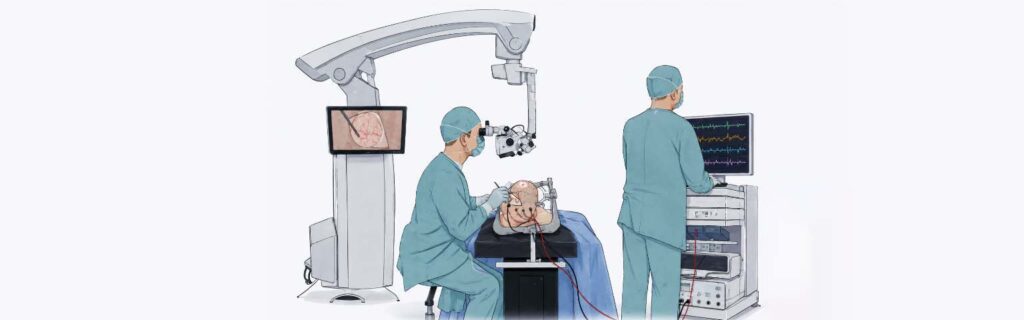
Neurosurgery has changed a lot over the past few decades, and a good chunk of that progress comes down to technology that lets surgeons see more, in real time, than they ever could before. One of the more important tools in that toolkit is Intraoperative Neurophysiological Monitoring — IONM for short — which tracks how the nervous system is functioning while surgery is actually happening.
Whether the surgeon is working on the brain, the spinal cord, or a peripheral nerve, protecting neurological function matters just as much as treating whatever brought the patient into the OR in the first place. IONM gives the surgical team a live read on that function, so nerve damage can often be caught and corrected before it becomes permanent.
What Is Intraoperative Neurophysiological Monitoring?
IONM tracks the nervous system’s electrical activity throughout surgery — signals from the brain, spinal cord, nerves, and muscles, monitored continuously as the operation unfolds.
A CT or MRI shows what things look like. IONM shows how things are working. That distinction matters, because it means the team can pick up on subtle shifts in nerve function before those shifts turn into lasting injury.
Why It Matters
The brain and spinal cord run on millions of nerve fibers, and between them they handle movement, sensation, speech, vision, balance, memory — most of what makes a person a person, functionally speaking.
During surgery, especially in tight or delicate areas, these structures can get stretched, compressed, or briefly cut off from blood flow. Without monitoring, nobody finds out until the patient wakes up.
With IONM running, the surgeon gets an alert the moment something changes, while there’s still time to do something about it.
How It Works
Before the operation starts, electrodes go on the scalp, limbs, or specific muscles, picking up electrical signals from the nervous system.
Once surgery is underway, a neurophysiologist watches those signals continuously, working in tandem with the neurosurgeon and anesthesiologist. If a signal weakens or drops out, that’s the cue that a nerve pathway might be under stress.
At that point, the team can:
- Pause
- Adjust instruments
- Ease off traction on a nerve
- Improve blood flow
- Reposition the patient
Small adjustments, made in time, are often what keep a complication from becoming a permanent deficit.
The Different Types of Monitoring
Not every technique watches the same part of the nervous system, and most complex surgeries use more than one at once.
Somatosensory Evoked Potentials (SSEPs) track the sensory pathways running from the limbs to the brain. A small electrical pulse goes into a peripheral nerve, and the response is picked up at the brain — a way of checking that the spinal cord and sensory pathways are still transmitting properly. SSEPs show up often in spine surgery, brain surgery, spinal cord tumor removal, and vascular neurosurgery.
Motor Evoked Potentials (MEPs) work the other direction — stimulation at the brain, response measured in the arms and legs — to check that the pathways controlling voluntary movement are intact. Any drop-off here is a red flag for motor pathway compromise, which is why MEPs matter so much in surgeries where muscle strength is on the line.
Electromyography (EMG) watches individual muscles for signs that a specific nerve is being irritated or injured. It’s a fixture in peripheral nerve surgery, skull base surgery, facial nerve surgery, and spine procedures near the nerve roots.
Electroencephalography (EEG) records the brain’s own electrical activity and is especially useful in surgery involving the vessels that supply the brain — a shift in the EEG pattern can flag reduced blood flow or oxygen delivery before it does real damage.
Brainstem Auditory Evoked Potentials (BAEPs) keep an eye on hearing pathways and brainstem function, and come up regularly in acoustic neuroma surgery, brainstem procedures, and posterior fossa surgeries.
Where IONM Gets Used
IONM has become close to standard in a wide range of neurosurgical procedures, including brain tumor surgery, spinal tumor surgery, cervical spine surgery, scoliosis correction, skull base surgery, epilepsy surgery, peripheral nerve surgery, brain aneurysm surgery, carotid artery surgery, spinal dysraphism surgery, and complex spinal deformity correction.
The specific type of IONM used depends on the surgical site, the neural structures at risk, and the complexity of the procedure.
What It Actually Buys the Patient
Fewer surprises. Neurological compromise gets caught while it can still be corrected, not after.
Lower risk of a lasting deficit. Catching nerve irritation early means fewer patients waking up with weakness, numbness, or sensory loss they didn’t have going in.
More precision. Surgeons get more room to work carefully around delicate structures without second-guessing every movement.
Better functional recovery. Protecting the nervous system while the rest of the procedure gets done improves the odds of a full recovery.
Real-time decisions. Immediate feedback means the surgeon can change course mid-procedure instead of finding out too late that something needed to change.
Is It Safe?
Yes. IONM is well-established and considered safe. The electrical stimulation involved is tightly controlled, delivered by trained specialists under standard safety protocols, and the patient — under anesthesia the whole time — doesn’t feel any of it. Complications tied directly to the monitoring itself are rare.
Where It Falls Short
IONM isn’t a replacement for surgical judgment. Signal quality can be thrown off by anesthesia, body temperature, blood pressure swings, pre-existing neurological conditions, or nerve damage that was already present before the surgery started. That’s why the readings never stand alone — they’re read alongside the patient’s clinical picture and what the surgeon is seeing directly.
It Takes a Team
None of this works without close coordination. A typical setup involves the neurosurgeon, a neurophysiologist, the anesthesiologist, OR nurses, and technicians, all in communication so that any change in the signals gets interpreted and acted on fast. That teamwork is a big part of why outcomes have improved as much as they have.
Where This Is Headed
The technology keeps getting better — faster signal processing, sharper accuracy, better nerve mapping, tighter integration with surgical navigation systems. As procedures get more complex, neuromonitoring in brain surgery and spine surgery alike will likely only become more central to how these operations are done safely.
In Short
IONM has changed what’s possible in brain and spine surgery by giving surgeons a live window into nervous system function instead of a wait-and-see approach. Problems that used to surface only after the patient woke up can now be caught — and often corrected — while the operation is still underway.
Consult Dr. Rajesh Reddy Sannareddy for Advanced Brain and Spine Care
Complex brain and spine surgery takes more than surgical skill alone — it takes the right technology backing it up. Dr. Rajesh Reddy Sannareddy, Consultant Brain, Spine & Endovascular Neurosurgeon and best neurosurgeon in Hyderabad, uses evidence-based techniques alongside advanced intraoperative monitoring to protect neurological function without compromising precision.
If you or someone you love has been advised to undergo brain or spine surgery, schedule a consultation with Dr. Rajesh Reddy Sannareddy to understand your condition and discuss what modern neurosurgical care can do for your outcome.