
Introduction
Of all the treatment decisions in cerebrovascular surgery, the choice between endovascular coiling and microsurgical clipping for a brain aneurysm is perhaps the most discussed — and the most misunderstood. Patients often arrive at their consultation hoping for a definitive answer: 'just tell me which one is better.' The honest answer is that neither is universally better. Both are effective treatments for brain aneurysms. The right choice depends on the aneurysm's anatomy, the patient's clinical condition, the urgency of treatment, and — critically — the expertise available.
What makes Dr. Rajesh Reddy Sannareddy's practice in Hyderabad genuinely different is that he is trained and experienced in both techniques. His Fellowship in Interventional Neuroradiology (FINR) from the University Hospital, Zurich provides deep endovascular expertise — coiling [standalone, balloon assisted, stent assisted], flow diversion [intrasaccular and extrasaccular]. His MCh and DNB in Neurosurgery (Double board certified) and over 5,000 neurosurgical procedures provide the microsurgical expertise for open aneurysm clipping. When he recommends one approach over the other, it is because that approach is genuinely better for that patient's specific aneurysm — not because it is the only one he can perform.
This page explains both treatments in depth, reviews the landmark clinical evidence, and provides a clear framework for understanding how the right treatment is chosen for each individual case.
What Each Treatment Does: The Core Principle
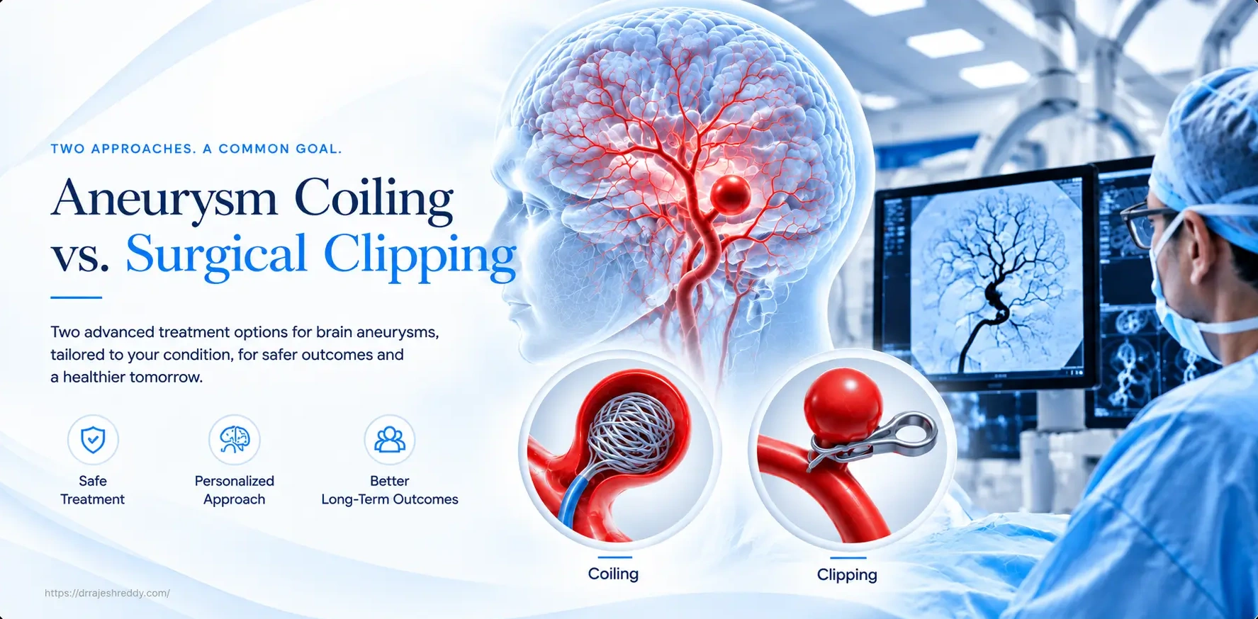
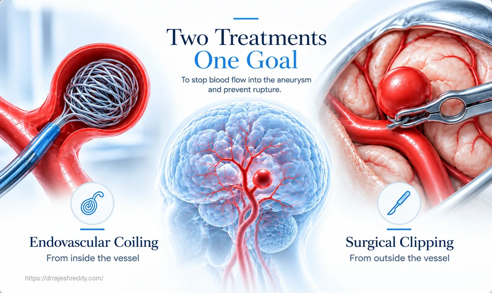
Both coiling and clipping aim to achieve the same goal: permanently exclude the aneurysm from the arterial circulation, eliminating the risk of rupture or rebleeding. They achieve this goal by fundamentally different routes.
Endovascular Coiling — From the Inside
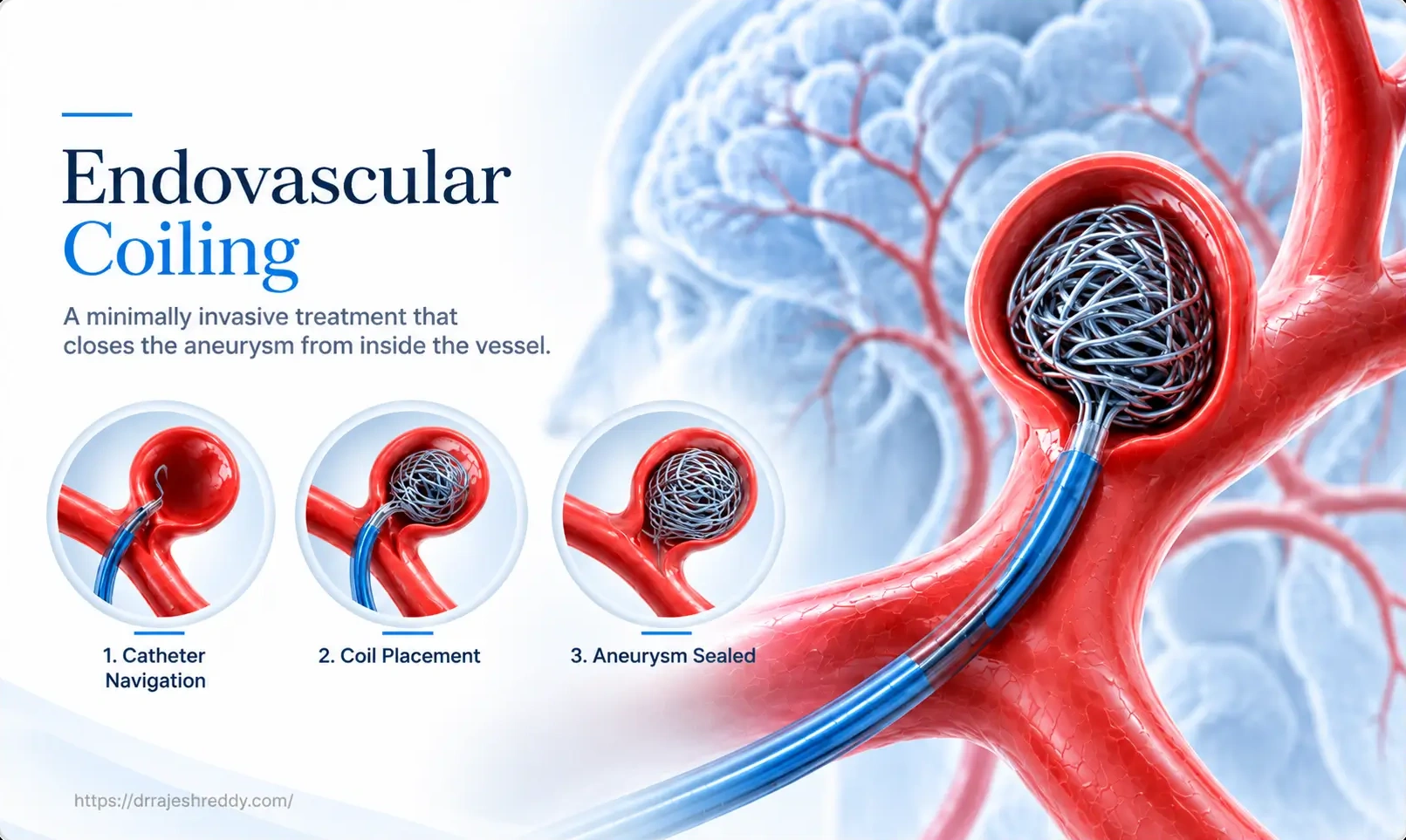
Endovascular coiling accesses the aneurysm from within the bloodstream. A catheter is introduced through a small puncture in the femoral artery in the groin and navigated under X-ray guidance through the aorta, carotid or vertebral arteries, and into the aneurysm sac itself. Soft platinum coils — thinner than a human hair — are delivered one by one into the aneurysm, progressively filling the sac. As the coils pack the aneurysm, they create a mesh that disrupts blood flow into the sac and promotes clot formation. Over hours to days, the aneurysm is progressively thrombosed and sealed from circulation.
No incision in the scalp. No removal of skull bone. No retraction of brain tissue. The only external evidence of the procedure is a small puncture site in the groin.
Microsurgical Clipping — From the Outside
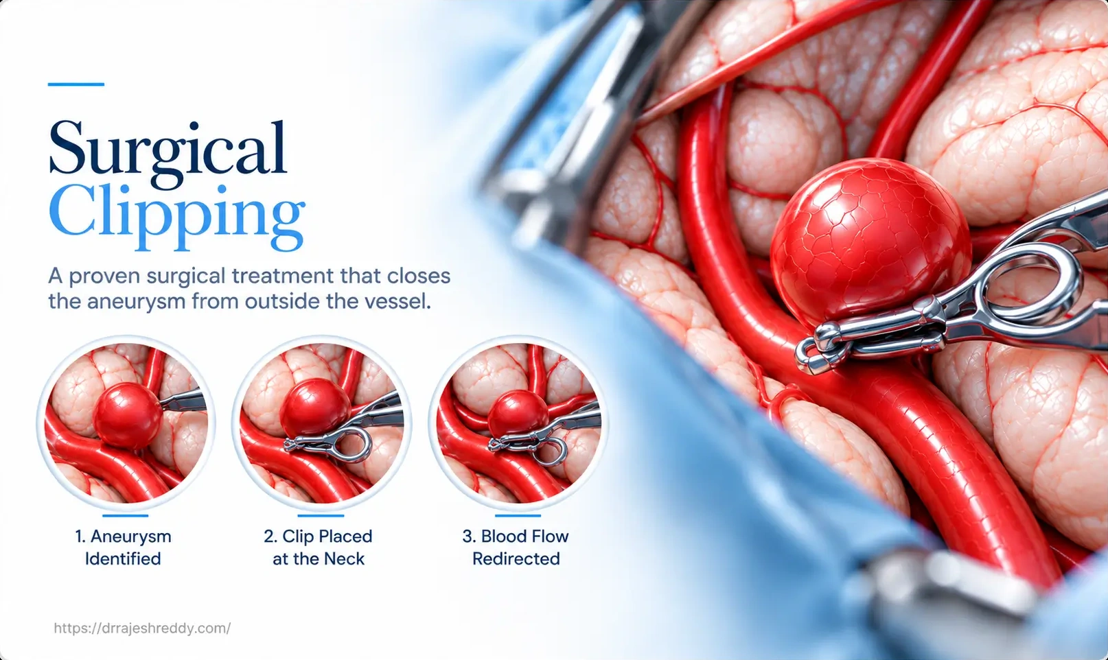
Microsurgical clipping accesses the aneurysm through an open craniotomy. The surgeon makes a scalp incision, removes a section of skull (the bone flap), and opens the dura to access the brain. Using a high-magnification operating microscope, the surgeon navigates along natural brain corridors — typically through the Sylvian fissure for anterior circulation aneurysms, or through the posterior fossa for basilar artery aneurysms — until the aneurysm and its parent artery are directly visualised.
A titanium clip — selected from a range of shapes and sizes — is then applied precisely across the aneurysm's neck, immediately and permanently closing it off from the parent artery. Intraoperative fluorescence angiography (using indocyanine green dye, ICG) is performed to confirm complete aneurysm occlusion and preservation of all surrounding vessels in real time, before the wound is closed.
Coiling vs. Clipping: A 14-Point Comparison
The table below compares coiling and clipping across the clinical and practical dimensions that matter most to patients:
| Feature | Endovascular Coiling | Microsurgical Clipping |
|---|---|---|
| Surgical access | Catheter through femoral artery in groin — no brain surgery | Open craniotomy — skull opening required |
| Anaesthesia duration | 1–3 hours typically | 3–8 hours depending on aneurysm complexity |
| Hospital stay | 1–3 days | 4–7 days |
| Recovery time | 1–2 weeks to normal activity | 4–8 weeks |
| Post-op pain | Mild — groin site discomfort only | Moderate — scalp wound and craniotomy discomfort |
| Immediate aneurysm occlusion | Progressive — coils fill and thrombose over hours | Immediate — clip applied directly, occlusion confirmed with ICG |
| Complete occlusion rate | 85–95% at 6 months (varies by technique) | 95–98% immediately; durable long-term |
| Retreatment rate | 15–20% at 5 years (recanalisation); higher in wide-neck or large aneurysms | < 2% at 10 years for completely clipped aneurysms |
| Best for ruptured aneurysms | Preferred in most ruptured cases (ISAT evidence) | Preferred when coiling unsuitable or haematoma present |
| MCA aneurysm | Technically possible but often difficult due to anatomy | Surgical anatomy often superior; widely favoured |
| Wide-neck aneurysm | Requires stent or balloon assistance; higher recurrence; Intrasaccular flow diverters have been developed, data pertaining to their long term efficacy yet to be published | Clip can be sized to any neck geometry |
| Young patient (< 50) | Good option; but retreatment may be needed over lifetime | Preferred — durable single-procedure cure over a lifetime |
| MRI compatibility post-procedure | Coils are MRI safe; no restrictions | Titanium clips are MRI safe; no restrictions |
| Follow-up imaging required | Mandatory DSA, CTA or MRA at 6 months and 18 months | CTA or MRA at 6 months; then 5-year intervals if stable |
What the Evidence Says: Key Clinical Trials
The debate between coiling and clipping has been shaped by several landmark clinical trials. Understanding the evidence — and its limitations — is essential for an informed treatment decision:
| Trial / Study | Population | Key Finding | Implication |
|---|---|---|---|
| ISAT (2002, 2005, 2015) | 2,143 ruptured aneurysm patients suitable for both treatments | Coiling: 23.5% vs Clipping: 30.9% dependency/death at 1 year. Benefit maintained at 10 years. | Coiling preferred for ruptured aneurysms suitable for both approaches |
| ISUIA (1998, 2003) | Unruptured aneurysms — natural history and treatment outcomes | Surgical clipping: 12.6% morbidity/mortality; endovascular: 9.8% at 1 year (unruptured) | Both safe for unruptured; anatomy determines choice |
| BRAT (2012) | Randomised clipping vs coiling for ruptured aneurysms | Coiling: better 1-yr outcomes; 38% crossover from coiling to clipping due to anatomy | Anatomy drives treatment selection; not all aneurysms suitable for coiling |
| CARAT (2006) | Recurrence after coiling | 20% recanalisation at 5 years in coiled aneurysms vs 1.8% after clipping | Coiled aneurysms require ongoing surveillance; retreatment in minority |
| Pipeline for Uncoilable Aneurysms (PUFS, 2013) | Large/giant uncoilable aneurysms | 86% complete occlusion with Pipeline at 180 days | Flow diversion transforms outcomes for complex aneurysms |
The ISAT trial is the most influential study comparing coiling and clipping, and its finding — better outcomes with coiling for ruptured aneurysms suitable for both treatments — has driven the global shift toward endovascular management of SAH. However, 38% of patients in the BRAT trial crossed over from the coiling arm to clipping due to anatomical unsuitability, underscoring the critical point that the best evidence applies to aneurysms suitable for both — and that anatomy ultimately drives the decision. The technical skill of the medical professional also plays an important role in outcome.
Why Aneurysm Anatomy Is the Most Important Factor
Neither clinical trials nor general preference determines what treatment is right for any individual patient. The aneurysm's anatomy — its size, neck width, shape, location, relationship to parent and branch arteries — is the single most important determinant of whether coiling or clipping is more appropriate.
| Aneurysm Characteristic | Favours Coiling | Favours Clipping | Either / Neutral |
|---|---|---|---|
| Neck width | Narrow neck (dome:neck ratio > 2) | Wide neck (dome:neck ratio < 1.5) | Intermediate neck with stent/balloon |
| Location | Basilar tip, PICA, PComm, AComm, ICA | MCA bifurcation, pericallosal | ACA, distal MCA |
| Size | < 10 mm (small to medium) | > 15 mm (large); any size if anatomy complex | 10–15 mm |
| Shape | Regular, saccular, well-defined neck | Irregular, lobulated, incorporated branches | Smooth but wide-necked |
| Ruptured + haematoma | Not suitable if haematoma needs evacuation | Preferred — clips aneurysm + evacuates haematoma in one procedure | — |
| Patient age | Elderly (> 65) — shorter life expectancy reduces retreatment risk | Young (< 50) — durable single-procedure cure preferred | 50–65 — individualised |
| Previous coiling (recurrent) | Re-coiling or flow diversion | Clip reconstruction of failed coiled aneurysm | Hybrid approaches |
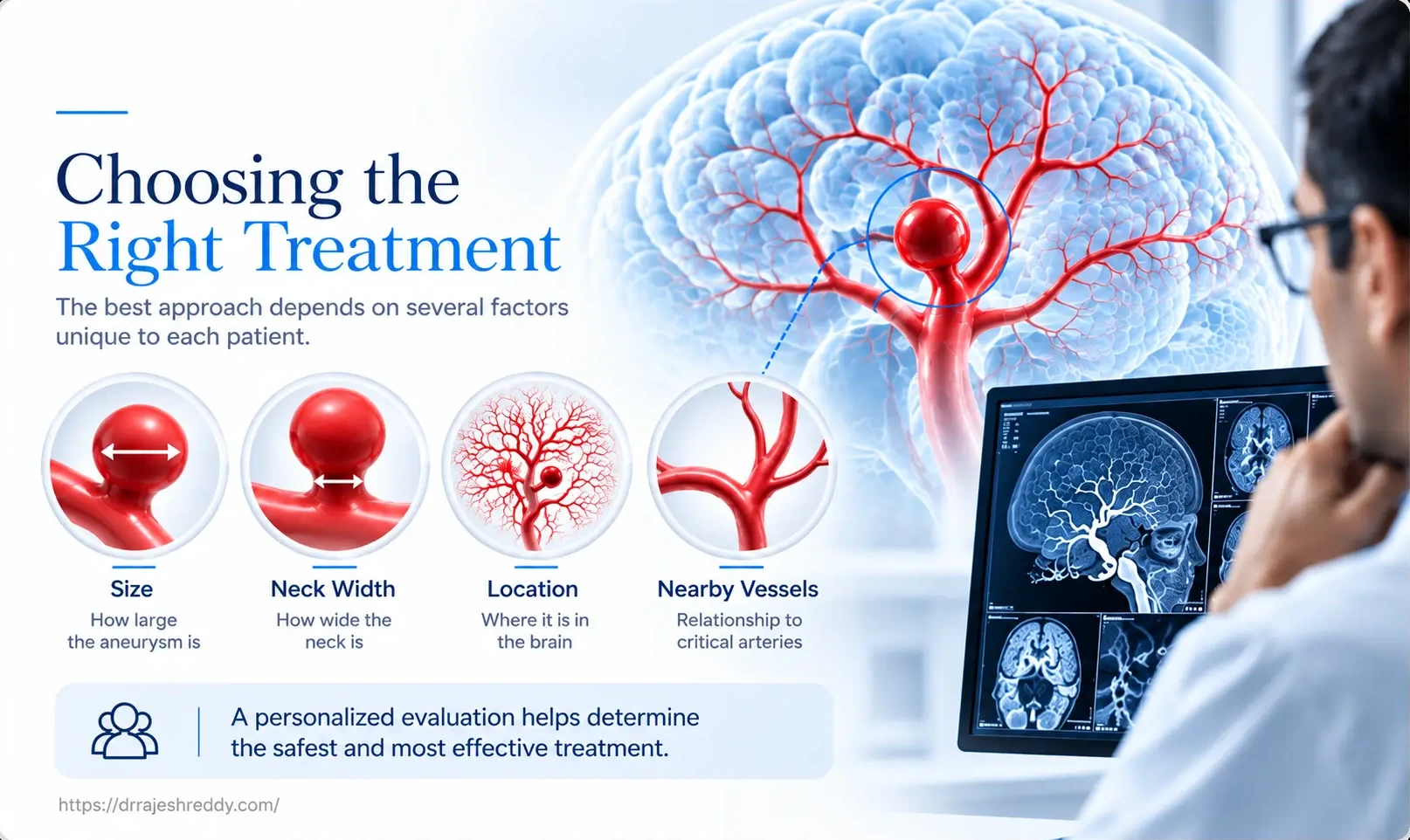
This is why Digital Subtraction Angiography (DSA) — not just CT angiography — is the mandatory investigation before any planned aneurysm treatment. DSA provides 3D rotational images of the aneurysm, measuring its dome diameter, neck width, dome-to-neck ratio, and the relationship of its neck to adjacent branch arteries with precision that CT cannot match.
Endovascular Coiling in Depth
The Basic Technique
Under general anaesthesia, a guide catheter is placed in the internal carotid or vertebral artery through a femoral puncture. A microcatheter — a flexible, steerable tube approximately 1.5 mm in diameter — is then navigated into the aneurysm sac under biplane fluoroscopic guidance. Soft platinum coils are detached one by one into the sac, each chosen for its size and shape to match the aneurysm's dimensions. The first coil (the 'framing coil') forms a basket within the aneurysm; subsequent coils ('filling coils' and 'finishing coils') progressively pack the sac until no further coils can be introduced without herniation into the parent artery.
Advanced Coiling Techniques
- Balloon-assisted coiling: A remodelling balloon is temporarily inflated across the aneurysm neck during coil delivery — preventing coil herniation into the parent artery and allowing tighter packing of wide-neck aneurysms. The balloon is deflated after each coil and removed at the end of the procedure.
- Stent-assisted coiling: A flexible stent [laser cut or braided] is deployed across the aneurysm neck before coiling, providing a scaffold that keeps coils within the sac. Particularly useful for wide-neck aneurysms. Requires dual antiplatelet therapy (aspirin + clopidogrel) for 3–6 months to prevent stent thrombosis.
- Intrasaccular device (WEB, Contour, Artisse, Seal): A nitinol mesh sphere deployed entirely within the aneurysm sac — disrupting inflow without requiring a stent or dual antiplatelet therapy. Particularly suited to wide-neck bifurcation aneurysms at the MCA, basilar tip, and AComm. A single-session outpatient-equivalent procedure.
Recanalisation — The Main Limitation of Coiling
The most significant limitation of endovascular coiling is recanalisation — where the coil mass compacts over time as the aneurysm remodels, allowing blood to re-enter the sac. This occurs in approximately 15–20% of coiled aneurysms at 5 years and is more common in larger aneurysms, wide-neck aneurysms, and those with incomplete initial occlusion. Recanalisation does not always mean rebleeding — most recanalised aneurysms can be retreated with further coiling or flow diversion — but it does mean that mandatory follow-up angiography (DSA, CTA or MRA at 6 months and 18 months) is non-negotiable after coiling.
Microsurgical Clipping in Depth
The Pterional Craniotomy — The Workhorse Approach
The vast majority of anterior circulation aneurysms — at the MCA, ICA, AComm, and PComm — are approached through a pterional craniotomy: a frontotemporal craniotomy that opens the Sylvian fissure, exposing the Circle of Willis at the skull base. This is the most commonly performed craniotomy in cerebrovascular surgery and provides excellent exposure to the majority of saccular aneurysms encountered in clinical practice.
The surgeon dissects the Sylvian fissure under microscope — separating the frontal and temporal lobes along a natural anatomical plane without retracting brain tissue — until the aneurysm, its neck, and the parent and branch arteries are fully visualised. Only then is the clip applied.
Clip Selection and Application
Titanium clips are available in dozens of shapes and sizes — straight, curved, fenestrated, bayonet, right-angle, miniature — each designed for specific anatomical situations. The surgeon trials one or more clips before final application, checking under the microscope that the clip spans the entire neck without incorporating any adjacent arteries or perforating vessels.
Intraoperative ICG fluorescence angiography is then performed: indocyanine green dye injected intravenously fluoresces under infrared light in the microscope, showing real-time blood flow through all vessels. A correctly placed clip shows no flow in the aneurysm sac (confirming occlusion) and unobstructed flow through all parent and branch arteries (confirming preservation). This step dramatically reduces the risk of inadvertent vessel compromise or incomplete clipping before wound closure.
Temporary Clipping — Managing Intraoperative Rupture
In complex aneurysms, the surgeon may apply a temporary clip to the parent artery before clipping the aneurysm — momentarily stopping flow to the aneurysm to reduce wall tension and make the final clip application safer. Temporary clips are removed within minutes of aneurysm clipping; the brain tolerates short periods (typically < 10 minutes) of temporary occlusion with minimal risk when performed under neuroprotective anaesthesia conditions.
Why Clipping Is More Durable
The titanium clip, once placed correctly, creates an immediate, mechanical, permanent seal of the aneurysm neck that does not depend on the body's clotting mechanism. Unlike coil masses — which can compact and recanalize over years — a well-placed clip does not change over time. The 10-year retreatment rate after surgical clipping for completely clipped aneurysms is below 2%, compared to 15–20% after coiling. For young patients who will live for decades after treatment, this durability is a significant advantage.
When Neither Coiling Nor Clipping Is the Answer: Flow Diversion
For a subset of aneurysms — large and giant aneurysms, fusiform aneurysms, wide-neck aneurysms where coiling has failed, and paraclinoid ICA aneurysms — neither conventional coiling nor surgical clipping provides the safest or most effective treatment. Flow diversion devices — principally the Pipeline Embolisation Device (PED), SILK, Surpass, Derivo, P64 and the FRED (Flow Re-Direction Endoluminal Device) — have transformed outcomes for this challenging group.
A flow diverter is a dense mesh stent deployed across the aneurysm neck within the parent artery. It redirects blood flow away from the aneurysm sac, causing progressive thrombosis over weeks to months, while simultaneously scaffolding the parent artery and forming a new smooth arterial wall that permanently excludes the aneurysm. Complete occlusion rates of 85–93% at 12 months make flow diversion the most effective treatment for complex aneurysms that cannot be reliably treated by coiling or clipping alone.
Flow diversion is not a substitute for coiling or clipping in straightforward aneurysms — it is a specialised tool for cases that would previously have been untreatable or required complex bypass surgery. Dr Rajesh Reddy’s exposure to complex cerebrovascular procedures at University Hospital, Zurich, Barrow Neurological Institute, Phoenix and University Hospital, Okayama in addition to training at high volume centres in Hyderabad provides the necessary knowledge, skill, judgement and precision in helping choose between microsurgical clipping, endovascular coiling, or, clinicoradiological follow, in selected cases.
How the Right Treatment Is Chosen: Dr. Rajesh Reddy's Decision Framework
When Dr. Rajesh Reddy reviews an aneurysm for treatment planning, the decision follows a systematic framework that integrates all available clinical and anatomical information:
Step 1: Characterise the Aneurysm on DSA
DSA with 3D rotational angiography provides the definitive anatomical map: aneurysm size, neck width, dome-to-neck ratio, relationship to parent and branch arteries, and presence of daughter sacs or irregularities. This is the non-negotiable first step.
Step 2: Determine Endovascular Suitability
Is the aneurysm suitable for coiling alone? If the dome-to-neck ratio is ≥ 2 and the neck is ≤ 4 mm, simple coiling is typically feasible. If the neck is wider, balloon or stent assistance is assessed. If the anatomy is unfavourable for coiling — particularly MCA bifurcation aneurysms where branch vessels arise from the aneurysm neck — clipping is considered. However, several intrasaccular devices have shown promising results in wide necked bifurcation aneurysms, expanding the horizon of endovascular treatment for MCA bifurcation aneurysms as well.
Step 3: Consider the Clinical Context
Is this a ruptured aneurysm? If so, what is the patient's clinical grade? Has there been an associated haematoma? Is the patient physiologically able to tolerate a prolonged general anaesthetic for open surgery? A poor-grade SAH patient may be better served by the physiologically less demanding coiling procedure. A patient with a large haematoma needs surgical decompression — which can be combined with clipping in a single procedure.
Step 4: Consider the Patient's Long-Term Interests
Age and life expectancy matter. A 70-year-old with significant comorbidities and a 6 mm MCA aneurysm will have a different calculus from a 35-year-old with the same aneurysm. The younger patient's lifetime retreatment risk after coiling may justify accepting the higher initial procedural burden of clipping for a more durable outcome.
Step 5: Discuss Openly with the Patient
Whenever the anatomy supports both approaches safely, the patient's informed preference is part of the decision. Some patients strongly prefer to avoid open surgery; others prefer the perceived permanence of clipping. Dr. Rajesh Reddy presents both options clearly — including retreatment rates, recovery, and follow-up requirements — and incorporates patient preference in the final plan.
Why Having Both Skills in One Surgeon Matters
In many centres, patients with a brain aneurysm are seen by either an endovascular interventionist or a neurosurgeon — not both. This means the treatment recommendation may be influenced by the specialist's training rather than the aneurysm's anatomy. A centre that primarily performs coiling will tend to recommend coiling; one that primarily performs clipping will tend toward surgery.
Dr. Rajesh Reddy's practice is structured differently. His double board certification in neurosurgery (MCh + DNB) and his Fellowship in Interventional Neuroradiology (FINR) from the University Hospital, Zurich — one of Europe's highest-volume cerebrovascular centres — mean that he can genuinely offer and perform both treatments. Every treatment recommendation is made based on the aneurysm's anatomy and the patient's clinical context — not on institutional preference or technical limitation.
Dr. Rajesh Reddy's Cerebrovascular Credentials
- MCh Neurosurgery (Exam Topper), NIMS Hyderabad — Double board certified (MCh + DNB Neurosurgery)
- FINR — Fellowship in Interventional Neuroradiology, University Hospital, Zurich
- Visiting Scholar — Barrow Neurological Institute, Phoenix, Arizona (one of the world's highest-volume cerebrovascular centres)
- Visiting Scholar — Okayama University, Japan; Klinikum Stuttgart, Germany
- Trained in: GDC coiling, balloon-assisted coiling, stent-assisted coiling, WEB device, Pipeline flow diversion, FRED, microsurgical clipping (pterional, orbitozygomatic, far-lateral approaches), ICG fluorescence angiography, temporary clipping
- Over 5,000 neurosurgical and endovascular procedures across 15+ years
- Clinical acumen and judgement gained through years of training with leading experts globally
Frequently Asked Questions
Get an Expert Second Opinion — Consult Dr. Rajesh Reddy
If you have been diagnosed with a brain aneurysm and want an expert opinion on whether coiling, clipping, or flow diversion is the right treatment for your specific aneurysm — from a surgeon trained and experienced in all three — Dr. Rajesh Reddy Sannareddy offers comprehensive cerebrovascular consultations in Hyderabad. Book in person or via the Dr. Reddy's Neuro Care app on Google Play.
Related Articles
Continue reading to explore specific aneurysm treatments in detail:
Brain Aneurysm Symptoms: When Is It an Emergency?
Thunderclap headache, third nerve palsy, warning signs, and when to seek immediate emergency care.
Read GuideRuptured vs. Unruptured Brain Aneurysm
Key differences in symptoms, emergency management, rupture risks, and treatment timelines.
Read GuideEndovascular Coiling in Hyderabad
Advanced catheter technique to treat brain aneurysms without open surgery.
Read GuideFlow Diversion Treatment
Flow diverter stents (WEB, Pipeline) for complex, wide-necked, and giant brain aneurysms.
Read GuideSubarachnoid Hemorrhage (SAH)
Causes, acute emergency management, vasospasm prevention, and critical care.
Read GuideLife After Brain Aneurysm Surgery
Recovery guide, timeline, returning to work, driving, and long-term angiography follow-up.
Read GuideArteriovenous Malformation (AVM) Treatment
Endovascular embolization, radiosurgery, and microsurgical excision for AVMs in Hyderabad.
Read GuideBrain Aneurysm Treatment (Pillar Page)
Visit our main pillar page for brain aneurysm treatment in Hyderabad by Dr. Rajesh Reddy.
View Pillar Page