
Introduction
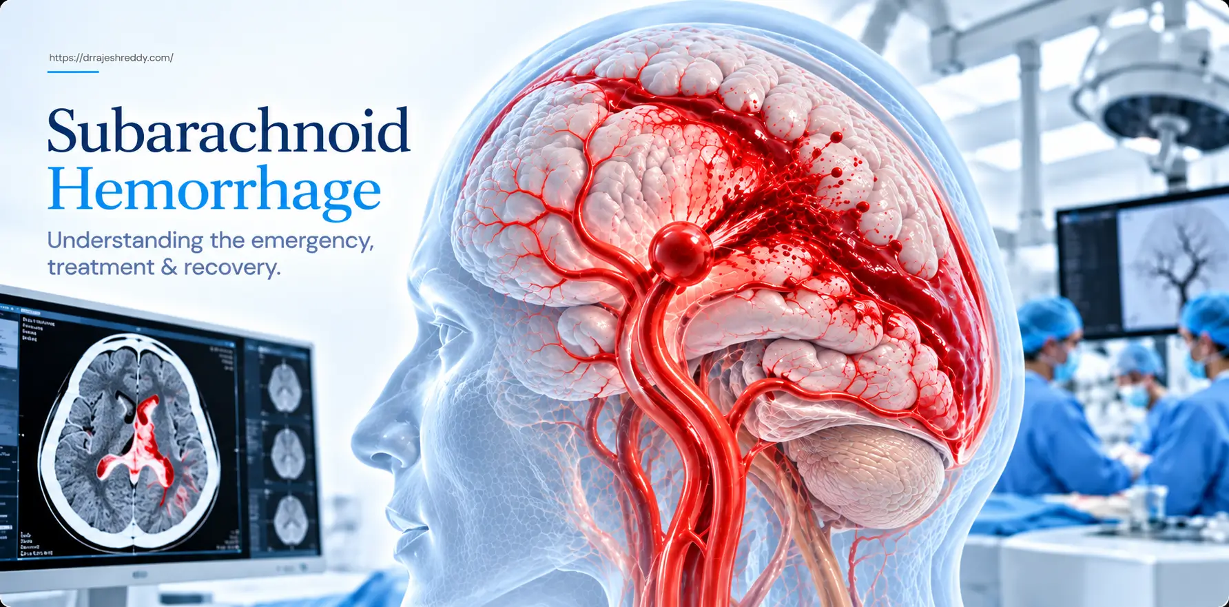
Subarachnoid hemorrhage — SAH — is among the most devastating neurological emergencies in medicine. In the seconds after an aneurysm ruptures, blood surges under arterial pressure into the subarachnoid space around the brain, causing an instantaneous, catastrophic headache and triggering a cascade of complications that can claim a patient's life or neurological function over the days and weeks that follow — even after the aneurysm itself has been treated.
For patients and families who encounter SAH — either as a sudden emergency or as a diagnosis on a scan — understanding what has happened, why it is so dangerous, and what treatment involves is essential for navigating the recovery ahead. This page provides a comprehensive, plain-language guide to SAH: what it is, why aneurysms rupture, how severity is assessed, what the emergency treatment involves, what complications to expect, and what recovery looks like.
Dr. Rajesh Reddy Sannareddy, Senior Consultant Endovascular Neurosurgeon in Hyderabad, manages the complete spectrum of SAH care — from emergency aneurysm clipping / coiling in the acute phase through vasospasm intervention, hydrocephalus management, and long-term neurovascular follow-up. His Fellowship in Interventional Neuroradiology (FINR) from the University Hospital, Zurich and visiting scholar experience at Barrow Neurological Institute, Phoenix give him direct exposure to the highest-volume SAH management programmes in Europe and North America.
What Is Subarachnoid Hemorrhage?
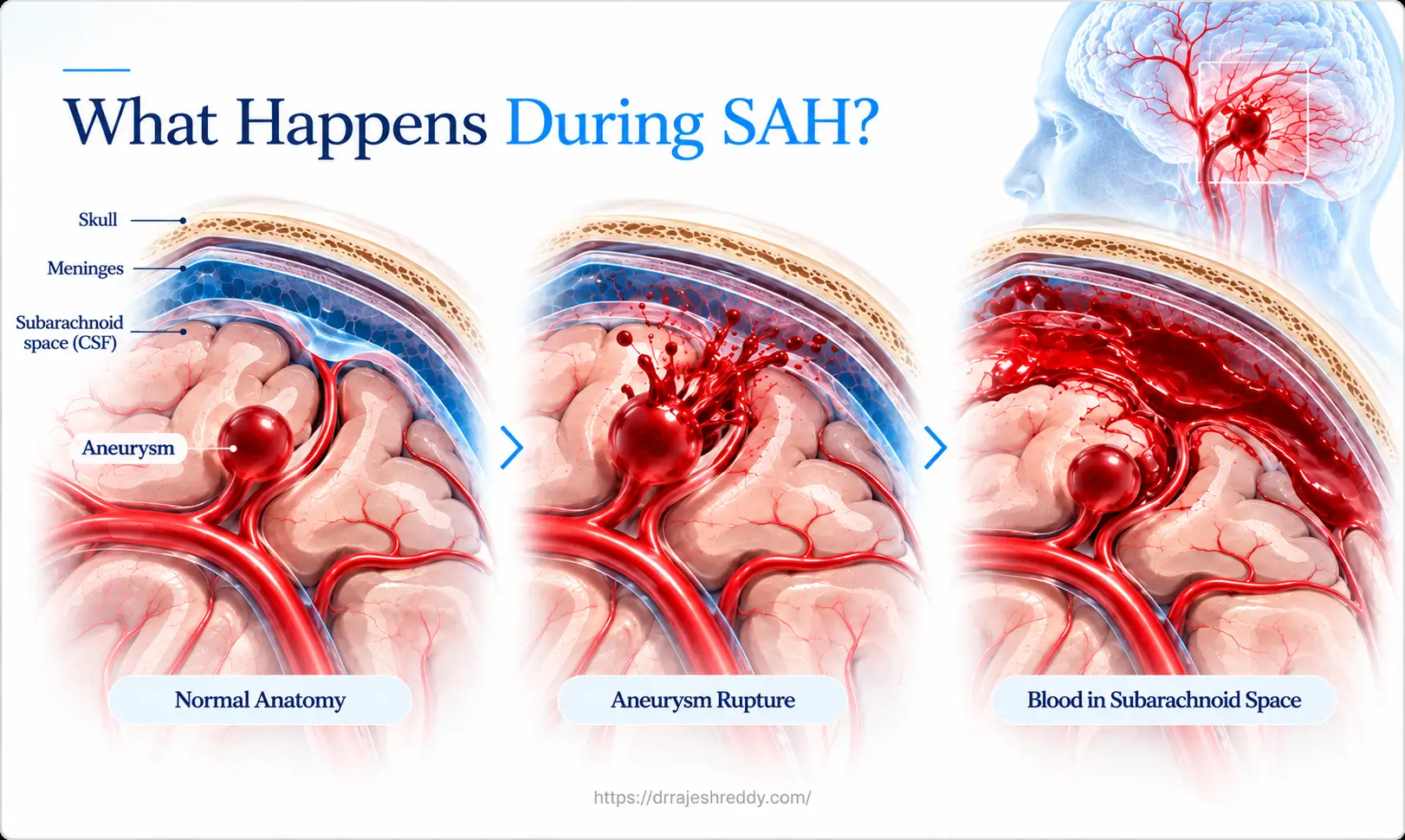
The brain is surrounded by three protective membranes — the dura, arachnoid, and pia mater — collectively called the meninges. Between the arachnoid and pia mater lies the subarachnoid space, a fluid-filled compartment through which cerebrospinal fluid (CSF) circulates and in which the major cerebral arteries run. Subarachnoid hemorrhage occurs when blood enters this space — most commonly when a brain aneurysm ruptures and releases arterial blood under high pressure into the subarachnoid space.
The consequences are immediate and severe. The sudden entry of blood causes an instantaneous, massive rise in intracranial pressure — producing the hallmark thunderclap headache — and triggers an intense inflammatory and vasoconstrictive response throughout the cerebral vasculature. Blood disperses through the CSF pathways, coating the brain surface and filling the basal cisterns, setting in motion the cascade of complications — re-bleeding, vasospasm, hydrocephalus, and brain injury — that define the clinical course of SAH.
How Common Is SAH?
SAH from aneurysm rupture affects approximately 10–12 per 100,000 people per year globally — approximately 6,000–8,000 new cases in India annually. It predominantly affects adults between 40 and 60 years of age, with women affected slightly more often than men. Despite advances in treatment, the overall mortality from SAH remains approximately 30–40% at 30 days, with approximately half of survivors experiencing long-term neurological or cognitive disability.
Causes of Subarachnoid Hemorrhage
The vast majority of non-traumatic SAH — approximately 85% — is caused by rupture of an intracranial aneurysm. The remaining causes include:
- Non-aneurysmal perimesencephalic SAH (~10%) — a benign variant where blood is confined to the region around the midbrain (perimesencephalic cisterns); no aneurysm is found on imaging; excellent prognosis with low risk of rebleeding
- Arteriovenous malformation (AVM) rupture (~3–5%) — tangles of abnormal blood vessels that bleed more commonly in younger patients
- Cerebral venous thrombosis — blockage of the brain's draining veins causing haemorrhagic infarction that can extend into the subarachnoid space
- Reversible cerebral vasoconstriction syndrome (RCVS) — thunderclap headache with arterial spasm; may cause SAH without an underlying aneurysm
- Trauma — head injury causing subarachnoid bleeding, particularly in the cortical sulci
- Rare causes — spinal AVM, intracranial arterial dissection, cocaine or stimulant use causing vessel rupture
When SAH is confirmed on CT, the immediate priority is identifying the source — typically through CT angiography (CTA) followed by digital subtraction angiography (DSA). If no aneurysm is found on initial imaging, a repeat DSA at 7–14 days is essential, as small aneurysms can be missed on first-pass imaging due to vasospasm or technical factors.
Symptoms of Subarachnoid Hemorrhage

The Thunderclap Headache — The Cardinal Symptom
The hallmark of aneurysmal SAH is the thunderclap headache — a sudden, explosive headache of maximal severity that reaches its peak within 1–2 seconds of onset. Patients consistently describe it in vivid terms: 'a gun going off in my head,' 'the worst pain of my life,' 'like an explosion behind my eyes.' This instantaneous maximal onset — not the severity alone — is the defining feature. A headache that builds over minutes to hours is unlikely to be SAH; one that is maximal at the very first moment is an emergency until proven otherwise.
Associated Symptoms
- Neck stiffness (meningism): Develops within 3–6 hours of bleeding as subarachnoid blood irritates the meninges. Patients resist neck flexion, and the Kernig and Brudzinski signs may be positive on examination.
- Nausea and vomiting: Often projectile, occurring within seconds to minutes of the headache — from the sudden rise in intracranial pressure.
- Photophobia and phonophobia: Extreme sensitivity to light and sound from meningeal irritation — similar to severe migraine, but in the context of a thunderclap headache.
- Loss of consciousness: Occurs at the time of rupture in approximately 45–50% of patients — caused by the sudden spike in ICP transiently stopping cerebral perfusion. Duration ranges from seconds (brief syncope) to prolonged coma in severe bleeds.
- Seizures: Occur in 10–20% of SAH patients at or shortly after rupture.
- Focal neurological deficit: Depending on the aneurysm location and extent of haemorrhage — weakness, speech difficulty, or cranial nerve palsies.
- Sentinel headache (warning bleed): In approximately 20–50% of cases, a milder headache — representing a minor leak — precedes the major SAH by days to weeks. This sentinel headache is the most important missed diagnosis in emergency medicine.
Diagnosing SAH: The Emergency Workup
CT Brain (Non-Contrast) — First Line
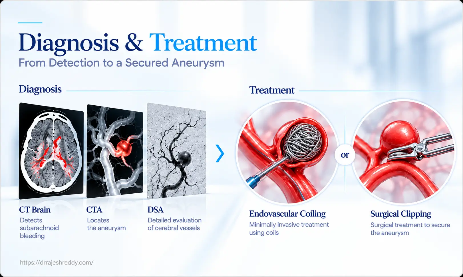
Non-contrast CT brain is the first-line investigation for suspected SAH. It is approximately 98% sensitive within the first 6 hours of symptom onset — detecting subarachnoid blood as hyperdense (white) material in the basal cisterns, sylvian fissures, and cerebral sulci. Sensitivity decreases progressively after 24 hours as blood is diluted and reabsorbed by the CSF — emphasising why immediate investigation is critical.
Lumbar Puncture — When CT Is Negative
If CT brain is normal but clinical suspicion of SAH remains high — particularly in patients presenting more than 6 hours after headache onset — a lumbar puncture (LP) is performed. CSF is examined for xanthochromia (yellow discolouration from haemoglobin breakdown products) and crenated Red Blood Cells, which persists for up to 2 weeks after SAH even when CT has normalised. A positive LP in the context of a thunderclap headache confirms SAH and mandates further vascular imaging.
CT Angiography (CTA) — Identifying the Aneurysm
Once SAH is confirmed, urgent CTA of the cerebral vessels is performed to identify the causative aneurysm, its location, size, and morphology. Modern multi-slice CTA has sensitivity approaching 97–99% for aneurysms ≥ 3mm and provides a rapid three-dimensional roadmap for treatment planning. In most centres, CTA has replaced diagnostic-only DSA as the first-line vascular investigation in acute SAH.
Digital Subtraction Angiography (DSA) — The Gold Standard and Treatment Platform
DSA remains the gold standard for definitive aneurysm characterisation and is the platform on which endovascular treatment is performed. In Dr. Rajesh Reddy's practice, DSA is performed as a combined diagnostic and therapeutic procedure: the aneurysm is fully characterised with 3D rotational angiography, and — when anatomy is suitable — treated with coiling or other endovascular devices in the same sitting, minimising the window of re-bleeding risk.
Grading SAH Severity: WFNS, Fisher & Hunt and Hess
Grading scales are used to assess SAH severity — each measuring different aspects of the clinical situation:
WFNS Clinical Grade — Assessing the Patient
The World Federation of Neurological Surgeons (WFNS) scale grades the patient's clinical condition based on level of consciousness (GCS) and presence of motor deficit. It guides prognosis and influences treatment decisions:
| WFNS Grade | GCS Score | Motor Deficit | Clinical State | Prognosis |
|---|---|---|---|---|
| Grade I | 15 | Absent | Alert, no deficit | Excellent — > 90% good outcome with prompt treatment |
| Grade II | 13–14 | Absent | Headache, neck stiffness, no deficit | Good — most recover well |
| Grade III | 13–14 | Present | Confusion or mild focal deficit | Moderate — variable recovery |
| Grade IV | 7–12 | Present or absent | Stupor, moderate-severe hemiparesis | Poor — high morbidity and mortality |
| Grade V | 3–6 | Present or absent | Deep coma, decerebrate posturing | Very poor — selective treatment |
Fisher Grade — Assessing the CT Scan
The Fisher grade assesses the amount and distribution of blood on the CT brain — which directly correlates with the risk of cerebral vasospasm:
| Fisher Grade | CT Finding | Vasospasm Risk |
|---|---|---|
| Grade 1 | No blood detected on CT | Low |
| Grade 2 | Diffuse thin SAH, no clots (< 1mm thick) | Moderate |
| Grade 3 | Localised clot or thick SAH (> 1mm) | High — highest risk of severe vasospasm |
| Grade 4 | Intracerebral or intraventricular haemorrhage (with or without SAH) | Moderate-high |
Fisher Grade 3 carries the highest risk of delayed cerebral ischaemia from vasospasm — the major source of secondary neurological injury after SAH. Knowing the Fisher grade on admission allows the treating team to anticipate and prepare for the vasospasm risk in each patient.
Hunt and Hess Clinical Grading — Assessing SAH Severity
The Hunt and Hess (H&H) scale is a clinical grading system used to assess the severity of subarachnoid haemorrhage (SAH) based on the patient's neurological condition and symptoms. It helps clinicians estimate prognosis and guide treatment decisions.
| Hunt and Hess Grade | Clinical Findings | Severity & Prognosis |
|---|---|---|
| Grade I | Asymptomatic or mild headache and slight neck stiffness | Mild SAH — generally favourable prognosis |
| Grade II | Moderate to severe headache, neck stiffness, no neurological deficit except possible cranial nerve palsy | Moderate severity — generally favourable outcome |
| Grade III | Drowsiness, confusion, or mild focal neurological deficit | Moderate-to-severe SAH — variable prognosis |
| Grade IV | Stupor, moderate-to-severe hemiparesis, possible early decerebrate rigidity, and autonomic disturbances | Severe SAH — increased risk of complications |
| Grade V | Deep coma, decerebrate rigidity, and moribund appearance | Very severe SAH — high risk of mortality |
How Hunt and Hess Differs from WFNS and Fisher
● WFNS Scale: Assesses clinical severity using the Glasgow Coma Scale (GCS) and motor deficits.
● Hunt and Hess Scale: Assesses clinical severity based on headache, neurological status, level of consciousness, and motor deficits.
● Fisher Scale: Assesses the amount and distribution of blood on CT brain and estimates the risk of cerebral vasospasm.
Emergency Treatment: Securing the Aneurysm
The single most important intervention after SAH is securing the ruptured aneurysm — eliminating the risk of re-bleeding, which carries a mortality of 70–80%. International guidelines recommend aneurysm treatment within 24–72 hours of admission for good-grade SAH (WFNS I–III). The two treatment options are:
Endovascular Coiling — Preferred in Most SAH Cases
The landmark ISAT trial (International Subarachnoid Aneurysm Trial) demonstrated significantly better neurological outcomes for coiling versus surgical clipping in SAH patients suitable for both treatments — 23.5% versus 30.9% dependency or death at one year. Coiling avoids craniotomy and prolonged general anaesthesia in an already critically ill patient, reducing physiological stress at a vulnerable time. For most aneurysm anatomies, coiling is the preferred first-line treatment for ruptured aneurysms.
Dr. Rajesh Reddy performs emergency coiling through his FINR-trained endovascular expertise — including simple coiling, balloon-assisted coiling for wide-neck aneurysms, and stent-assisted coiling in select cases — as a combined diagnostic DSA and treatment procedure to minimise time to aneurysm security.
Microsurgical Clipping — When Surgery Is Preferred
Surgical clipping is preferred when: the aneurysm anatomy is not suitable for coiling (wide neck, complex morphology, incorporated branch vessels); the patient has a large intracerebral haematoma requiring surgical evacuation — where clipping and haematoma drainage can be performed in a single operation; or the aneurysm is at the MCA bifurcation where surgical anatomy is often superior to catheter access. Dr. Rajesh Reddy's combined endovascular and microsurgical training means this decision is made on clinical grounds.
The choice between coiling and clipping for SAH is made urgently — typically within hours of the initial imaging — by Dr. Rajesh Reddy reviewing the CTA and DSA anatomy, the patient's clinical grade, and the most likely approach to achieve safe, complete aneurysm security as rapidly as possible.
Managing the Aftermath: SAH Complications
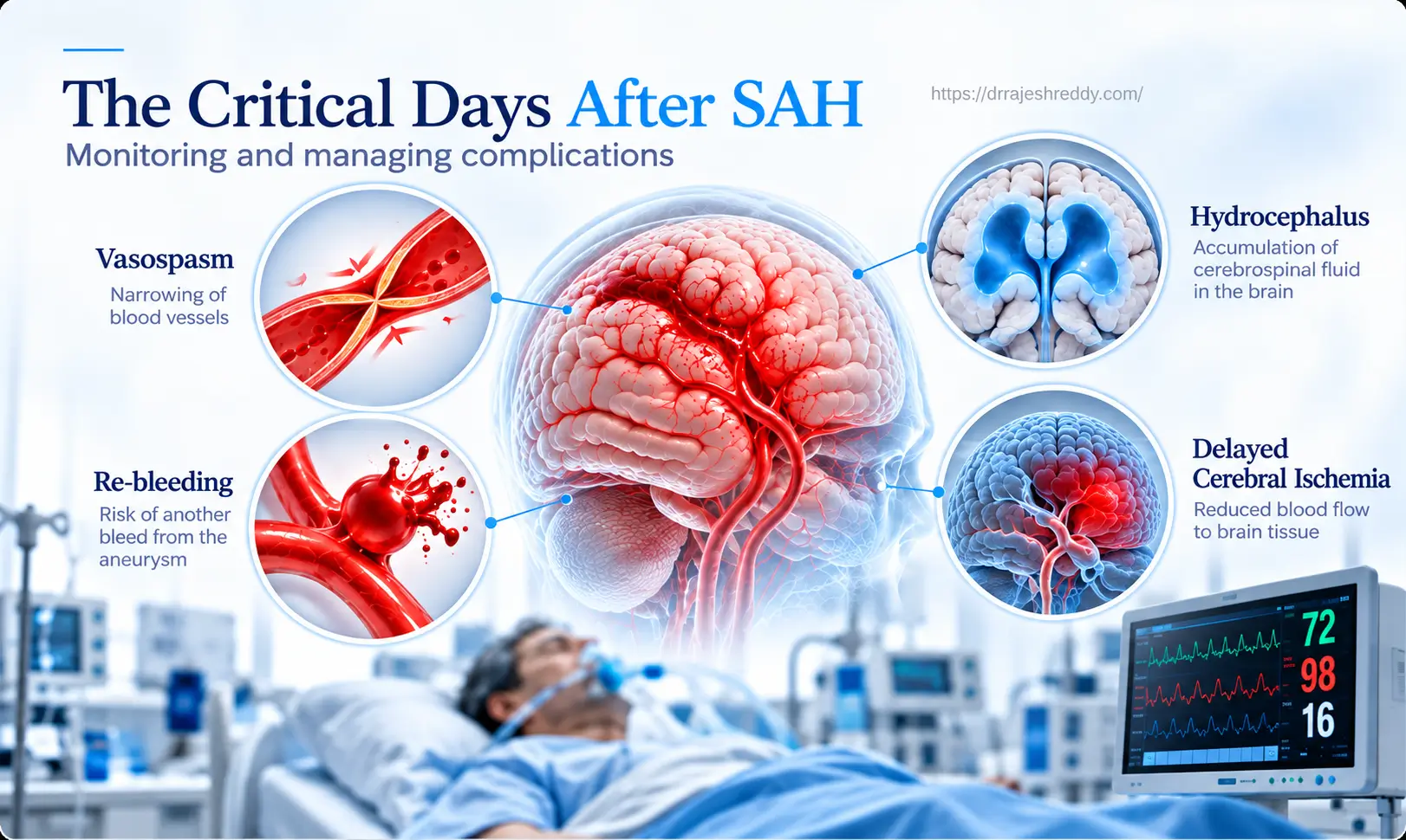
Treating the aneurysm is only the beginning. The weeks after SAH are dominated by a sequence of potentially devastating complications that require intensive, expert management:
| Complication | Timing | Frequency | Management |
|---|---|---|---|
| Re-bleeding | Highest first 24 hrs (4%); 1–2%/day until aneurysm secured | 20–30% if untreated within 2 weeks | Secure aneurysm within 24–72 hrs; strict BP control; bed rest |
| Cerebral Vasospasm | Days 4–14 (peak day 7–10) | 30–70% angiographic; 20–30% symptomatic | Nimodipine; euvolaemia; TCD monitoring; endovascular rescue |
| Acute Hydrocephalus | Hours to days | 15–20% | External ventricular drain (EVD); later VP shunt if persistent |
| Delayed Cerebral Ischaemia (DCI) | Days 4–14 | 20–30% | Induced hypertension; endovascular angioplasty / vasodilators if refractory |
| Hyponatraemia | Days 3–14 | 30–50% | Careful fluid management; fludrocortisone; hypertonic saline if severe |
| Neurogenic Cardiac Dysfunction | First 24–48 hrs | 25–30% ECG changes | Cardiology review; echo; avoid aggressive fluid restriction |
| Seizures | At rupture or delayed | 10–20% | Anti-epileptic prophylaxis; EEG monitoring in comatose patients |
| Chronic Hydrocephalus | Weeks to months | 15–20% | VP shunt placement |
Vasospasm — The Delayed Danger
Cerebral vasospasm — progressive narrowing of the cerebral arteries in response to blood breakdown products in the subarachnoid space — is the leading cause of death and disability in SAH patients who survive the initial rupture. It typically develops between days 4–14 after bleeding, peaking around day 7–10, and can cause delayed cerebral ischaemia (DCI) — effectively a stroke — even in patients whose aneurysm has been successfully treated.
Medical management includes oral nimodipine (started on admission and continued for 21 days), maintenance of adequate blood pressure and fluid balance (euvolaemia), and transcranial Doppler (TCD) monitoring — which tracks blood flow velocities in the cerebral arteries as a surrogate for vasospasm severity.
When vasospasm becomes symptomatic — the patient develops new neurological deficits despite optimal medical management — endovascular rescue therapy is performed: intra-arterial infusion of vasodilators (Nimodipine, Verapamil, Milrinone) directly into the spastic vessel through a microcatheter, or balloon angioplasty of severely narrowed arterial segments. Dr. Rajesh Reddy's endovascular expertise is directly applicable to this often-overlooked but critically important aspect of SAH management.
Hydrocephalus
Blood in the subarachnoid space obstructs the arachnoid granulations that reabsorb CSF, causing communicating hydrocephalus. Acute hydrocephalus — developing within the first 24–72 hours — is managed with an external ventricular drain (EVD), a bedside procedure placing a small catheter into the lateral ventricle to relieve pressure. Approximately 15–20% of SAH patients develop chronic hydrocephalus requiring a permanent ventriculoperitoneal (VP) shunt, typically inserted 3–6 weeks after the bleed once the CSF has cleared of blood products.
Cardiac and Systemic Complications
SAH triggers a massive catecholamine surge that affects multiple organ systems. ECG changes — T-wave inversions, QT prolongation, ST changes — are seen in up to 90% of SAH patients and do not indicate primary cardiac disease. Neurogenic stunned myocardium — transient left ventricular dysfunction — occurs in 10–15% and may require inotropic support. Neurogenic pulmonary oedema can develop suddenly. Hyponatraemia from cerebral salt wasting or SIADH is among the most common and clinically significant systemic complications, requiring careful electrolyte management to avoid exacerbating cerebral oedema.
ICU Care After SAH: What to Expect
Good-grade SAH patients (WFNS I–III) who have had their aneurysm secured sometimes spend 14–21 days in the neurosurgical ICU or High Dependency Unit — not because of the aneurysm treatment itself, but because of the vasospasm monitoring and management required during the critical period (days 4–14). During this time:
- Continuous neurological monitoring — hourly neurological checks; any new deficit triggers immediate CT and angiography assessment
- Transcranial Doppler daily — monitoring middle cerebral artery flow velocities for early vasospasm detection
- Nimodipine 60 mg every 4 hours — continued for the full 21-day course; the only proven medical prophylaxis for vasospasm-related DCI
- Strict euvolaemia — maintaining adequate circulating volume to optimise cerebral perfusion through narrowed vasospastic vessels
- Electrolyte management — daily sodium, potassium, and magnesium monitoring; hyponatraemia corrected promptly
- Physiotherapy — early passive and active mobilisation commences from day 1–2 to prevent DVT and muscle deconditioning
- Psychological support — fear, anxiety, and delirium are common in the ICU phase; a calm, reassuring environment and clear family communication are part of the care
Recovery After SAH: What to Expect
Recovery from SAH is a prolonged process — very different from recovery after elective aneurysm treatment. The severity of recovery depends primarily on the initial WFNS grade and the occurrence of complications, particularly vasospasm-related DCI.
Physical Recovery
- Total hospital stay: Average 7 to 14 days for good-grade SAH without major complications; longer for poor-grade or complicated cases
- Return to independent daily activities: 1 - 3 months for good-grade SAH
- Return to work: approximately 50–70% of good-grade SAH survivors return to previous employment, typically over 3-6 months
- Chronic hydrocephalus (15–20%): managed with VP shunt placement — usually resolves walking and cognitive difficulties rapidly after shunting
Cognitive and Psychological Recovery
Even patients who make a good physical recovery after SAH frequently experience what is now recognised as post-SAH syndrome — a constellation of cognitive and psychological symptoms that are often invisible to outside observers but significantly impact quality of life:
- Fatigue — the most universal and persistent symptom; often described as a profound, different quality of tiredness from anything experienced before
- Memory and concentration difficulties — word-finding problems, difficulty multitasking, slower information processing
- Headaches — often persistent for months; typically managed with simple analgesics and gradual return to activity
- Depression and anxiety — affect up to 50% of SAH survivors; proactive psychological support, not just reactive treatment, is part of good post-SAH care
- Personality and mood changes — subtle shifts in personality, emotional regulation, and social behaviour that may be noticed by family before the patient