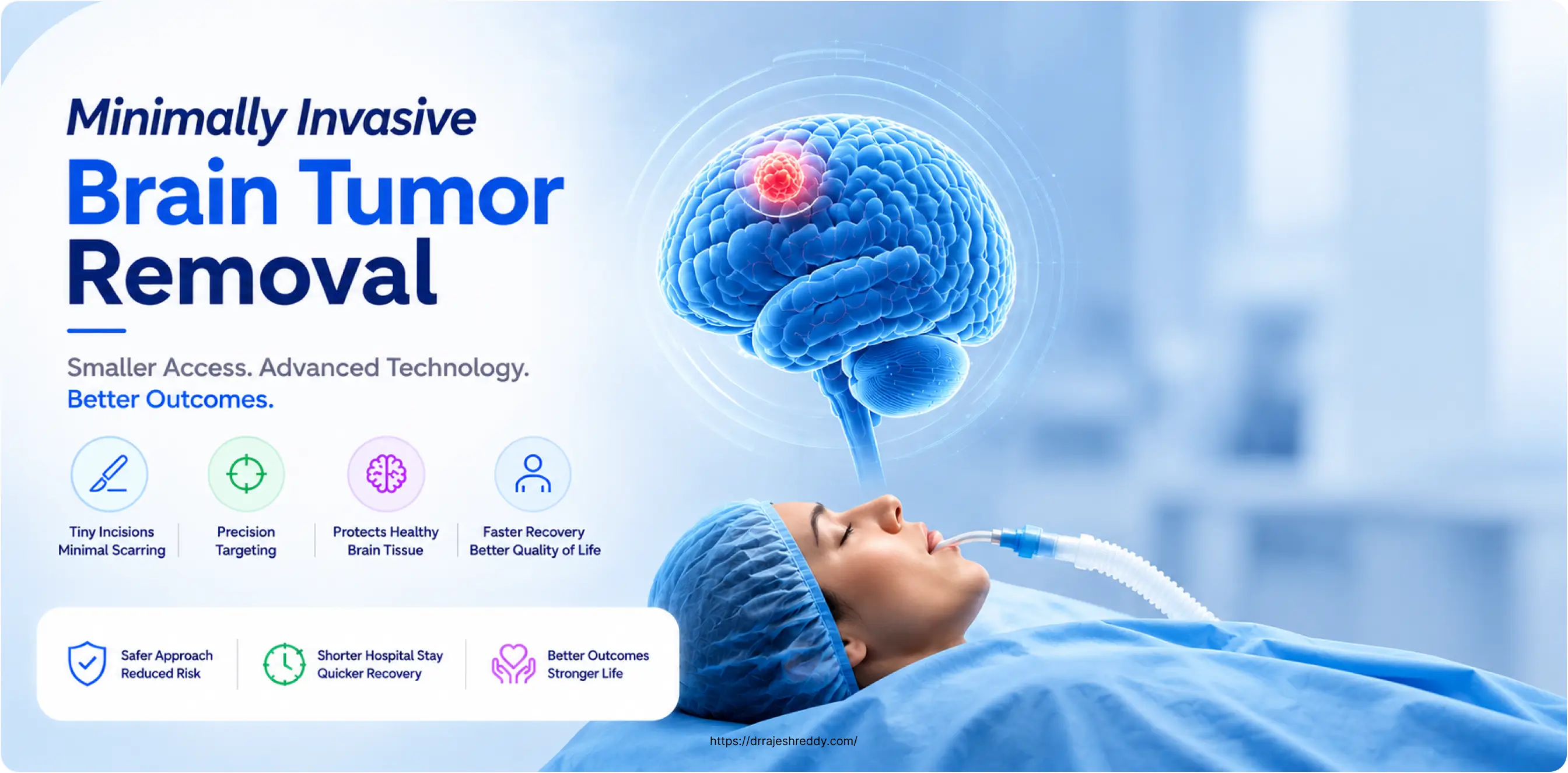
Introduction & Principles of Minimally Invasive Neurosurgery
When most people imagine brain tumor surgery, they picture a large scalp incision, a significant section of skull removed, and weeks of difficult recovery. This image — rooted in the neurosurgery of decades past — is increasingly at odds with what modern minimally invasive brain surgery actually looks like. Today, many brain tumors that once required large open craniotomies can be removed through incisions measured in centimetres rather than inches, through natural body corridors with no incision at all, or treated entirely without surgery using precisely targeted radiation.
Minimally invasive brain tumor surgery is not a single technique — it is a family of approaches united by the principle of achieving the surgical goal (tumor removal or control) while minimising disruption to the scalp, skull, brain, and surrounding structures. The result: less pain, less blood loss, shorter hospital stays, faster recovery, and often better cosmetic outcomes — without compromising the completeness of tumor removal.
Dr. Rajesh Reddy Sannareddy, Senior Consultant Neurosurgeon in Hyderabad, offers the full spectrum of minimally invasive brain tumor techniques — from endoscopic pituitary surgery through the nose to keyhole craniotomies, neuroendoscopic ventricular surgery, and stereotactic radiosurgery. His training at the University Hospital, Zurich and visiting scholar programmes at Barrow Neurological Institute (Phoenix), Okayama University (Japan), and Klinikum Stuttgart (Germany) have given him direct exposure to the most advanced minimally invasive neurosurgical techniques practised globally.
What Does 'Minimally Invasive' Mean in Brain Tumor Surgery?
In conventional open craniotomy, access to the brain requires a large scalp incision (typically 8–15 cm), removal of a correspondingly large section of skull bone (the bone flap), retraction of brain tissue to create a working corridor to the tumor, and closure of all layers at the end of the procedure. This approach provides excellent exposure and is appropriate for large, complex, or deep tumors — but carries the costs of significant tissue disruption, longer operating time, and extended recovery.
Minimally invasive approaches reduce one or more of these steps:
- Smaller scalp incision — keyhole craniotomies use 2–3 cm incisions rather than 8–15 cm
- Smaller bone opening — a burr hole or mini-craniotomy (2–3 cm) rather than a large bone flap
- No incision at all — endoscopic transsphenoidal surgery operates through the nostril; stereotactic radiosurgery requires no surgical access
- Less or no brain retraction — endoscopes and angled instruments navigate around brain structures rather than pushing them aside
- Shorter operative time — smaller access corridors mean less tissue to open and close
Minimally invasive does not mean less effective. For appropriately selected tumors and patients, the extent of tumor removal achieved with minimally invasive techniques is equivalent to — and sometimes superior to — what is achieved with open surgery. The key is patient and tumor selection: the right technique for the right tumor in the right patient.
Minimally Invasive Brain Tumor Techniques: A Complete Overview
The table below summarises the full range of minimally invasive approaches available for brain tumor removal, the incision or access method used, the tumor types best suited to each, and the key advantage over open surgery:
Key Minimally Invasive Approaches
Tailored corridors for minimal tissue disruption
Supraorbital (Eyebrow)
Eyebrow crease incision for anterior skull base & frontal tumors.
Transsphenoidal (Nose)
Through nostrils with zero external incision for pituitary adenomas.
Transventricular
Single burr hole endoscopic access for colloid cysts & intraventricular lesions.
Retrosigmoid Keyhole
Behind-the-ear mini-craniotomy for acoustic neuromas & posterior fossa lesions.
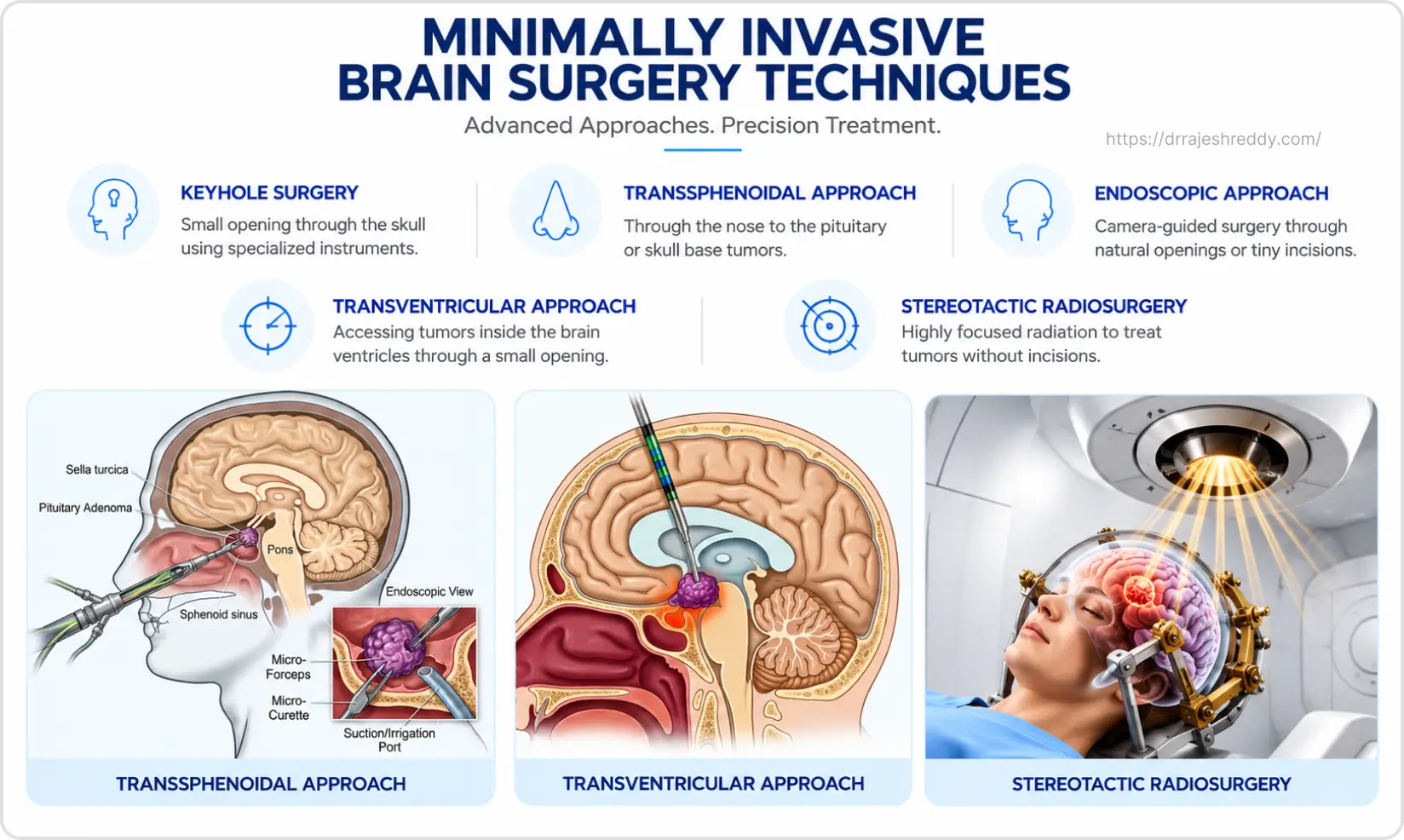
| Technique | Access / Incision | Best Suited Tumors | Key Advantage Over Open Surgery |
|---|---|---|---|
| Endoscopic Keyhole Craniotomy | 2–3 cm scalp incision; small burr hole or mini-craniotomy | Small metastases, superficial gliomas | Minimal brain retraction; faster recovery; smaller scar |
| Endoscopic Transsphenoidal Surgery | Through nostrils — zero external incision | Pituitary adenomas, craniopharyngiomas, select skull base tumors | No scalp incision; no craniotomy; 2–3 day hospital stay |
| Endoscopic Transventricular Surgery | Small burr hole; endoscope navigated through ventricle | Colloid cysts, intraventricular tumors, ependymomas | Avoids open brain retraction; treats hydrocephalus simultaneously |
| Supraorbital Keyhole Craniotomy | Small incision within eyebrow; 2 cm craniotomy | Anterior skull base tumors, olfactory groove meningiomas, small frontal tumors | Incision hidden in eyebrow; excellent cosmetic outcome; direct skull base access |
| Retrosigmoid / Retromastoid Keyhole | Small incision behind ear; 2–3 cm craniotomy | Vestibular schwannomas, posterior fossa meningiomas, trigeminal nerve tumors | Excellent access to posterior fossa; nerve-preserving approach |
| Stereotactic Radiosurgery (SRS) | No incision — focused radiation beams | Small meningiomas, acoustic neuromas, brain metastases, residual tumor | Completely non-invasive; outpatient procedure; no recovery period |
| Neuroendoscopy + Neuronavigation | Endoscope guided by real-time 3D imaging | Deep-seated tumors; complex anatomy | Sub-millimetre accuracy; avoids eloquent pathways |
Each Technique in Depth
1. Endoscopic Keyhole Craniotomy
The keyhole craniotomy replaces the large bone flap of traditional open surgery with a precisely placed mini-craniotomy of 2–3 cm. A high-definition endoscope or operating microscope is introduced through this small opening, providing magnified visualisation of the tumor and surrounding structures. Advanced angled endoscopes (30°, 45°, 70°) allow the surgeon to look around corners within the surgical cavity — accessing tumor tissue that would require much larger openings in traditional surgery.
Neuronavigation provides real-time GPS guidance throughout, ensuring that the small craniotomy is placed in exactly the optimal position for the specific tumor. Blood loss is substantially reduced, and the scalp incision heals to a small, often barely visible scar.
Best for: Small to medium metastases, superficial gliomas, and select skull base tumors accessible via supraorbital or retrosigmoid approaches.
2. Supraorbital Keyhole Craniotomy (Eyebrow Approach)
The supraorbital keyhole craniotomy is one of the most elegant minimally invasive approaches in neurosurgery. A 3–4 cm incision is placed within the natural crease of the eyebrow — virtually invisible when healed. A small 2 cm craniotomy is made in the orbital rim, providing direct access to the anterior skull base, the region above the pituitary, the optic nerves, and the frontal lobes.
This approach is particularly valuable for tumors at the anterior skull base — olfactory groove meningiomas, tuberculum sellae meningiomas, and craniopharyngiomas — that would otherwise require a large frontal craniotomy with significant brain retraction. Patients often remark that they cannot find the scar within weeks of surgery.
Best for: Anterior skull base tumors, olfactory groove meningiomas, small frontal lobe tumors, and select suprasellar tumors not accessible via the transsphenoidal route.
3. Endoscopic Transsphenoidal Surgery (Through the Nose)
The most impactful minimally invasive advance in neurosurgery of the past two decades, the endoscopic transsphenoidal approach removes pituitary tumors — and increasingly, other skull base tumors — entirely through the patient's nostrils. No scalp incision. No craniotomy. No visible change to the patient's appearance whatsoever.
A 4mm high-definition endoscope is introduced through one nostril, navigated through the sphenoid sinus to the sella turcica (the bony housing of the pituitary gland), and the tumor is removed under direct endoscopic visualisation. The approach has been extended to reach tumors beyond the sella — craniopharyngiomas, clival chordomas, and some cavernous sinus tumors — using the expanded endoscopic endonasal approach (EEEA).
Best for: All pituitary adenomas (micro and macro), craniopharyngiomas, clival tumors, and select cavernous sinus lesions.
4. Endoscopic Transventricular Surgery
The brain's ventricular system — a network of fluid-filled cavities within the brain — provides a natural corridor to tumors growing within or adjacent to the ventricles. A rigid endoscope is navigated through a small burr hole into the ventricle, allowing tumors to be visualised, biopsied, or removed under direct camera guidance.
Colloid cysts of the third ventricle — which can cause sudden, life-threatening hydrocephalus — are ideally suited to endoscopic resection. The endoscope navigates through the lateral ventricle into the third ventricle, where the cyst is opened, drained, and its wall removed — all through a single burr hole, with no brain retraction and minimal recovery time.
Best for: Colloid cysts, intraventricular meningiomas, small ependymomas, and ventricular tumors causing hydrocephalus.
5. Retrosigmoid / Retromastoid Keyhole Approach
The retrosigmoid keyhole approach accesses the posterior fossa — the region behind the brainstem — through a 3 cm incision and mini-craniotomy behind the ear, hidden within the natural hairline. This corridor provides excellent access to vestibular schwannomas (acoustic neuromas), posterior fossa meningiomas, and tumors along the trigeminal or facial nerve.
The goal in vestibular schwannoma surgery via this approach is maximum tumor removal while preserving facial nerve function and, where possible, residual hearing. For tumors under 2.5–3 cm, the combination of keyhole microsurgery for larger tumors and stereotactic radiosurgery for smaller ones provides a full treatment spectrum.
Best for: Vestibular schwannomas, posterior fossa meningiomas, trigeminal schwannomas, epidermoid cysts, and petroclival tumors.
6. Stereotactic Radiosurgery (SRS) — Surgery Without a Scalpel
Stereotactic radiosurgery is the ultimate expression of minimally invasive brain tumor treatment — no incision, no anaesthesia, no hospital admission, and no recovery period. Multiple precisely focused radiation beams converge on the tumor from different angles, delivering a high dose of radiation to destroy tumor cells while sparing surrounding brain tissue.
SRS is not conventional radiotherapy — it delivers a very high dose in a single session (or occasionally 2–5 fractions for larger tumors) with sub-millimetre targeting accuracy. The result is effective tumor control — the tumor shrinks or stabilises over months — for a range of tumor types.
Best for: Meningiomas < 3 cm (particularly cavernous sinus), acoustic neuromas < 3 cm, brain metastases (1–4 lesions), and residual or recurrent tumor after surgical resection.
7. Fluorescence-Guided Minimally Invasive Resection
When minimally invasive keyhole surgery is used for high-grade gliomas, 5-ALA or Sodium Fluorescein fluorescence guidance compensates for the reduced surgical field by making tumor tissue visually distinct from normal brain — glowing pink or yellow-green respectively under specific light filters. This allows the surgeon to maximise tumor removal through the small keyhole opening by precisely identifying tumor margins that would otherwise require a larger exposure to see reliably.
Best for: High-grade gliomas (GBM, Grade III) where a keyhole approach is planned and maximum resection is the goal.
Minimally Invasive vs. Traditional Open Surgery: A Direct Comparison
Patients frequently want to understand what they gain — and what they might give up — by choosing a minimally invasive approach. The comparison below addresses this directly:
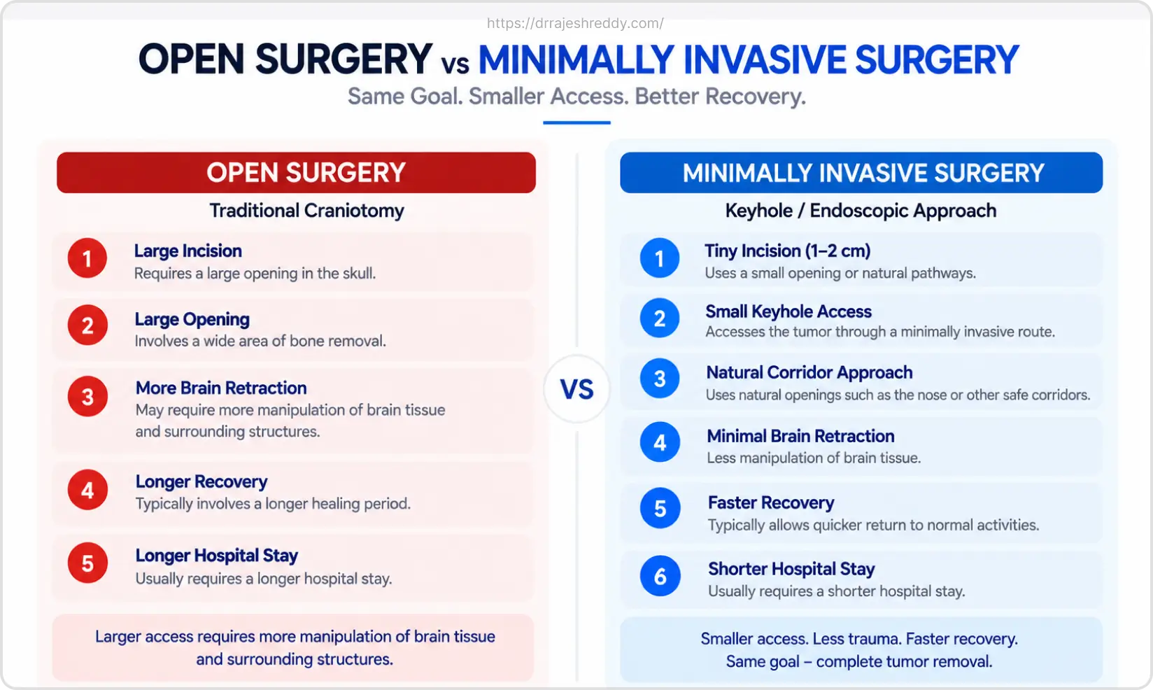
| Feature | Minimally Invasive Approach | Traditional Open Craniotomy |
|---|---|---|
| Incision size | 1–3 cm (keyhole) or none (endoscopic / SRS) | 6–15 cm scalp incision; larger bone flap removal |
| Brain retraction | Minimal to none — natural corridors used | Significant retraction of brain tissue required |
| Hospital stay | 2–5 days for most procedures | 5–10 days |
| Post-operative pain | Mild — manageable with simple analgesics | Moderate to significant — requires stronger analgesia |
| Return to normal activity | 1–4 weeks depending on procedure | 4–8 weeks |
| Blood loss | Significantly lower | Higher — particularly for large tumors |
| Cosmetic outcome | Minimal or invisible scar | Visible scalp scar; possible skull contour change |
| Suitable for all tumors? | No — large tumors, complex anatomy may require open surgery | Yes — applicable to all tumor types and locations |
| Tumour removal | Equivalent for appropriate tumor types and sizes | Required for large or complex tumors |
| Technology required | High — neuronavigation, HD endoscopy, fluorescence guidance | Lower — operating microscope; standard craniotomy instruments |
The choice between minimally invasive and open surgery is not about patient preference alone — it is a clinical decision based on tumor size, location, type, and the surgical goal. Dr. Rajesh Reddy recommends the approach that offers the safest and most complete tumor removal for each individual patient. For many tumors, that is now a minimally invasive approach. For others, open surgery remains the right choice — and both are performed with the same commitment to precision and safety.
Which Brain Tumors Can Be Removed Minimally Invasively?
The table below summarises which tumor types are well-suited to minimally invasive approaches, what those options are, and when traditional open surgery is still the preferred choice:
| Tumor Type | Minimally Invasive Option Available | When Open Surgery Is Still Preferred |
|---|---|---|
| Pituitary adenoma | Endoscopic transsphenoidal — first choice for virtually all cases | Very large tumors with significant lateral cavernous sinus extension |
| Small to medium meningioma | Keyhole craniotomy or stereotactic radiosurgery | Large meningiomas > 4–5 cm; parasagittal with sinus involvement |
| Colloid cyst | Endoscopic transventricular resection — preferred approach | Rarely requires open surgery |
| Vestibular schwannoma | Retrosigmoid keyhole; SRS for tumors < 3 cm | Large tumors > 3 cm with brainstem compression |
| Brain metastasis (single, small) | Keyhole craniotomy + SRS for residual/adjuvant | Multiple metastases; large tumors > 3 cm causing mass effect |
| Low-grade glioma (non-eloquent) | Keyhole craniotomy with neuronavigation | Large gliomas requiring extensive resection in deep locations |
| Craniopharyngioma | Endoscopic extended transsphenoidal — preferred for most | Tumors with significant lateral extension beyond endoscopic reach |
| Intraventricular tumors | Neuroendoscopy — preferred for colloid cysts, small ependymomas | Large or solid ventricular tumors requiring extensive resection |
The Technology Behind Minimally Invasive Brain Surgery
Minimally invasive neurosurgery is made possible by a convergence of technologies that together compensate for the reduced visual field and working space of small access corridors:
High-Definition Endoscopy
Modern 4K endoscopes provide a magnified, panoramic, high-resolution view of the surgical field through an access corridor as small as 4 mm. Angled endoscopes (30°, 45°, 70°) allow the surgeon to look around corners within the surgical cavity — visualising structures that would be out of sight in a standard straight-line view. This visualisation quality often exceeds what the operating microscope provides through a traditional craniotomy.
Neuronavigation
Neuronavigation systems (StealthStation, BrainLab) provide real-time, 3D GPS-like guidance by registering the patient's pre-operative MRI or CT scan to their actual head position on the operating table. The surgeon can see a continuously updated display of instrument position relative to the tumor, critical structures, and planned surgical corridors — with sub-millimetre accuracy. This is particularly important in minimally invasive surgery, where the reduced visual field makes spatial orientation more demanding.
Diffusion Tensor Imaging (DTI) and Connectomics Integration
DTI tractography maps the brain's white matter pathways and is loaded into the neuronavigation system for use during minimally invasive resection. Connectomics — the mapping of neural networks — provides an additional layer, ensuring that not just individual functional areas but the critical connections between them are protected. This integration is particularly valuable in keyhole surgery near eloquent cortex, where the surgical margins are tighter.
Fluorescence Guidance
5-ALA and Sodium Fluorescein allow high-grade glioma cells to be visually identified during resection — compensating for the reduced visual field of keyhole surgery by making the tumor-brain boundary visible under specific light wavelengths. This technology has been shown to significantly improve the extent of GBM resection in multiple international studies.
Intraoperative Neurophysiological Monitoring (IONM)
Motor evoked potentials (MEPs), somatosensory evoked potentials (SSEPs), and cranial nerve monitoring provide continuous, real-time assessment of neurological function during minimally invasive resection — providing a safety net that is particularly valuable when working through a small corridor near functional brain areas or cranial nerves.
Why Choose Dr. Rajesh Reddy for Minimally Invasive Brain Tumor Surgery?
Minimally invasive brain tumor surgery demands more from a surgeon, not less — it requires mastery of advanced instruments, fluency with endoscopic technique in confined spaces, and the clinical judgement to know when a minimally invasive approach is appropriate and when it is not. Dr. Rajesh Reddy's training and practice span the full range of minimally invasive neurosurgical techniques:
Minimally Invasive Expertise — Dr. Rajesh Reddy
- Endoscopic transsphenoidal pituitary surgery — 4K HD endoscopy, angled scopes, neuronavigation, nasoseptal flap reconstruction
- Keyhole craniotomy — supraorbital (eyebrow), retrosigmoid, pterional, and interhemispheric keyhole approaches
- Endoscopic transventricular surgery — colloid cysts, intraventricular tumors, endoscopic third ventriculostomy
- Fluorescence-guided resection — 5-ALA and Sodium Fluorescein for high-grade glioma keyhole resection
- Stereotactic radiosurgery coordination — for meningiomas, acoustic neuromas, and metastases where SRS is the optimal approach
- FINR (Zurich) — endovascular skills enabling pre-operative embolisation of vascular tumors before minimally invasive resection
- Visiting Scholar — Barrow Neurological Institute, Phoenix; Okayama University, Japan; Klinikum Stuttgart, Germany — global exposure to advanced minimally invasive skull base and brain tumor techniques
- DTI / connectomics integration in surgical planning — protecting neural networks, not just individual functional areas
- Clinical acumen and judgement gained through years of training with leading experts globally
Perhaps most importantly, Dr. Rajesh Reddy brings the judgement to know when minimally invasive surgery is the right choice and when open surgery will achieve a better outcome — and the honesty to recommend the latter even when the former is more technologically impressive.
Recovery After Minimally Invasive Brain Tumor Surgery
One of the most tangible benefits of minimally invasive brain tumor surgery is the accelerated recovery compared to traditional open craniotomy:
Endoscopic Pituitary Surgery
- Hospital stay: 2–3 days
- Return to normal activity: 1–2 weeks
- Nasal congestion and mild headache for 1–2 weeks; no scalp pain
- No activity restrictions after 2 weeks except nose blowing (4 weeks) and strenuous exercise (4–6 weeks)
Keyhole Craniotomy (Supraorbital, Retrosigmoid)
- Hospital stay: 3–5 days
- Return to normal activity: 2–4 weeks
- Mild headache and scalp numbness around the small incision; resolves over 2–4 weeks
- Driving typically from week 4–6 after medical clearance
Endoscopic Transventricular Surgery (Colloid Cyst)
- Hospital stay: 2–4 days
- Return to normal activity: 1–3 weeks
- Mild headache for a few days; minimal wound discomfort from small burr hole incision
Stereotactic Radiosurgery
- No hospital admission — outpatient procedure
- Return to normal activity: same day or next day
- Mild scalp tenderness at frame pin sites (if frame-based) for 1–2 days
- No surgical recovery — tumour control assessed on follow-up MRI at 3–6 months