Introduction
A diagnosis of a brain tumor is one of the most disorienting moments a person and their family can experience. In the hours after receiving that news, patients are often left with an overwhelming number of questions: Is it cancerous? Does it need surgery? What will surgery involve? Will life ever return to normal? These are questions that deserve clear, honest, and expert answers —not generic information, but guidance rooted in deep clinical experience.
Dr. Rajesh Reddy Sannareddy, Senior Consultant Neurosurgeon in Hyderabad, has built a career on providing exactly that. Having been part of over 5,000 neurosurgical procedures, he offers a level of experience that few neurosurgeons in the region can match.
This page is a comprehensive resource for patients and families who want to understand brain tumors — what they are, how they are diagnosed, what surgical options exist, and what recovery looks like. If you have been referred to a neurosurgeon, or if you are exploring your options after a recent MRI finding, this guide will help you make sense of the path ahead.
What Is a Brain Tumor?
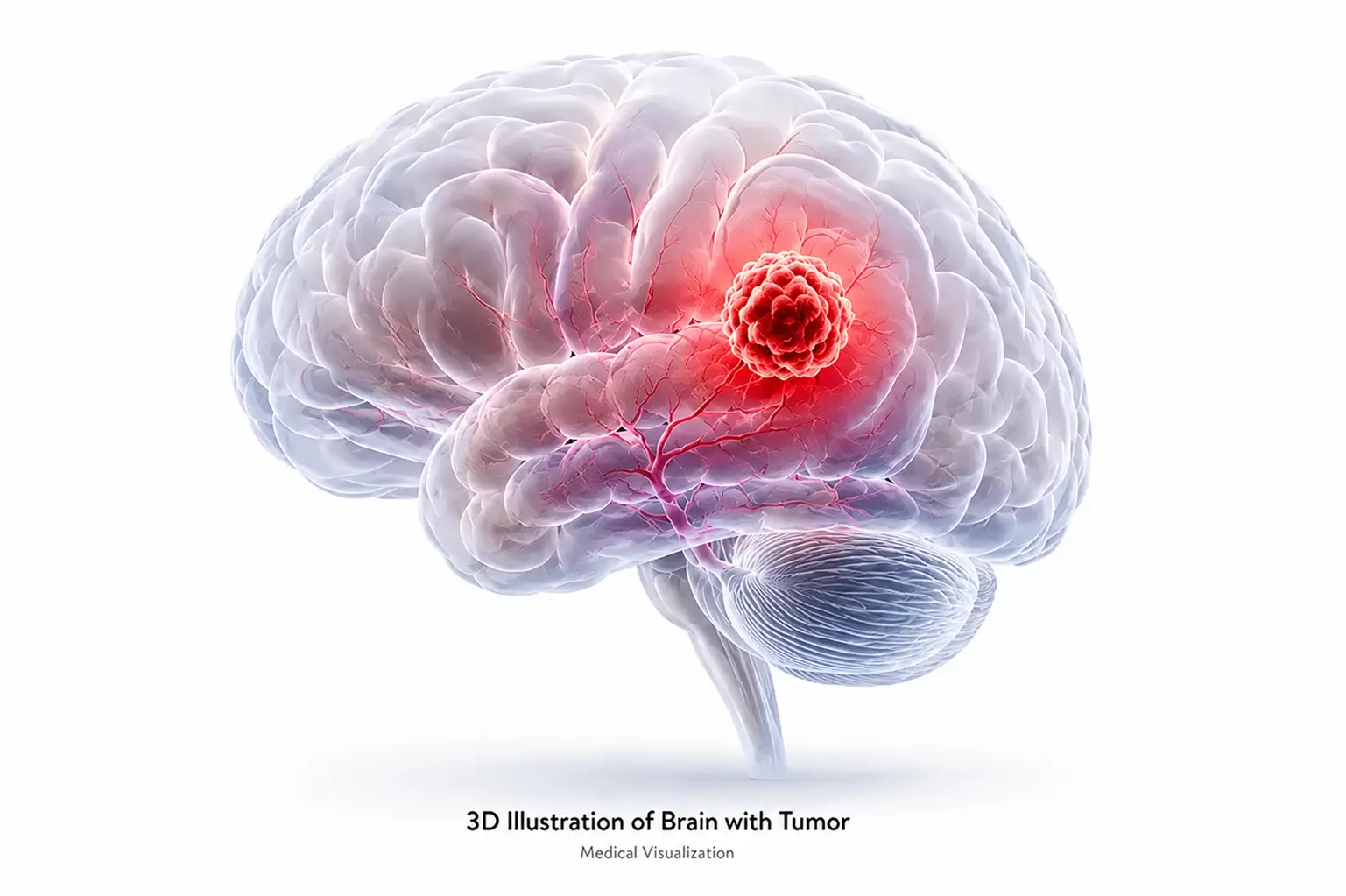
A brain tumor is an abnormal mass of cells that grows within the brain or the structures surrounding it — including the meninges (the protective membranes encasing the brain), the cranial nerves, the pituitary gland, and the skull base. Tumors can arise from the brain tissue itself, or they can spread to the brain from cancers in other parts of the body.
It is important to understand from the outset that not all brain tumors are cancers, and not all brain tumors are immediately life-threatening. The prognosis and treatment pathway depend heavily on the type, grade, size, and location of the tumor.
Primary vs. Secondary (Metastatic) Brain Tumors
Primary brain tumors originate in the brain itself. They can arise from neurons, glial cells (the supportive cells of the brain), meninges, cranial nerves, or the pituitary gland. Examples include gliomas, meningiomas, pituitary adenomas, medulloblastomas, and acoustic neuromas.
Secondary (metastatic) brain tumors originate from cancer cells that have spread from elsewhere in the body — commonly from the lung, breast, kidney, colon, or skin (melanoma). Metastatic brain tumors are actually more common than primary brain tumors and may present as single or multiple lesions.
Benign vs. Malignant Brain Tumors
Benign tumors are non-cancerous. They grow slowly, have well-defined borders, and do not invade surrounding brain tissue. However, benign does not mean harmless — even a benign tumor can cause serious neurological problems by pressing on critical brain structures. Meningiomas, pituitary adenomas, and acoustic neuromas are common benign brain tumors.
Malignant tumors are cancerous. They grow more rapidly, often invade adjacent brain tissue, and can be life-threatening. Glioblastoma multiforme (GBM) is the most aggressive primary brain tumor and carries a poor prognosis without prompt, multimodal treatment. Surgical resection is a key first step in managing malignant brain tumors.
Important: A brain tumor diagnosis requires a full assessment by an experienced neurosurgeon before any treatment decisions are made. Imaging alone does not always determine whether a tumor is benign or malignant — tissue diagnosis (biopsy or surgical specimen) is often necessary.
Types of Brain Tumors Treated by Dr. Rajesh Reddy
Dr. Rajesh Reddy manages the full spectrum of intracranial tumors. The table below summarises the most common tumor types, their characteristics, and the surgical approach Dr. Reddy typically employs:
| Tumor Type | Location in Brain | Benign / Malignant | WHO Grade | Dr. Reddy's Approach |
|---|---|---|---|---|
| Glioblastoma (GBM) | Brain parenchyma | Malignant | Grade IV | Microsurgical / Awake craniotomy plus adjuvant therapy |
| Low-Grade Glioma (LGG) | Brain parenchyma | Slow-growing | Grade I–II | Microsurgical / Awake craniotomy plus adjuvant therapy |
| Meningioma | Brain surface / skull base | Usually benign | Grade I–III | Microsurgical resection / Radiosurgery / Embolisation |
| Pituitary Adenoma | Pituitary gland | Benign | — | Endoscopic transsphenoidal / Medical management for select prolactinomas / Radiosurgery |
| Medulloblastoma | Cerebellum | Malignant | Grade IV | Microsurgical + adjuvant therapy |
| Vestibular Schwannoma | Acoustic nerve (CN VIII) | Benign | Grade I | Microsurgical / Radiosurgery |
| Craniopharyngioma | Skull base / sellar region | Benign | Grade I | Endoscopic / Microsurgical |
| Ependymoma | Ventricles / spinal canal | Variable | Grade II–III | Microsurgical resection plus adjuvant therapy |
| Epidermoid Cyst | Skull base / cisterns / ventricles | Benign | — | Microsurgical excision |
| Colloid Cyst | Ventricle | Benign | — | Microscopic / Endoscopic |
| Trigeminal Schwannoma | Trigeminal nerve | Benign | Grade I | Microsurgery / Radiosurgery |
| Metastatic Brain Tumor | Variable (any lobe) | Malignant | — | Microsurgical / Stereotactic biopsy / Radiosurgery |
A Note on Skull Base Tumors
Skull base tumors — including craniopharyngiomas, chordomas, meningiomas at the skull base, and vestibular schwannomas — present a particular surgical challenge due to their proximity to critical cranial nerves, blood vessels, and the brainstem. Dr. Rajesh Reddy's training in both microsurgical and endoscopic skull base surgery, combined with his endovascular expertise, means he can address these complex cases with techniques that prioritise function preservation alongside tumor removal.
Brain Tumor Symptoms: Recognising the Warning Signs
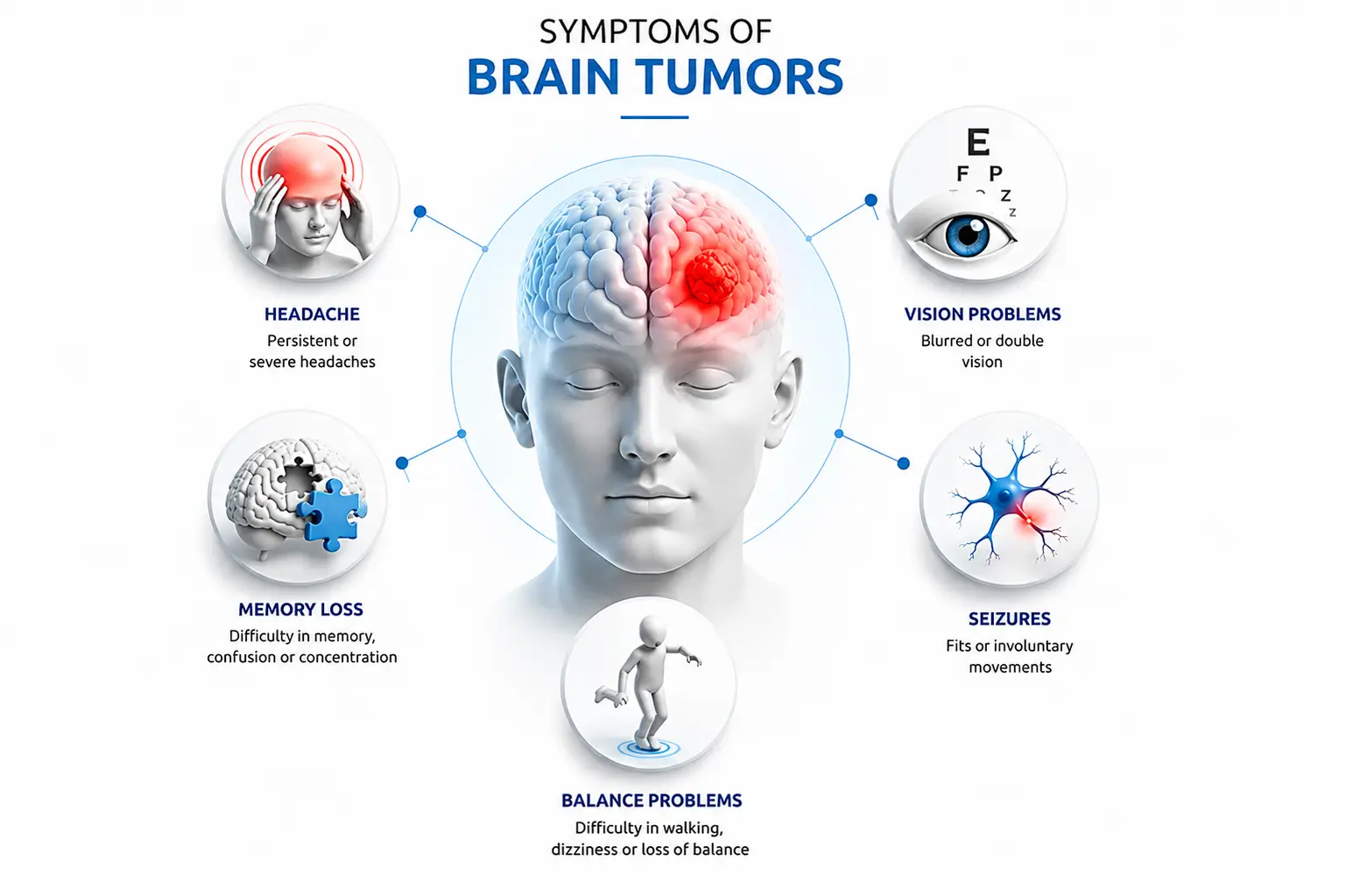
Brain tumor symptoms arise from two mechanisms: the effect of the tumor on the brain tissue it occupies or invades, and the indirect effect of increased pressure within the skull (raised intracranial pressure). Symptoms vary based on the tumor's size, type, and location.
General Symptoms (Due to Raised Intracranial Pressure)
- ▪ Persistent, progressive headaches — especially severe in the morning, or waking the patient from sleep
- ▪ Nausea and vomiting, particularly in the morning or with changes in head position
- ▪ Visual disturbances — blurred vision, double vision (diplopia), or loss of peripheral vision
- ▪ Cognitive changes — difficulty concentrating, memory problems, confusion, or personality changes
- ▪ Fatigue and general decline in function unexplained by other causes
Location-Specific Symptoms
Seizures — A Key Red Flag
New-onset seizures in an adult who has no prior history of epilepsy should always prompt an urgent MRI of the brain to rule out an underlying tumor. Seizures can be the first and only presenting symptom for some brain tumors, particularly low-grade gliomas, meningiomas, and metastases.
Emergency warning: If you or a family member develops sudden severe headache (often described as 'the worst headache of my life'), new seizures, rapidly progressive neurological weakness, or sudden loss of vision or speech, seek emergency medical care immediately. These may be signs of a rapidly growing tumor or a complication such as hemorrhage into a tumor mimicking a stroke.
How Is a Brain Tumor Diagnosed?
Accurate diagnosis is the foundation of effective brain tumor treatment. Dr. Rajesh Reddy follows a structured diagnostic pathway that combines clinical assessment with advanced neuroimaging and, when needed, tissue analysis.
Step 1: Neurological Examination
Assessments cover cognitive functions, speech, cranial nerves (vision, eyeball movements, hearing, swallowing), muscle strength, sensations, reflexes, coordination and gait. This clinical mapping helps localise the problem and guides imaging decisions.
Step 2: Advanced Neuroimaging
The following table outlines the key diagnostic investigations used in brain tumor diagnosis:
| Test | What It Shows | When It Is Used |
|---|---|---|
| MRI Brain (with contrast) | Size, precise location, relationship to critical structures, edema, enhancement pattern | First-line investigation for all suspected brain tumors |
| Functional MRI (fMRI) | Maps eloquent brain areas — speech, motor, vision — relative to tumor location | Pre-surgical planning for tumors near functional areas |
| CT Scan (with/without contrast) | Bony involvement, acute bleeds, calcifications within the tumor | Emergency settings; skull base tumors; post-operative check |
| MR Spectroscopy | Differentiates high-grade from low-grade tumors; radiation necrosis check | Ambiguous lesions on standard MRI; post-treatment surveillance |
| PET Scan (FDG / FDOPA) | Metabolic activity of the tumor; identifies hotspots for biopsy targeting | High-grade glioma staging; recurrence vs. treatment change |
| Intraoperative MRI (iMRI) | Real-time imaging during surgery to assess extent of resection | Complex glioma resections; eloquent area tumors |
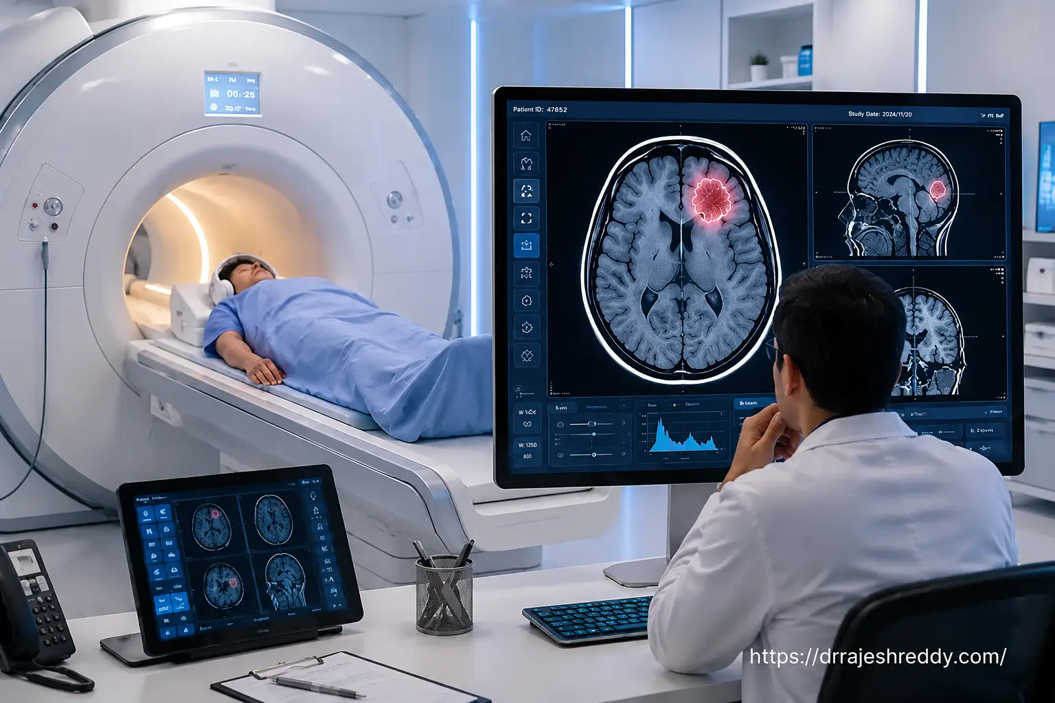
Tractography / Diffusion Tensor Imaging (DTI): An advanced MRI technique that maps the white matter tracts — the fibre pathways connecting different regions of the brain. When a tumor lies near or within these critical tracts (such as the corticospinal tract for motor function or the arcuate fasciculus for language), DTI allows the surgeon to plan around them, reducing the risk of functional deficit.
Step 3: Blood Tests & Hormonal Assessment
For suspected pituitary tumors, a comprehensive hormonal panel is essential to assess whether the tumor is secreting hormones (prolactin, ACTH, growth hormone) and to coordinate endocrinological and surgical plans.
Step 4: Tissue Diagnosis & Molecular Profiling
The 2021 WHO Classification of Central Nervous System Tumors integrates molecular markers — including IDH mutation status, MGMT promoter methylation, 1p/19q codeletion, and EGFR amplification — into grading, directly influencing chemotherapy selection and prognosis.
Surgical Techniques and Approaches
The goal of brain tumor surgery is to remove as much of the tumor as safely possible — 'maximum safe resection' — while preserving neurological function and quality of life.
| Technique | Access Method | Best For | Key Advantage |
|---|---|---|---|
| Microsurgical Craniotomy | Small skull opening, operating microscope | Gliomas, meningiomas, metastases, skull base tumors | High-magnification precision; maximum tumor control |
| Endoscopic Keyhole Surgery | Small burr hole or natural corridor (nose/eye) | Pituitary tumors, ventricular tumors, skull base lesions | Minimal tissue disruption; faster recovery |
| Awake Craniotomy | Craniotomy with patient awake intraoperatively | Tumors in speech / motor / language cortex | Real-time neurological mapping; function preservation |
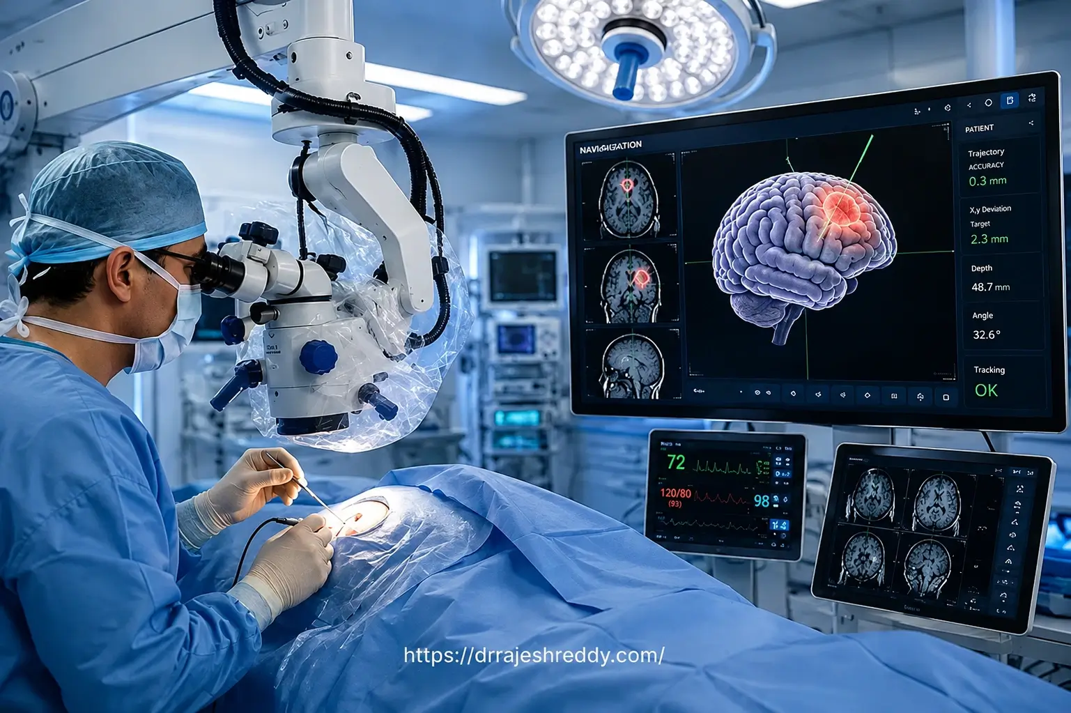
| Neuronavigation-Guided | GPS-like 3D imaging guidance during any approach | Deep-seated or irregularly shaped tumors | Sub-millimetre surgical accuracy |
| Fluorescence-Guided Resection | Fluorescent dye highlights tumor tissue intraoperatively | High-grade gliomas (GBM) | Distinguishes tumor from normal brain in real time |
| Stereotactic Radiosurgery | Focused radiation beams (no incision) | Small residual tumors; acoustic neuromas; metastases | Non-invasive; treats eloquent or inaccessible lesions |
Understanding Key Techniques
Microsurgical Craniotomy: The foundation of brain tumor surgery. Under up to 40x magnification, Dr. Rajesh Reddy meticulously separates the tumor from surrounding brain tissue, protecting blood vessels and nerve fibres throughout. Patients are typically walking within 24–48 hours of surgery.
Endoscopic Keyhole Surgery: Reaches pituitary and skull base lesions entirely through the nostrils (transsphenoidal), navigating without scalp incisions or brain retraction. Patients typically go home within 2–3 days.
Awake Craniotomy: Used near eloquent speech or motor areas. Electrical stimulation maps the brain's functional areas in real time as the patient interacts, creating a personalized safety margin.
Fluorescence-Guided Resection: Administering oral 5-ALA before surgery makes tumor cells glow pink under specialized microscope light, separating active tumor margins from healthy tissue.
Risks of Brain Tumor Surgery: A Balanced Assessment
Brain tumor surgery is a major neurosurgical procedure. Dr. Rajesh Reddy believes in transparent, thorough pre-operative counselling so that every patient and family enters the operating theatre with realistic expectations.
General Surgical Risks
- ▪ Anaesthesia-related complications (rare with modern techniques)
- ▪ Bleeding (intracranial haemorrhage) during or after surgery
- ▪ Infection —wound infection or, rarely, meningitis or brain abscess
- ▪ Deep vein thrombosis (DVT) or pulmonary embolism — preventable with early mobilisation and prophylaxis
- ▪ CSF (cerebrospinal fluid) leak — more common after skull base or posterior fossa surgery
Neurological Risks (Dependent on Tumor Location)
- ▪ Temporary or permanent weakness in an arm or leg (if tumor is near motor cortex)
- ▪ Speech or language difficulties (if tumor is near Broca's or Wernicke's areas)
- ▪ Visual field defects (if tumor is near the optic pathways)
- ▪ Memory or personality changes (if tumor is in the frontal or temporal lobe)
- ▪ Seizures — risk may increase transiently after surgery; managed with anti-epileptic medication
How Dr. Rajesh Reddy Minimises Risk
- ▪ Clinical acumen and Judgement gained through years of training with leading experts.
- ▪ Neuronavigation for precise, GPS-guided tumor localisation throughout surgery
- ▪ Intraoperative neurophysiological monitoring (motor evoked potentials, somatosensory evoked potentials) to detect nerve stress in real time
- ▪ Connectomics integrated with pre-surgical fMRI and DTI to plan surgical approaches that protect critical neural networks alongside maximising tumor removal
- ▪ Awake craniotomy mapping for tumors near eloquent cortex
- ▪ Pre-operative functional MRI to plan the safest surgical corridor
- ▪ Fluorescence guidance (5-ALA) to maximise resection accuracy in GBM
- ▪ Collaboration with neuro-anaesthesia, neuro-ICU, and neurophysiology teams for comprehensive perioperative management
Why Choose Dr. Rajesh Reddy?
Choosing the right neurosurgeon is the single most important decision a brain tumor patient will make. Here is why patients trust Dr. Rajesh Reddy Sannareddy with their care:
Credentials & Training
Expertise Across the Spectrum
Dr. Rajesh Reddy is among a small group of neurosurgeons in India who combine expertise across microsurgical, endoscopic, and endovascular neurosurgery. This means that for complex cases — such as a brain tumor that has caused a secondary vascular problem, or a skull base tumor requiring pre-operative embolisation to reduce blood loss during surgery — Dr. Rajesh Reddy can manage the entire treatment pathway himself, without the need for multiple specialists.
The Role of Connectomics in Modern Brain Tumor Surgery
Connectomics refers to the comprehensive mapping of the brain's structural and functional connectivity networks. In the context of brain tumor surgery, connectomic analysis helps the surgeon understand not just where individual eloquent areas are located, but how they are interconnected — allowing for a more nuanced assessment of which white matter pathways are at risk during resection. By integrating connectomic data with pre-surgical fMRI and DTI, Dr. Rajesh Reddy plans surgical approaches that protect not just discrete functional areas, but the broader networks that underlie complex cognitive and motor abilities.
Comprehensive, Patient-Centred Care
Dr. Rajesh Reddy understands that a brain tumor diagnosis affects the entire family. His approach to patient care is built on clear communication, compassionate counselling, and a commitment to involving patients and families in every treatment decision. Every case is individualised — no two brain tumors are identical, and neither are the patients who carry them.
Recovery Timeline & Rehabilitation
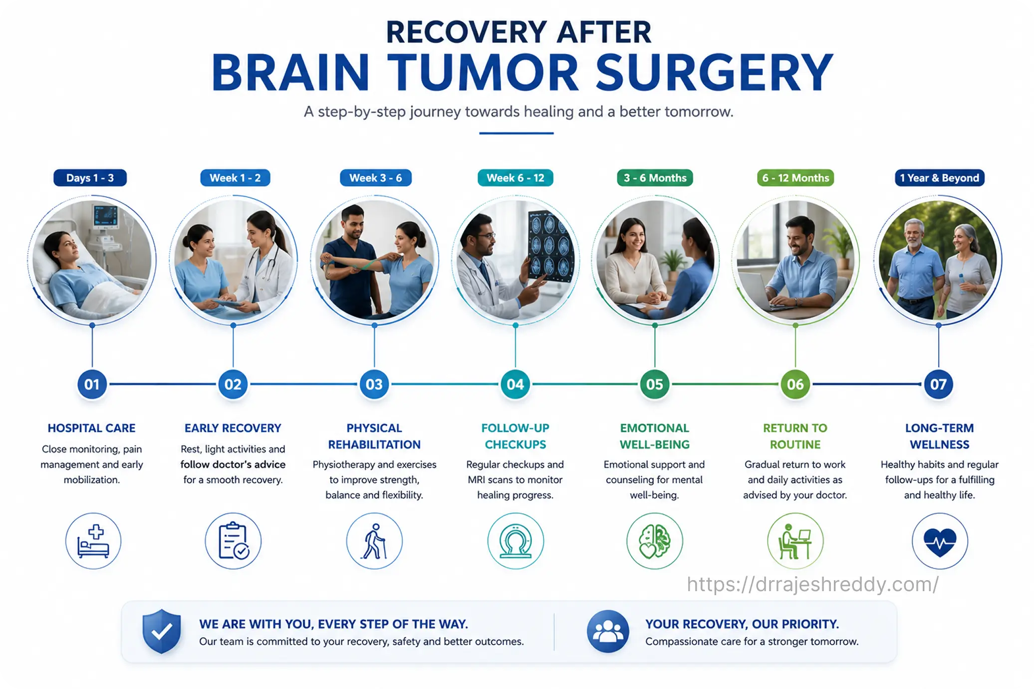
Recovery from brain tumor surgery is a process that unfolds over weeks to months. Understanding what to expect at each phase helps patients and families prepare practically and emotionally.
| Phase | What Happens | Patient Milestones |
|---|---|---|
| Day 0–1 (ICU) | Neurological monitoring, pain management, anti-swelling steroids, anti-seizure prophylaxis. MRI / CT checks. | Wakes from anaesthesia; neurological status assessed hourly |
| Day 2–3 (Ward) | IV medications tapered to oral; early physiotherapy begins; wound check | Sits up, takes fluids, short walks with support |
| Day 4–10 (Discharge) | Wound inspection; discharge planning and medication education, Suture/Stapler removal | Independent in basic activities; discharge criteria met |
| Week 2–4 (Rest) | Rest at home; avoid driving, strenuous activity, heavy lifting. Tumor board discussion | Fatigue improves; headaches reduce; resuming light activity |
| Week 6 (Follow-up) | MRI brain check, radiation + chemotherapy planning as per protocol | Neurological assessment; rehabilitation planning if needed |
| Month 3 (Surveillance) | Clinical review, imaging checks | Most benign tumor patients return to normal daily activities |
| Month 6 and beyond | Regular MRI surveillance (every 3–6 months for malignant; annually for benign) | Return to work evaluated; long-term quality-of-life planning |
Factors That Influence Recovery Speed
- ▪ Age and baseline health —younger, fitter patients generally recover faster
- ▪ Tumor type and location — surgery near eloquent cortex may require rehabilitation
- ▪ Extent of resection — complete removal vs. debulking (partial removal)
- ▪ Malignant vs. benign —malignant tumors require additional oncology treatment that affects the overall recovery timeline
- ▪ Presence of medical comorbidities —Diabetes mellitus, ailments related to lungs, heart, kidney, liver can affect healing and recovery pace
- ▪ Presence of complications —infection, bleeding, or CSF leak prolongs hospital stay and recovery
Rehabilitation Support
Depending on any post-operative neurological deficits, Dr. Rajesh Reddy's team coordinates with rehabilitation specialists for:
- ▪ Physiotherapy — to rebuild strength, coordination, and mobility affected by motor pathway tumors
- ▪ Speech and Language Therapy — for patients with speech, language, or swallowing difficulties after temporal or frontal lobe surgery
- ▪ Occupational Therapy — to support the return to daily activities and, ultimately, work
- ▪ Neuropsychological support — for patients experiencing cognitive changes or mood disturbances after surgery
Beyond Surgery: Multimodal Treatment for Malignant Brain Tumors
For malignant brain tumors — particularly high-grade gliomas like GBM — surgery is the first step in a multimodal treatment protocol. Maximum safe surgical resection significantly improves the response to subsequent treatments.
Radiation Therapy (Radiotherapy)
Standard of care for GBM and other high-grade tumors following surgery. Typically delivered as fractionated external beam radiotherapy over 6 weeks. Stereotactic radiosurgery (SRS) — highly focused single or few-fraction radiation — may be used for smaller residual tumors, metastases, or recurrent disease.
Chemotherapy
Temozolomide (TMZ) is the standard chemotherapy agent for GBM, administered concurrently with radiation and then as adjuvant treatment. MGMT promoter methylation — a molecular marker tested on the surgical specimen — predicts response to TMZ and helps personalise chemotherapy decisions.
Tumour Treating Fields (TTFields)
A relatively newer treatment modality that uses alternating electric fields delivered via scalp electrodes to disrupt tumor cell division. Used in combination with adjuvant TMZ for GBM, TTFields have been shown to improve progression-free and overall survival.
Targeted Therapy and Immunotherapy
For select patients with specific molecular targets (BRAF mutation, IDH mutation), targeted therapeutic agents are available. Immunotherapy is an active area of clinical research for brain tumors and may be accessible through clinical trial participation. Dr. Rajesh Reddy works closely with neuro-oncologists to discuss emerging treatment options for eligible patients.
Post-Surgery Care and Follow-Up
- Close neurological monitoring in a dedicated neurosurgical ICU setting during the immediate post-operative period
- Access to in-house physiotherapy, speech therapy, and occupational therapy teams
- Digital prescriptions for convenient access and renewal
- Digitally saved medical records and appointment booking accessible through the Dr. Reddy's Neuro Care app — available on Google Play and App Store.
- Video consultation facility for follow-up appointments — particularly useful for outstation patients who have travelled from other states for surgery
- Coordinated care with medical oncology, radiation oncology, and endocrinology for comprehensive tumor management
Clinical Insights: Video Guides
Dr. Rajesh Reddy Sannareddy shares regular educational videos and Shorts on his YouTube channel to help patients and families understand brain tumors, advanced surgical techniques, and postoperative recovery guidelines.