
Introduction & What Is a Meningioma?
One of the most important — and most reassuring — things a patient can hear after a meningioma diagnosis is this: not every meningioma needs surgery. Unlike most brain tumors, meningiomas are usually benign, slow-growing, and in many cases can be safely monitored without immediate intervention. When treatment is required, options range from focused radiation without a single incision to precisely planned microsurgical removal — and choosing the right path depends on a careful assessment of the tumor's size, location, behaviour, and the patient's individual circumstances.
This page explains the full spectrum of meningioma treatment options — from active surveillance to stereotactic radiosurgery to open microsurgical resection — and helps patients understand when each approach is appropriate, what it involves, and what outcomes they can realistically expect.
Dr. Rajesh Reddy Sannareddy, Senior Consultant Neurosurgeon in Hyderabad, treats meningiomas across all locations and grades. His dual expertise in microsurgical and endovascular neurosurgery — including pre-operative tumour embolisation — means he can manage even the most complex skull base and highly vascular meningiomas with a comprehensive, individualised treatment approach.
What Is a Meningioma?
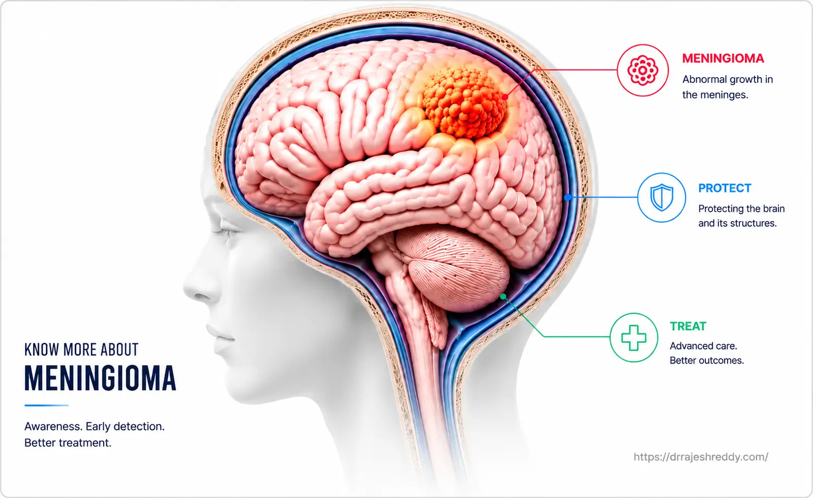
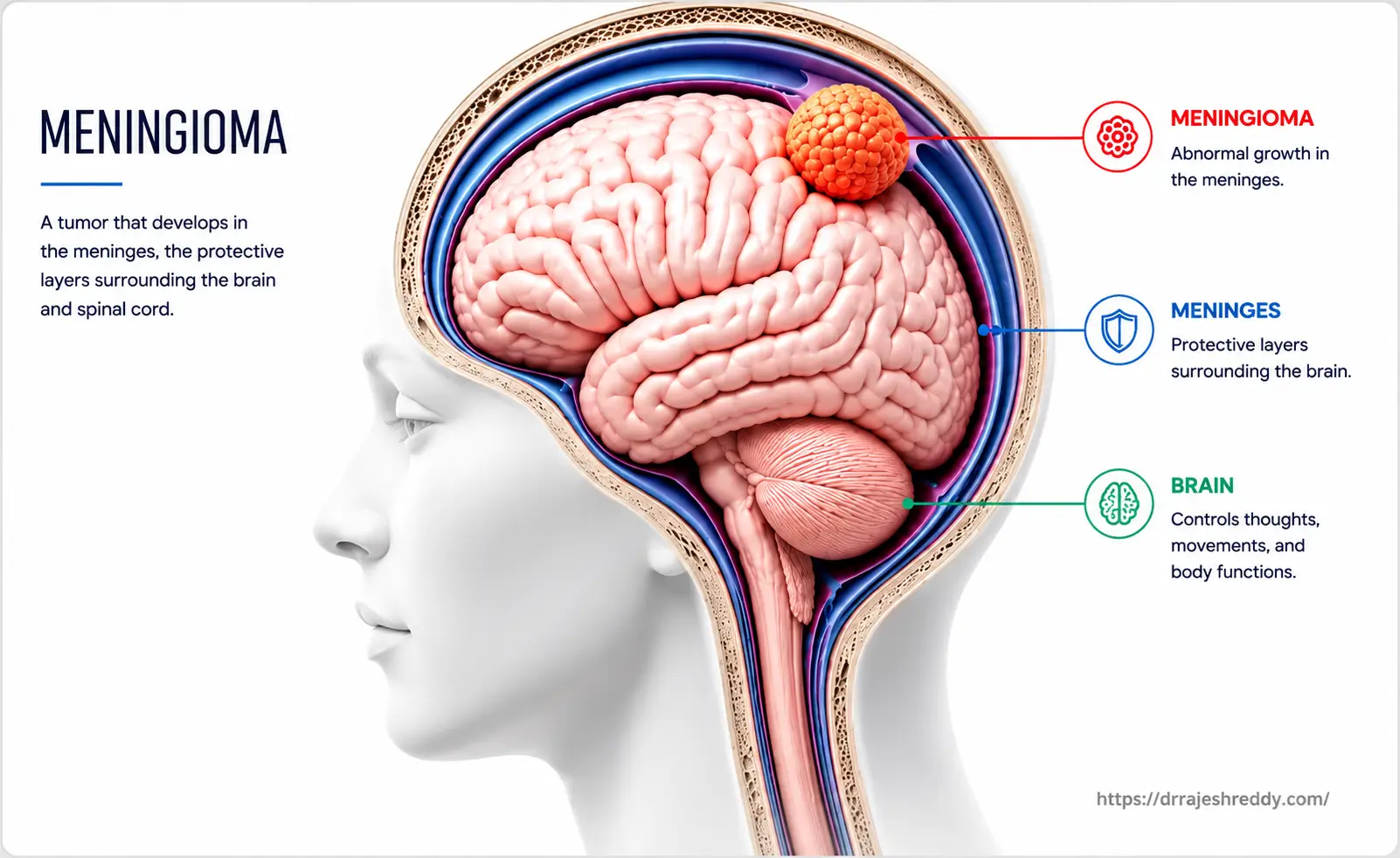
A meningioma is a tumor that arises from the meninges — the three-layered protective membrane that covers the brain and spinal cord. The meninges consist of three layers: the dura mater (outermost), the arachnoid mater (middle), and the pia mater (innermost). Meningiomas typically arise from the arachnoid cap cells of the arachnoid mater and grow inward, compressing rather than invading the brain.
Meningiomas are the most common primary brain tumor overall, accounting for approximately 37% of all primary brain tumors diagnosed. They are:
- More common in women than men (approximately 2:1 ratio)
- Most frequently diagnosed in middle-aged and older adults (peak incidence in the 6th and 7th decades)
- Usually benign (WHO Grade I) — approximately 80% of all meningiomas
- Occasionally atypical (WHO Grade II) — approximately 15–18%; higher recurrence risk
- Rarely malignant (WHO Grade III) — approximately 1–3%; aggressive behaviour, requires adjuvant treatment
Most meningiomas are discovered incidentally — found on an MRI or CT scan done for an unrelated reason, such as investigation of headaches or after a minor head injury. Many patients are understandably alarmed by the word 'tumor,' but the majority of incidentally discovered meningiomas are small, slow-growing, and can be safely monitored without immediate surgery.
Where Do Meningiomas Occur? Location and Its Impact on Treatment
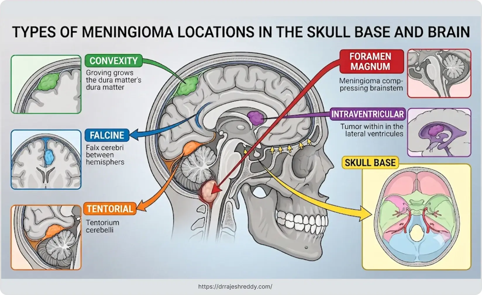
Meningiomas can arise anywhere along the meningeal lining — on the surface of the brain (convexity), at the skull base, within the ventricular system, or along the spinal cord. The location profoundly influences symptoms, surgical accessibility, and which treatment approach is most appropriate:
| Location | Frequency | Key Clinical Features |
|---|---|---|
| Convexity (brain surface) | Most common | Seizures, focal weakness, headaches; most surgically accessible |
| Parasagittal / Falcine | Common | Leg weakness (bilateral if large); may involve the superior sagittal sinus |
| Sphenoid Wing | Common | Visual loss, proptosis (eye bulging), headaches |
| Skull Base — Anterior Fossa | Moderate | Anosmia (loss of smell), personality change, visual disturbance |
| Skull Base — Posterior Fossa | Moderate | Hearing loss, facial numbness, balance problems; near cranial nerves |
| Cavernous Sinus | Less common | Eye movement problems, facial pain/numbness; highly vascular, complex surgery |
| Olfactory Groove | Less common | Loss of smell, personality change, frontal lobe syndrome |
| Petroclival | Rare | Multiple cranial nerve deficits; one of the most surgically challenging locations |
| Intraventricular | Rare | Hydrocephalus, headaches, cognitive changes |
| Suprasellar | Less common | Visual field defects (bitemporal), headache behind eyes, hormonal disturbances; often misdiagnosed as pituitary tumor |
| Foramen Magnum | Rare | Neck pain, progressive upper & lower limb weakness/numbness, gait disturbance; symptoms mimic cervical spine disease |
| Spinal | Less common | Back pain, limb weakness, bladder/bowel disturbance |
Skull base meningiomas — including those in the cavernous sinus, petroclival region, and posterior fossa — are among the most technically demanding neurosurgical cases. They require a surgeon with specific skull base expertise and, in many cases, the ability to perform pre-operative embolisation to reduce intraoperative blood loss. Dr. Rajesh Reddy's combined microsurgical and endovascular training makes him particularly well-suited to managing these complex cases.
Meningioma Symptoms: From Incidental to Severe
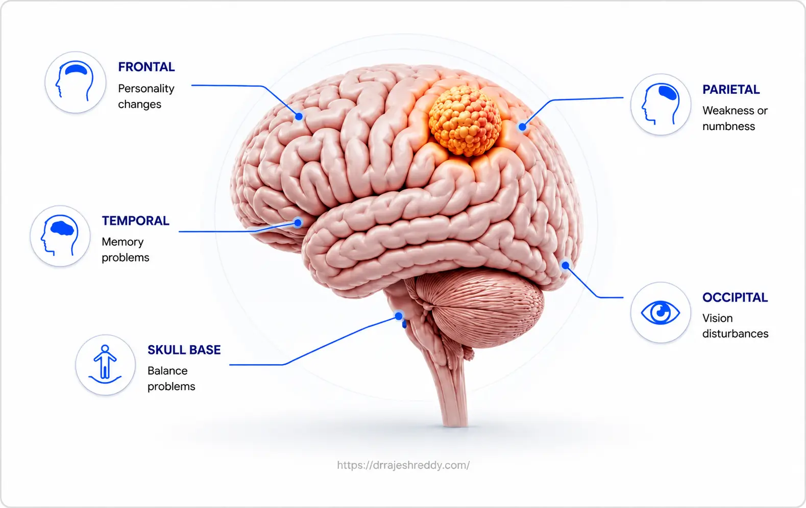
Meningioma symptoms depend entirely on the tumor's size and location. Many meningiomas cause no symptoms at all and are found by chance. When symptoms do occur, they reflect the compression of adjacent brain tissue, cranial nerves, or major venous sinuses:
General Symptoms From Raised Intracranial Pressure
- Progressive headaches — typically dull, pressure-like, worsening over weeks to months
- Nausea and vomiting — particularly in the morning
- Visual blurring or papilloedema on fundoscopic examination
Location-Specific Symptoms
Convexity meningioma: Seizures (often the only symptom for years), progressive weakness or numbness on one side of the body.
Parasagittal meningioma: Leg weakness — bilateral if the tumor involves both sides of the superior sagittal sinus; bladder urgency in some cases.
Sphenoid wing meningioma: Progressive visual loss in one eye, protrusion of the eyeball (proptosis), or headache behind the eye.
Olfactory groove meningioma: Loss of smell (anosmia) — often not noticed by the patient — followed by personality change and frontal lobe syndrome as the tumor enlarges.
Cavernous sinus meningioma: Drooping of the eyelid (ptosis), double vision, facial pain or numbness — from compression of cranial nerves III, IV, V, and VI.
Posterior fossa meningioma: Hearing loss, tinnitus, facial weakness or numbness, balance disturbance — from cranial nerve involvement.
Suprasellar meningioma: Progressive visual loss and visual field defects (typically bitemporal) from compression of the optic chiasm; headache behind the eyes; hormonal disturbances if the pituitary stalk is involved. Often misdiagnosed as a pituitary tumor — MRI characterisation is essential to distinguish the two.
Foramen magnum meningioma: Neck pain and suboccipital headache; progressive weakness and numbness starting in the arms and spreading to the legs; difficulty with fine hand movements; balance and gait disturbance. These tumors are notoriously difficult to diagnose early as their symptoms mimic cervical spine disease — any patient with progressive upper and lower limb neurological findings should have MRI of both the cervical spine and the posterior fossa.
Diagnosing a Meningioma
Meningiomas have a characteristic appearance on MRI that experienced neurosurgeons recognise readily. Key diagnostic features include:
MRI with Contrast — The Gold Standard
Meningiomas show intense, homogeneous enhancement with gadolinium contrast on MRI. The 'dural tail sign' — a linear enhancement of the dura adjacent to the tumor — is highly suggestive of meningioma. MRI also defines the tumor's relationship to adjacent brain, venous sinuses, cranial nerves, and major arteries — all critical for surgical planning.
CT Scan
CT is useful for assessing bony involvement — meningiomas can cause hyperostosis (thickening and remodelling) of the adjacent skull, which is visible on CT but not on MRI. CT angiography maps the tumor's blood supply and its relationship to major vessels — essential planning information for vascular meningiomas.
Cerebral Angiography
For highly vascular meningiomas — particularly skull base tumors supplied by meningeal and dural arteries — formal cerebral angiography (DSA) is performed to map the tumor's vascularity in detail. This is also the procedure through which pre-operative embolisation is performed, if indicated.
Is a Biopsy Needed Before Treatment?
For most meningiomas, the MRI appearance is so characteristic — a dural-based, homogeneously enhancing mass with a dural tail — that a pre-operative tissue biopsy is not required and has no role. The diagnosis is confirmed histologically when the tumor is surgically removed. Treatment decisions (surgery vs. SRS vs. observation) are made on the basis of imaging characteristics, clinical symptoms, and patient factors — not biopsy.
Meningioma Treatment Options: A Full Comparison
The treatment of meningioma is not one-size-fits-all. Dr. Rajesh Reddy evaluates each case across multiple dimensions — tumor size, location, grade, growth rate, patient age, neurological status, and comorbidities — before recommending a treatment approach. The table below outlines the full spectrum of options:
| Treatment Option | Best For | What It Involves | Key Limitation |
|---|---|---|---|
| Active Surveillance (Watch & Wait) | Small, asymptomatic, incidental meningiomas | Regular MRI every 6–12 months; no immediate intervention | Requires ongoing monitoring; not suitable if symptoms present or rapid growth seen |
| Microsurgical Resection | Symptomatic, accessible, or growing meningiomas | Craniotomy; tumor removed under operating microscope | Hospital stay 3–7 days; recovery 4–8 weeks |
| Stereotactic Radiosurgery (SRS) | Small to medium tumors (< 3 cm); residual tumor; elderly or high surgical risk patients | Single-session focused radiation; no incision; day procedure | Not suitable for large tumors; slow response (months to years) |
| Fractionated Radiotherapy | Large tumors near critical structures; atypical/malignant meningiomas post-surgery | Multiple radiation sessions over 5–6 weeks | Longer treatment course; potential radiation side effects |
| Pre-operative Embolisation / Standalone Embolisation | Highly vascular meningiomas before surgery; also in select patients as standalone tumour growth control (mechanism similar to SRS) | Angiography-guided occlusion of tumor blood supply 24–48 hrs before surgery; latest studies show standalone embolisation can prevent tumour growth in select patients | As pre-surgical adjunct, reduces blood loss significantly; as standalone — tumour remains in situ; not applicable for all locations |
Active Surveillance: When Watching Is the Right Choice
Not every meningioma needs immediate treatment. Current evidence and international guidelines support active surveillance (also called watch-and-wait) for:
- Small meningiomas (typically < 3 cm) that are asymptomatic and discovered incidentally
- Elderly patients or those with significant comorbidities for whom the risks of surgery outweigh the benefits
- Tumors in surgically challenging locations (e.g., cavernous sinus) where surgery carries high cranial nerve risk and the tumor is not causing symptoms
- Patients who decline intervention after informed counselling
During active surveillance, MRI is performed at regular intervals — typically at 3 months after diagnosis, then annually for several years, and less frequently thereafter if the tumor shows no growth. Studies show that approximately 50–60% of small, incidentally discovered meningiomas show no significant growth over 5 years. However, tumors that do show growth — or that develop new symptoms — are reassessed for intervention.
Active surveillance is not passive neglect — it is a deliberate, evidence-based strategy for meningiomas that do not require immediate treatment. Regular follow-up imaging is essential. If a tumor shows growth or new symptoms develop, the treatment plan is revised accordingly.
Stereotactic Radiosurgery: Treating Meningioma Without Open Surgery
Stereotactic radiosurgery (SRS) — delivered by platforms such as Gamma Knife, CyberKnife, or linear accelerator-based systems — is one of the most important advances in meningioma management. Despite the name, it is not conventional surgery. It uses multiple highly focused beams of radiation that converge precisely on the tumor, delivering a high dose to the target while sparing surrounding tissue. There is no incision, no anaesthesia (or only mild sedation), and no hospital admission in most cases.
When Is SRS the Right Choice for Meningioma?
- Small to medium-sized meningiomas — typically less than 3 cm in maximum diameter — in patients who have no significant pressure-related symptoms. Where a tumor under 3 cm is causing symptoms from mass effect (visual loss, focal weakness, seizures), surgery is generally preferable to SRS even within this size range, as SRS does not provide immediate decompression
- Tumors in surgically inaccessible locations — particularly cavernous sinus, petroclival, and posterior fossa meningiomas where open surgery carries significant cranial nerve risk
- Residual or recurrent tumor after previous surgery
- Elderly patients or those with medical comorbidities who are not good surgical candidates
- Patients who prefer a non-surgical approach and whose tumor characteristics are suitable
How Effective Is SRS for Meningioma?
For WHO Grade I meningiomas treated with SRS, long-term tumour control rates (defined as no growth or shrinkage on imaging) are excellent: approximately 90–95% at 5 years and 85–90% at 10 years. Many tumors shrink after SRS, though this response is gradual — occurring over months to years. SRS does not remove the tumor; it stops it from growing by damaging the DNA of rapidly dividing tumor cells.
Cranial nerve preservation rates after SRS for cavernous sinus meningiomas are generally superior to open surgery — which is one of the main reasons SRS is preferred for tumors in this location when the tumor is of an appropriate size.
Radiation for Grade II and III Meningiomas
For atypical (Grade II) and anaplastic (Grade III) meningiomas, post-operative radiation is an important part of the treatment plan. These higher-grade tumors have significantly higher recurrence rates even after complete resection, and adjuvant radiation — using standard fractionated external beam radiotherapy or SRS for smaller residual volumes — is recommended. The decision on radiation modality and framing is made in the multidisciplinary tumour board, tailored to the extent of resection, tumor grade, and the patient's overall condition.
Fractionated Stereotactic Radiotherapy (FSRT)
For larger meningiomas that are too big for single-session SRS, or for tumors very close to critical structures (optic nerves, brainstem), fractionated stereotactic radiotherapy delivers the radiation dose in multiple smaller sessions (typically 25–30 fractions over 5–6 weeks). This allows a higher total dose to be given safely to larger targets near sensitive structures.
Microsurgical Meningioma Resection: When Surgery Is the Answer
For symptomatic meningiomas, large tumors, tumors showing rapid growth, or cases where a histological diagnosis is required, microsurgical resection is the definitive treatment. The goal is Simpson Grade I or II resection — complete removal of the tumor including its dural attachment — which carries the lowest recurrence risk.
The Simpson Grading System: Measuring Completeness of Resection
The extent of meningioma resection is described using the Simpson grading system. The grade achieved directly correlates with the risk of recurrence:
| Simpson Grade | Extent of Resection | 10-Year Recurrence Risk |
|---|---|---|
| Grade I | Complete removal including dural attachment and any involved bone | ~9% |
| Grade II | Complete tumor removal; dural attachment cauterised (coagulated) | ~19% |
| Grade III | Complete tumor removal; dural attachment not resected or coagulated | ~29% |
| Grade IV | Subtotal removal — tumor left behind intentionally | ~44% |
| Grade V | Biopsy only | Near 100% |
Achieving Simpson Grade I resection is the surgical ideal, but is not always possible when the tumor involves a major venous sinus, cranial nerves, or critical arteries. In these cases, intentional subtotal resection (Simpson Grade IV) combined with post-operative SRS for the residual tumor may offer the best balance of tumour control and safety.
Pre-operative Embolisation: Reducing Surgical Risk for Vascular Meningiomas
Meningiomas — particularly skull base tumors — have a rich blood supply from dural arteries. Large, highly vascular tumors can cause significant blood loss during surgery, increasing operative risk. Pre-operative embolisation addresses this by occluding the tumor's feeding arteries 24–48 hours before surgery. Performed through a small arterial access point in the groin under X-ray guidance, embolisation significantly reduces intraoperative bleeding and can make a complex tumour resection substantially safer.
Emerging Role in Tumour Growth Control: Latest studies have shown that in a select group of patients — particularly those with small asymptomatic meningiomas who are not candidates for surgery or SRS — standalone embolisation (without subsequent surgery) can prevent tumour growth by devascularising the tumour and depriving it of its blood supply. The mechanism is similar to SRS: the goal is tumour growth arrest rather than immediate removal. This is an evolving area and patient selection for standalone embolisation is discussed on a case-by-case basis at Dr. Rajesh Reddy's multidisciplinary tumour board.
Dr. Rajesh Reddy's fellowship training in Interventional Neuroradiology at the University Hospital, Zurich — and his endovascular expertise — allows him to perform pre-operative meningioma embolisation himself, providing seamless integration between the embolisation and surgical phases of treatment. This is a significant advantage over centres where these two procedures are performed by different specialists.
Neuronavigation and Intraoperative Monitoring
All meningioma surgeries performed by Dr. Rajesh Reddy utilise neuronavigation — a real-time 3D GPS-like imaging system that tracks the surgeon's instruments relative to the patient's pre-operative MRI throughout the procedure. For tumors near cranial nerves — particularly in the skull base — intraoperative neurophysiological monitoring (cranial nerve EMG, auditory evoked potentials) provides real-time feedback on nerve function, allowing the surgeon to adjust the dissection before a deficit occurs rather than after.
Atypical and Malignant Meningiomas (WHO Grade II and III)
Approximately 15–20% of meningiomas are atypical (WHO Grade II) or malignant (WHO Grade III). These tumors have higher rates of recurrence after surgery and require a more aggressive treatment approach:
WHO Grade II (Atypical) Meningioma: Higher mitotic activity than Grade I. Recurrence risk after gross total resection is approximately 30–40% at 5 years. Post-operative radiotherapy is increasingly recommended even after complete resection, particularly for tumors in locations prone to recurrence.
WHO Grade III (Malignant) Meningioma: Rare (approximately 1–3% of all meningiomas) but behaves aggressively — invading brain tissue and recurring despite surgery and radiation. Requires maximum surgical resection followed by adjuvant radiotherapy. Chemotherapy has limited efficacy; clinical trial participation should be considered.
The management of atypical and malignant meningiomas is determined in a tumour board meeting involving neurosurgery, radiation oncology, and medical oncology, with treatment plans tailored to the individual patient's tumor grade, location, extent of resection, and performance status.
Recovery After Meningioma Treatment
After Microsurgical Resection
- Hospital stay: typically 3–7 days depending on tumor size, location, and any post-operative neurological changes
- ICU monitoring for the first 24–48 hours; neurological observations every 2–4 hours
- Post-operative MRI within 24–72 hours to assess extent of resection
- Steroids tapered over 1–2 weeks to manage brain swelling
- Most patients with convexity meningiomas return to normal activities within 4–6 weeks
- Skull base meningioma patients may have a longer recovery depending on cranial nerve involvement
After Stereotactic Radiosurgery
- Day procedure — patients go home the same day or the following morning
- Mild headache and fatigue in the first 1–2 weeks; managed with steroids and analgesics
- First follow-up MRI at 3–6 months; then annually
- Tumour response (shrinkage or stabilisation) develops gradually over months to years
- Return to normal activities within days of the procedure
After Active Surveillance
- Regular MRI appointments as scheduled — typically at 3 months, then annually
- Any new or worsening symptoms should prompt an earlier review
- Lifestyle is unrestricted — meningioma surveillance does not require activity limitation