
Introduction
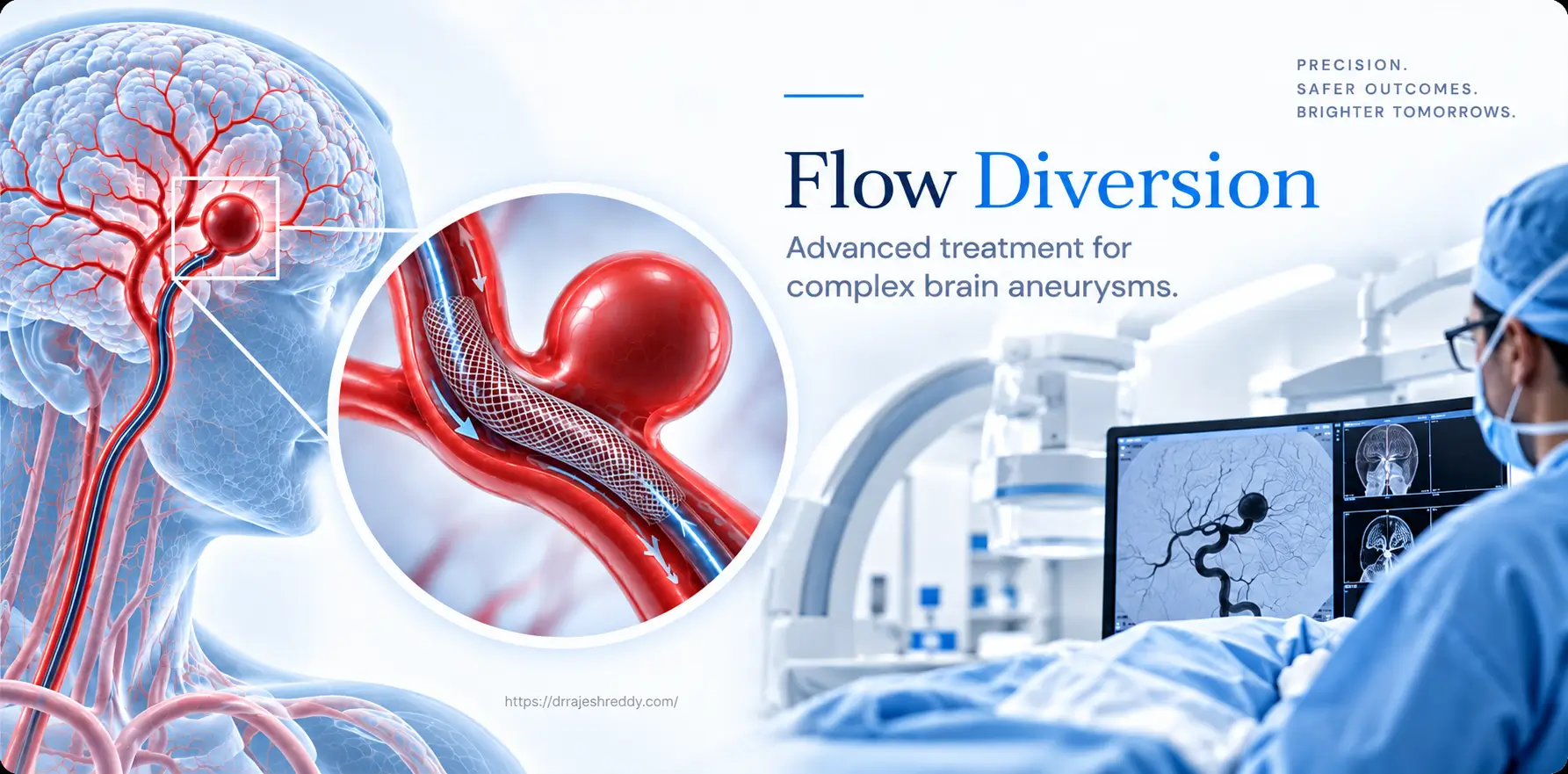
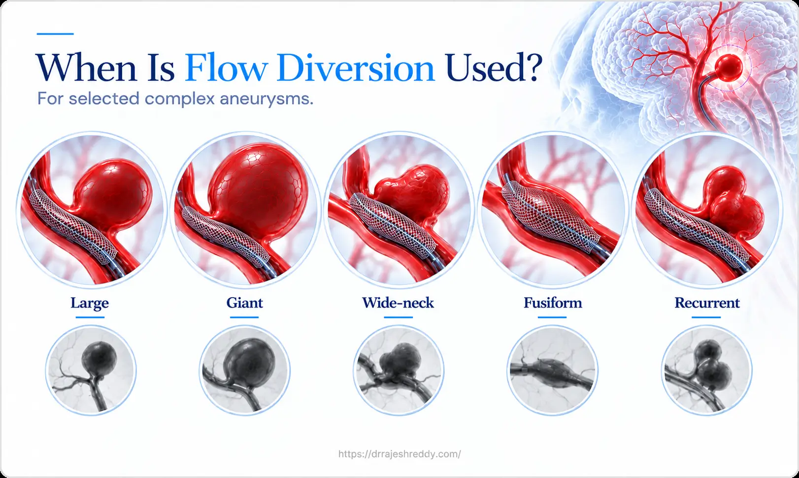
For most brain aneurysms — small, saccular, with a well-defined neck — endovascular coiling or surgical clipping provides effective, durable treatment. But a significant subset of aneurysms do not fit neatly into either category. Large and giant aneurysms whose sheer volume makes complete coil packing impractical. Fusiform aneurysms with no defined neck that cannot be coiled or clipped conventionally. Wide-neck aneurysms where coils herniate into the parent artery. Giant partially thrombosed aneurysms causing TIAs, seizures, or progressive weakness by pressing on adjacent brain. For these complex cases, flow diversion has transformed what is achievable.
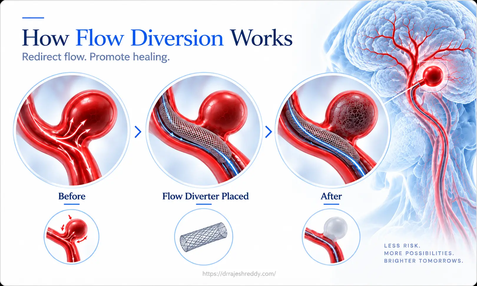
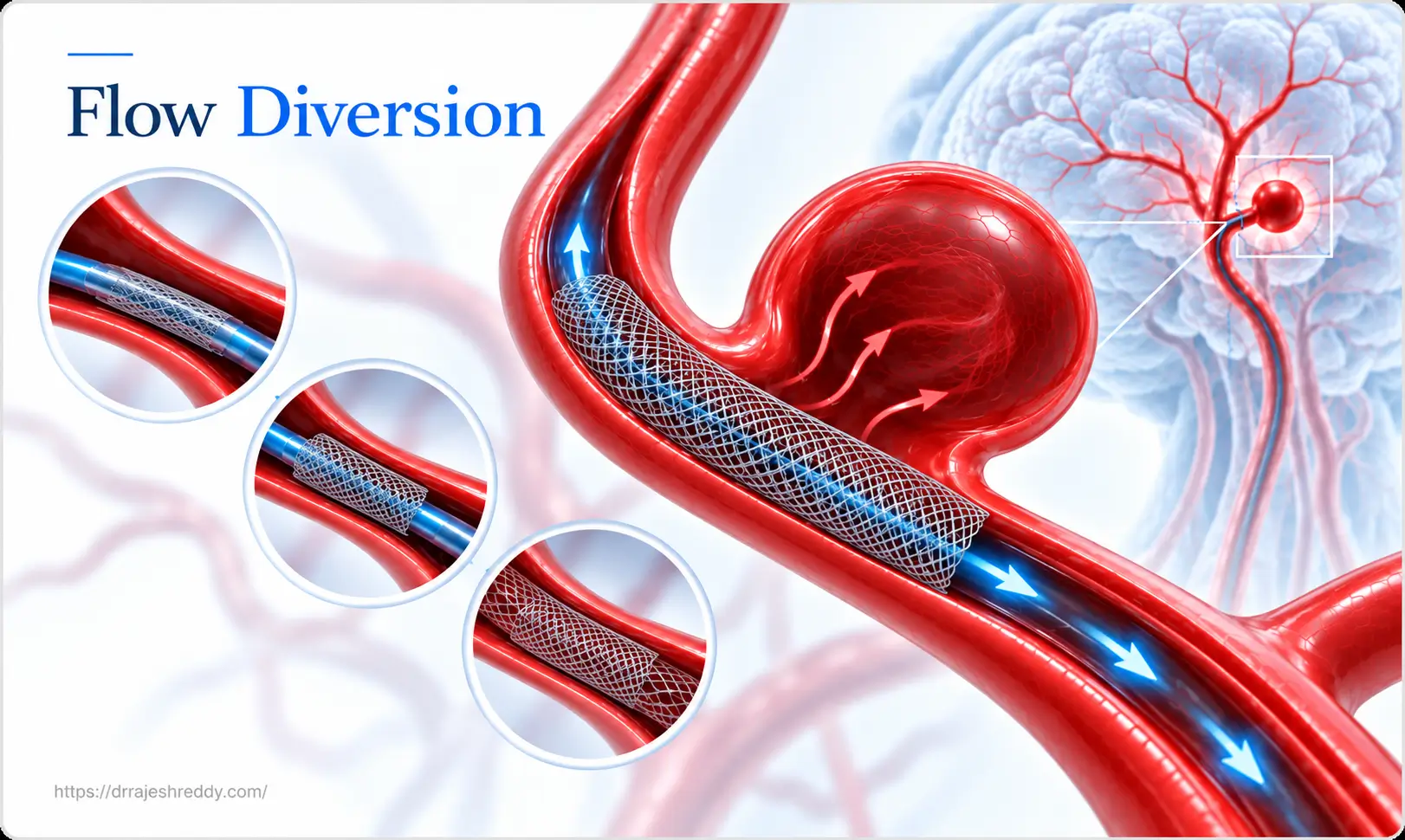
Flow diversion is a fundamentally different concept from conventional coiling. Rather than filling the aneurysm sac with coils from the inside, a flow diverter is deployed in the parent artery — across the aneurysm neck — and works from the outside in. The dense mesh of the device redirects blood flow away from the aneurysm, causes progressive thrombosis within the sac over weeks to months, and simultaneously scaffolds the parent artery to form a new, smooth arterial wall that permanently excludes the aneurysm. The result is a biological cure, not a mechanical plug.
Dr. Rajesh Reddy Sannareddy, Senior Consultant Endovascular Neurosurgeon in Hyderabad, is trained in the full range of flow diversion devices — including the Pipeline Embolisation Device, FRED, Surpass, SILK, p64, and Derivo — with hands-on experience from his Fellowship in Interventional Neuroradiology (FINR) at the University Hospital, Zurich and visiting scholar exposure at Barrow Neurological Institute, Phoenix. He is one of the few neurosurgeons in Hyderabad with dedicated training in these advanced endovascular techniques.
What Is Flow Diversion and How Does It Work?
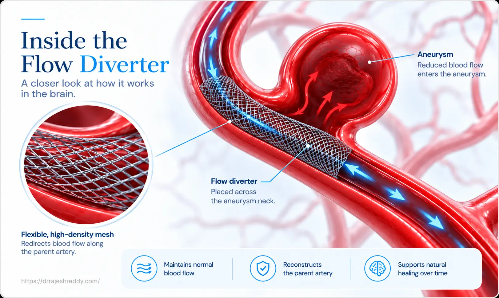
A flow diverter is a high-porosity braided mesh stent deployed within the parent artery — the artery from which the aneurysm arises — across the aneurysm neck. Unlike conventional stents, flow diverters have very high metal surface coverage — typically 30–35% of the luminal wall — achieved by braiding dozens of fine metal wires into a mesh tube.
The Two Mechanisms of Flow Diversion
- Haemodynamic flow redirection: The high metal coverage acts as a flow barrier at the aneurysm neck. Inflow into the aneurysm sac is reduced by approximately 60–80% immediately after deployment. Blood that previously entered the sac with each heartbeat is redirected along the stent into the distal artery, no longer pushing against the aneurysm wall.
- Progressive aneurysm thrombosis and neointimal healing: With reduced flow in the sac, blood stagnates and progressively clots. Over weeks to months, the thrombus organises and is replaced by connective tissue. Simultaneously, endothelial cells migrate across the stent struts at the aneurysm neck, forming a new arterial wall that permanently seals the aneurysm from the circulation — a healing process, not just a mechanical blockage.
Flow Diversion Devices: The Full Spectrum
Several flow diversion devices are available. Dr. Rajesh Reddy's training includes experience with the following extrasaccular flow diverters:
| Device | Mechanism | Best Suited For | Key Feature |
|---|---|---|---|
| Pipeline Embolisation Device (PED) | High-porosity braided cobalt-chromium mesh across aneurysm neck | Large/giant ICA aneurysms; paraclinoid ICA; fusiform; recurrent wide-neck | Most studied; occlusion rates 85–93% at 12 months; FDA and CE approved |
| FRED (Flow Re-Direction Endoluminal Device) | Dual-layer braided nitinol; inner high-mesh layer over neck | Similar to Pipeline; also wide-neck MCA and basilar aneurysms | Visible inner/outer layers on fluoroscopy; easier deployment confirmation |
| Surpass Streamline | Braided cobalt-chromium; higher metal coverage than Pipeline | Large aneurysms; complex neck geometry | High mesh density; single device covers wider neck widths |
| SILK Flow Diverter | Low-profile braided nitinol; high mesh coverage | Wide-neck fusiform and saccular aneurysms | Very flexible; useful for tortuous anatomy |
| p64 Flow Modulation Device | Braided nitinol; retrievable and repositionable | Wide-neck aneurysms; need for precise placement | Fully retrievable before detachment — high safety margin |
| Derivo Embolisation Device | Braided nitinol with anti-thrombogenic surface | Wide-neck saccular and fusiform aneurysms | Surface modification may reduce thromboembolism risk |
| Pipeline Flex with Shield Technology | PED with phosphorylcholine anti-thrombogenic coating | Patients who may not tolerate standard dual antiplatelet therapy | Coating reduces platelet adhesion; potential for modified antiplatelet regimen |
The Pipeline Embolisation Device — The Most Studied Flow Diverter
The Pipeline consists of 48 cobalt-chromium and platinum-tungsten wires braided into a self-expanding cylindrical mesh. The Pipeline Flex — the current generation — incorporates a resheathing capability: the partially deployed device can be recaptured and repositioned if initial placement is suboptimal, significantly improving deployment safety and accuracy. The Pipeline Flex with Shield Technology adds a phosphorylcholine surface coating that reduces platelet adhesion and may allow modified antiplatelet regimens in selected patients.
When Is Flow Diversion the Right Treatment?
Flow diversion is not a first-line treatment for all aneurysms — it is a specialised tool for cases where conventional approaches are inadequate or associated with unacceptably high recurrence rates:
| Aneurysm Type | Why Flow Diversion Is Preferred | Occlusion Rate |
|---|---|---|
| Large aneurysm (10–24 mm) | Coiling alone has high recanalisation; sac volume makes complete coil packing impractical; flow diverter treats entire neck in one device | 85–90% at 12 months |
| Giant aneurysm (≥ 25 mm) | Surgery carries very high morbidity; coiling alone invariably recanalises; flow diversion achieves progressive occlusion without entering the sac | 73–86% at 6 months; continues improving to 90%+ at 24 months |
| Fusiform aneurysm | No defined neck — cannot be coiled or clipped conventionally; flow diverter lines the parent artery, excluding the fusiform segment | Variable; depends on length of fusiform segment |
| Wide-neck saccular aneurysm | Dome:neck ratio < 1.5; coils herniate into parent artery; stent-assisted coiling has higher recurrence; flow diverter treats neck without entering sac | 80–90% at 12 months |
| Paraclinoid ICA aneurysm | Optimal anatomical fit for Pipeline; alternative is complex skull base surgery | 85–93% at 12 months (best results in class) |
| Recurrent aneurysm after coiling | Coil mass compacted; re-coiling has high re-recurrence; flow diverter treats residual neck and prevents further recanalisation | 75–85% at 12 months |
| Blister / Pseudoaneurysm | Thin fragile wall — clipping risks tearing; coiling has poor geometry; flow diverter supports the parent artery wall and promotes healing | Moderate; often requires supplemental coiling |
| Giant partially thrombosed aneurysm | Embolic TIAs, seizures, or progressive hemiparesis from mass effect; flow diversion causes progressive thrombosis and sac shrinkage, reducing mass effect and eliminating embolic source | Variable; sac shrinkage continues over 12–24 months |
The Flow Diversion Procedure: What Happens
Pre-Procedure: Dual Antiplatelet Preparation
Flow diversion requires mandatory dual antiplatelet therapy before the procedure. Aspirin 150 mg and clopidogrel 75 mg or Ticagrelor 180 mg/day are started 5–7 days before planned deployment. Platelet function testing (VerifyNow P2Y12 assay) is performed on the day of procedure in selected cases to confirm adequate clopidogrel effect — poor responders may require prasugrel or ticagrelor. Inadequate antiplatelet effect at deployment significantly increases the risk of in-stent thrombosis.
DSA and 3D Angiography — Sizing the Device
A high-quality 3D rotational DSA is essential for flow diverter sizing. The diameter and length of the device must precisely match the parent artery — too large and it will not fully appose the vessel wall; too small and it will be undersized at the neck. Device sizing software provides measurements of the parent artery diameter proximal and distal to the aneurysm, neck width, and the length of artery requiring coverage.
Device Deployment
Under general anaesthesia, a guide catheter is positioned in the internal carotid or vertebral artery. A microcatheter is navigated past the aneurysm neck into the distal parent artery. The flow diverter is deployed by slowly retracting the microcatheter — allowing the mesh to expand and conform to the arterial wall across the aneurysm neck. Correct apposition is confirmed with contrast injections and DSA. An immediately post-deployment DSA confirms neck coverage and visible flow reduction in the sac. Intraprocedural Dyna CT or Vaso CT softwares help in assessing apposition of stent to the vessel wall.
Adjunctive Coiling
For aneurysms more than 1.5 cm where spontaneous thrombosis after flow diversion alone would be slow, adjunctive coiling within the sac may be performed at the same sitting. The coils partially fill the sac and accelerate thrombosis, reducing the risk of delayed aneurysm rupture during the progressive occlusion phase and also helping to decompress the mass effect more rapidly.
Outcomes and Evidence for Flow Diversion
Occlusion Rates
- PUFS trial (Pipeline for Uncoilable or Failed Aneurysms): 73% complete occlusion at 6 months and 86.8% at 180 days for large/giant uncoilable ICA aneurysms — the landmark study that led to FDA approval
- IntrePED registry: 5.6% major stroke or death rate in 793 patients across 21 centres — establishing real-world safety data
- PREMIER trial (Pipeline for ICA aneurysms 7–12 mm): 76.9% complete occlusion at 12 months — expanding Pipeline indication beyond large/giant aneurysms
- Multiple single-centre series: 85–93% complete occlusion at 12 months for paraclinoid ICA aneurysms — the best flow diversion results reported for any anatomical location
Sac Shrinkage After Flow Diversion
One of the most clinically rewarding outcomes of successful flow diversion — particularly for large and giant aneurysms causing mass effect — is progressive shrinkage of the aneurysm sac over months to years. As the organised thrombus reabsorbs and the sac collapses, symptoms from compression — visual loss, cranial nerve palsies, headache, hemiparesis — gradually improve. For giant partially thrombosed aneurysms, sac shrinkage also eliminates the embolic source responsible for TIAs and reduces seizure frequency. This shrinkage is a unique advantage of flow diversion over coiling, where the sac volume is maintained by the coil mass.
Transient Worsening After Flow Diversion
A distinctive phenomenon after flow diversion of large and giant aneurysms is transient worsening of mass effect symptoms in the weeks after treatment. As the aneurysm sac thromboses, the clot may initially swell — temporarily increasing volume and worsening compression on adjacent structures (optic nerve, cranial nerves, brainstem, or adjacent cortex). This perianeurysmal oedema is managed with short courses of oral corticosteroids and typically resolves as the thrombus organises and shrinks. Patients should be counselled that symptoms may transiently worsen before improving — this is expected and does not indicate treatment failure.
Risks of Flow Diversion: An Honest Assessment
Flow diversion is a more complex procedure than standard coiling and carries specific risks that patients must understand:
- Thromboembolic stroke: The most significant complication — approximately 3–6% across major series. Pre-procedural dual antiplatelet therapy and platelet function testing are the primary preventive measures.
- Delayed aneurysm rupture: A paradoxical but rare complication (approximately 1–2%) — particularly in large and giant aneurysms — during the progressive thrombosis phase. Adjunctive coiling may reduce this risk.
- Perforator artery occlusion: Small perforating arteries covered by the device mesh may rarely be affected — approximately 1–3% in published series. Modern flow diverters are designed to preserve perforator flow.
- In-stent stenosis or thrombosis: Occurs in approximately 3–5% of cases. Stenosis is usually asymptomatic; acute thrombosis requires emergency endovascular management.
- Incomplete occlusion: Approximately 10–15% of aneurysms do not achieve complete occlusion at 24 months — requiring assessment for supplemental coiling or a second flow diverter.
Antiplatelet Therapy and Follow-Up After Flow Diversion
Antiplatelet Protocol
Dual antiplatelet therapy — aspirin (75–150 mg) and clopidogrel (75 mg) or Ticagrelor 180 mg/day — is required for 6 months after flow diverter deployment. After 6 months, clopidogrel is stopped and aspirin continued for at least 3–5 years or indefinitely (duration at the discretion of the treating doctor). Stopping either medication without consulting Dr. Rajesh Reddy risks in-stent thrombosis. Any planned surgical procedure during the dual antiplatelet period — including dental surgery — must be discussed in advance.
Follow-Up Angiography Schedule
- 6 months: DSA — primary assessment of aneurysm occlusion; confirms device apposition and absence of in-stent stenosis
- 12 months: DSA or MRA or CTA — second assessment; most aneurysms that will achieve complete occlusion have done so by this point
- 24 months: MRA or CTA — confirms sustained occlusion; identifies late incomplete occlusion requiring retreatment
- Annual MRA, CTA or Selective DSA thereafter: for select cases of large/giant aneurysms — long-term surveillance to confirm ongoing sac shrinkage and absence of recanalisation
Why Choose Dr. Rajesh Reddy for Flow Diversion in Hyderabad?
Flow diversion requires a level of endovascular expertise and judgement that goes beyond standard coiling. Correct device sizing, precise deployment, management of deployment challenges, and recognition of complications are skills built through dedicated training and case volume.
Flow Diversion Expertise — Dr. Rajesh Reddy
- FINR — Fellowship in Interventional Neuroradiology, University Hospital, Zurich, a high-volume European cerebrovascular centre
- Visiting Scholar — Barrow Neurological Institute, Phoenix (one of the highest-volume cerebrovascular centres in North America); Okayama University, Japan; Klinikum Stuttgart, Germany
- Double board certified neurosurgeon (MCh + DNB) — surgical understanding of cerebrovascular anatomy underpins every endovascular decision
- Trained in the full spectrum: simple coiling, balloon-assisted coiling, stent-assisted coiling, Flow Diversion — treatment recommendation based on aneurysm anatomy, not available technique
- Several complex neurosurgical and endovascular procedures across a 15+ year career
- Clinical acumen and judgement gained through years of training with leading global experts
Frequently Asked Questions
Complex Aneurysm? Consult Dr. Rajesh Reddy for Flow Diversion in Hyderabad
If you have been told your aneurysm is large, giant, wide-necked, fusiform, or 'difficult to treat' — or if a previous coiling procedure has recurred — Dr. Rajesh Reddy Sannareddy offers flow diversion consultations with access to the full range of intrasaccular and extrasaccular flow diverter devices. Book in person or via the Dr. Reddy's Neuro Care app on Google Play.
Related Articles
Continue reading to understand the full spectrum of aneurysm treatment options:
Brain Aneurysm Symptoms: When Is It an Emergency?
Thunderclap headache, warning signs and when immediate emergency care is needed.
Read GuideRuptured vs. Unruptured Brain Aneurysm: Key Differences
Symptoms, emergency management and treatment considerations.
Read GuideAneurysm Coiling vs. Surgical Clipping
Understand the differences between endovascular coiling and microsurgical clipping.
Read GuideEndovascular Coiling for Brain Aneurysm in Hyderabad
Minimally invasive detachable coil embolisation procedure guide.
Read GuideSubarachnoid Hemorrhage (SAH): Causes and Treatment
Causes, emergency management, vasospasm prevention and critical care.
Read GuideLife After Brain Aneurysm Surgery: Recovery Guide
Recovery timeline, antiplatelet protocols, and rehabilitation guide.
Read GuideArteriovenous Malformation (AVM) Treatment Hyderabad
Diagnosis and treatment options for cerebral arteriovenous malformations.
Read GuideBrain Aneurysm Treatment in Hyderabad (Pillar Page)
Explore the main pillar page for endovascular & surgical aneurysm treatments.
Read Pillar Page