Introduction & The Pituitary Gland
Pituitary tumors are among the most misunderstood diagnoses in neurosurgery — not because they are rare (they account for roughly 15% of all intracranial tumors) but because their symptoms are hormonal rather than neurological. Patients with a pituitary tumor may spend months or years consulting gynaecologists for menstrual irregularity, endocrinologists for unexplained weight gain, or ophthalmologists for progressive vision loss — before a pituitary MRI finally connects the dots.
The further good news is that pituitary tumor surgery has undergone a revolution in the past two decades. The traditional approach — opening the skull to access the pituitary from above — has been largely replaced by the endoscopic transsphenoidal approach: operating through the patient's own nostrils, via the sphenoid sinus, to reach the pituitary gland at the skull base. No scalp incision. No craniotomy. No brain retraction. Most patients go home within 2–3 days.
Dr. Rajesh Reddy Sannareddy, Senior Consultant Neurosurgeon in Hyderabad, performs endoscopic pituitary surgery with high-definition endoscopy, neuronavigation, and intraoperative fluoroscopy. His training at the University Hospital, Zurich and visiting scholar experience at Barrow Neurological Institute, Phoenix — a global reference centre for skull base and pituitary surgery — informs every aspect of his pituitary surgical practice.
The Pituitary Gland: The Brain's Master Hormone Controller
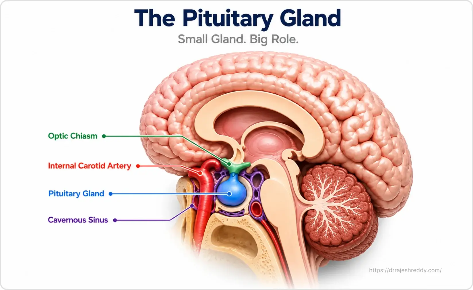
The pituitary gland is a small, pea-sized structure — barely 1 cm in diameter — located at the base of the brain in a bony cavity called the sella turcica (Latin for 'Turkish saddle'). Despite its tiny size, the pituitary gland is often called the 'master gland' of the body because it produces and regulates hormones that control virtually every major endocrine system.
Hormones Produced by the Pituitary
- Prolactin (PRL) — stimulates milk production after childbirth; regulates reproductive function
- Growth Hormone (GH) — regulates body composition, bone growth, and metabolism
- Adrenocorticotrophic Hormone (ACTH) — stimulates the adrenal glands to produce cortisol
- Thyroid-Stimulating Hormone (TSH) — controls thyroid hormone production
- Luteinising Hormone (LH) and Follicle-Stimulating Hormone (FSH) — regulate reproductive function in both sexes
- Antidiuretic Hormone (ADH / Vasopressin) — regulates water balance; produced in the hypothalamus, released by posterior pituitary
- Oxytocin — regulates uterine contractions and milk ejection
The pituitary gland sits directly below the optic chiasm — where the optic nerves from both eyes cross — and is flanked on each side by the cavernous sinuses, which contain the carotid arteries and the cranial nerves controlling eye movement. These anatomical relationships explain why pituitary tumors cause visual field defects when they grow upwards, and eye movement problems when they grow sideways.
Types of Pituitary Tumors: Functioning vs. Non-Functioning
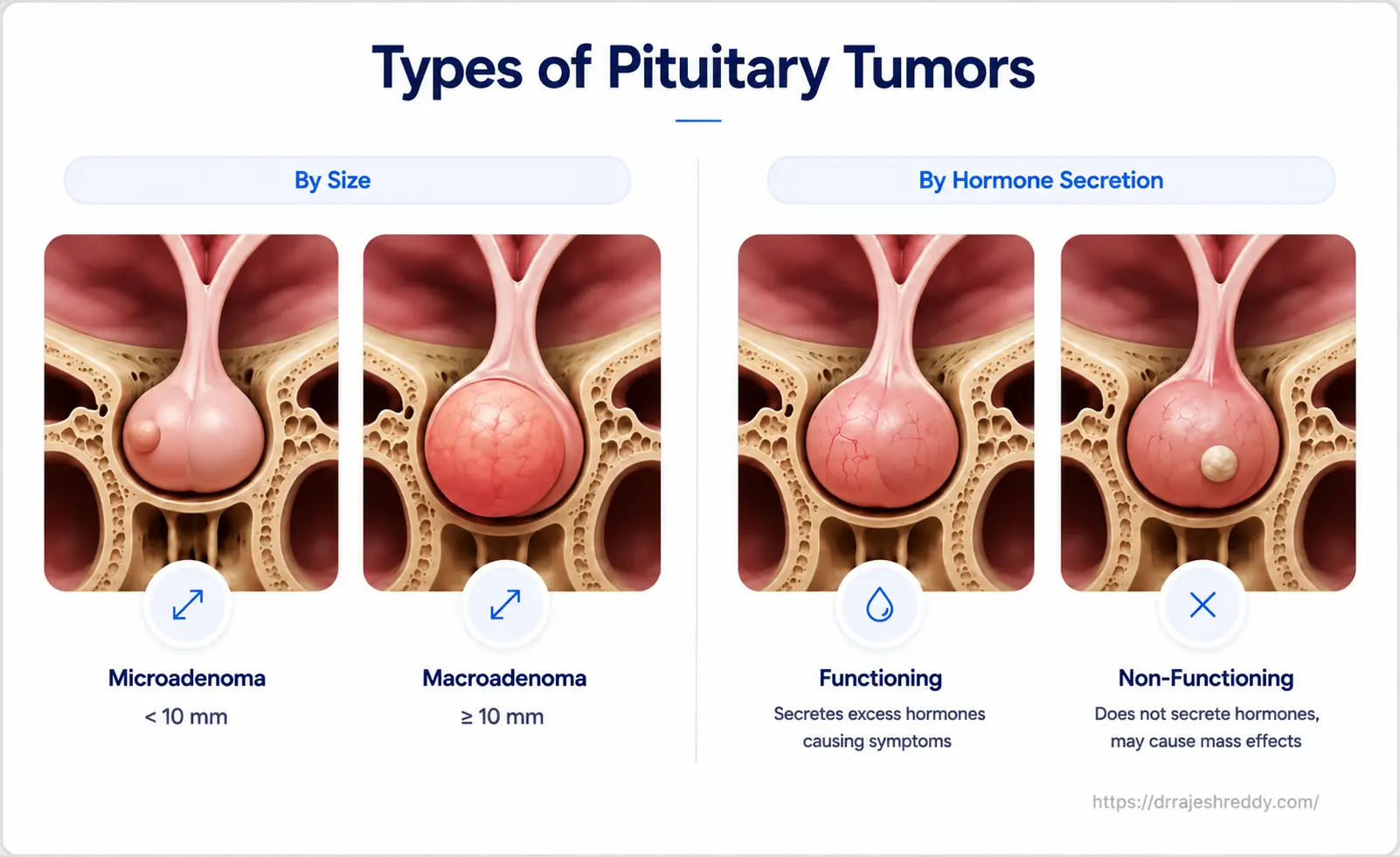
Pituitary adenomas are benign tumors of the pituitary gland. They are classified by size and by whether they secrete hormones. Understanding this classification is essential, because it determines the symptoms, the diagnosis, and critically — the treatment approach.
By Size
Microadenoma: Less than 10 mm in diameter. Often found incidentally or during workup for hormonal symptoms. Treated medically or surgically depending on the hormone secreted.
Macroadenoma: 10 mm or larger. More likely to cause mass effect — compressing the optic chiasm, invading the cavernous sinus, or causing pituitary insufficiency. Almost always requires surgery.
By Hormone Secretion: A Full Reference Table
| Tumor Type | Hormone Secreted | Clinical Syndrome | First-Line Treatment |
|---|---|---|---|
| Prolactinoma | Prolactin (PRL) | Irregular periods, galactorrhoea, infertility (women); erectile dysfunction, infertility (men) | Dopamine agonist medication (cabergoline / bromocriptine) — surgery if medication fails |
| Corticotroph Adenoma | ACTH | Cushing's disease — weight gain, stretch marks, hypertension, diabetes, mood changes | Endoscopic transsphenoidal surgery — first-line in most cases |
| Somatotroph Adenoma | Growth Hormone (GH) | Acromegaly — enlarged hands, feet, jaw; sweating; joint pain; carpal tunnel | Endoscopic transsphenoidal surgery — first-line; somatostatin analogues if surgery incomplete |
| Thyrotroph Adenoma | TSH | Hyperthyroidism — palpitations, weight loss, tremor; rare cause of hyperthyroidism | Endoscopic transsphenoidal surgery |
| Gonadotroph Adenoma | FSH / LH (or none effectively) | Usually non-functioning; presents as macroadenoma with visual loss and headaches | Endoscopic transsphenoidal surgery |
| Non-Functioning Adenoma (NFA) | None | Mass effect — bitemporal visual field loss, headache, pituitary insufficiency | Endoscopic transsphenoidal surgery if symptomatic or growing |
| Craniopharyngioma | None (not a true adenoma) | Visual loss, hormonal deficiency, diabetes insipidus, cognitive changes | Endoscopic / microsurgical resection; may need radiation |
Pituitary Tumor Symptoms: Why They Are Often Missed
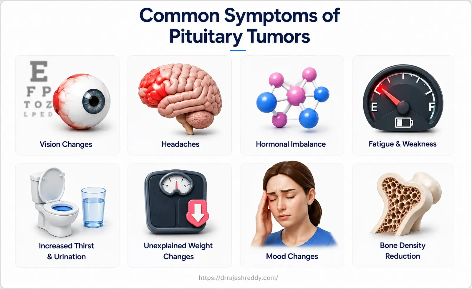
Pituitary tumor symptoms fall into two categories that often develop so gradually that patients adapt to them before recognising they are abnormal:
Hormonal (Endocrine) Symptoms — The Most Commonly Missed
Prolactinoma: Irregular or absent menstrual periods; unexpected milk discharge from the breasts (galactorrhoea) without pregnancy; difficulty conceiving in women. In men: reduced libido, erectile dysfunction, infertility, and rarely — gynaecomastia (breast enlargement). Because these symptoms overlap with many other causes, patients are often investigated and treated for other conditions before the prolactinoma is identified.
Cushing's Disease (ACTH excess): Progressive weight gain predominantly around the abdomen and face ('moon face'); purple stretch marks (striae) on the abdomen, thighs, and arms; easy bruising; high blood pressure; elevated blood sugar (diabetes); muscle weakness; mood disturbances including depression. The diagnosis requires specific cortisol testing — not just a blood glucose check.
Acromegaly (GH excess): Gradual enlargement of the hands and feet (patients notice rings and shoes no longer fitting); coarsening of facial features; enlarged jaw, brow, and nose; excessive sweating; joint pain; carpal tunnel syndrome; obstructive sleep apnoea. These changes are so gradual that patients — and their families — often fail to notice them until they compare old photographs.
Mass Effect Symptoms — Pressing on Surrounding Structures
- Bitemporal hemianopia: The classic visual field defect of pituitary macroadenoma — loss of the outer (temporal) half of the visual field in both eyes, caused by compression of the optic chiasm directly above the pituitary. Patients often describe walking into door frames or difficulty seeing objects to their side.
- Headache: Dull, often retro-orbital (behind the eyes); caused by pressure within the sella turcica or by tumour extension beyond it.
- Pituitary insufficiency (hypopituitarism): When a macroadenoma compresses normal pituitary tissue, it can suppress production of any or all pituitary hormones — leading to fatigue, weight changes, cold intolerance (hypothyroidism), reduced libido, and adrenal insufficiency.
- Diabetes Insipidus: Excessive thirst and very high urine output, caused by compression or damage to the posterior pituitary or hypothalamus.
Clinical Advice:
If you have been investigated for infertility, irregular periods, unexplained weight gain, or progressive visual loss without a clear cause — ask your doctor specifically whether a dedicated pituitary MRI has been performed. Many pituitary tumors are diagnosed only after months of investigation through other specialties.
How Is a Pituitary Tumor Diagnosed?
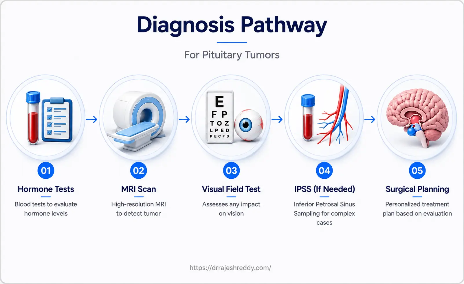
A pituitary tumor diagnosis requires the integration of clinical assessment, dedicated neuroimaging, and a comprehensive hormonal evaluation. The table below outlines the key investigations Dr. Rajesh Reddy uses in the pre-operative workup:
| Test | What It Measures | Why It Matters |
|---|---|---|
| Serum Prolactin | Prolactin level | Elevated prolactin suggests prolactinoma — treated with medication first, not surgery |
| IGF-1 and GH suppression test | Growth hormone excess | Confirms acromegaly; baseline for post-operative cure assessment |
| 24-hr urinary free cortisol / LNSC / 1mg DST | Cortisol excess | Confirms Cushing's disease before surgical planning |
| Morning serum cortisol, ACTH | Adrenal axis function | Screens for adrenal insufficiency from pituitary compression |
| TSH, Free T4 | Thyroid axis | TSH-secreting tumour screen; screens for secondary hypothyroidism |
| LH, FSH, Testosterone / Oestradiol | Gonadal axis | Hypogonadism from pituitary compression; baseline for recovery |
| Inferior Petrosal Sinus Sampling (IPSS) | ACTH lateralisation | Performed in Cushing's disease to confirm pituitary source and lateralise before surgery |
| MRI Pituitary (dedicated 3T, dynamic) | Tumour size, location, cavernous sinus invasion, optic chiasm relationship | Essential for surgical planning; thin-slice dynamic sequences identify microadenomas |
| Visual Field Testing (Humphrey perimetry) | Bitemporal or other field defects | Baseline before surgery; monitors optic chiasm decompression after surgery |
Inferior Petrosal Sinus Sampling (IPSS)
Inferior Petrosal Sinus Sampling (IPSS) is a specialised endovascular procedure recommended for patients with biochemically confirmed Cushing's disease when the pituitary MRI is normal or equivocal — which occurs in up to 40% of cases. It involves sampling blood from the inferior petrosal sinuses (small veins draining the pituitary) after stimulation with CRH, to confirm that the ACTH excess is of pituitary origin and to lateralise the source to the left or right side of the gland. This information directly guides the surgeon during endoscopic exploration. Dr. Rajesh Reddy's endovascular training equips him to perform this procedure himself, providing an integrated diagnostic and surgical pathway for Cushing's disease patients.
Endoscopic Transsphenoidal Pituitary Surgery: How It Works
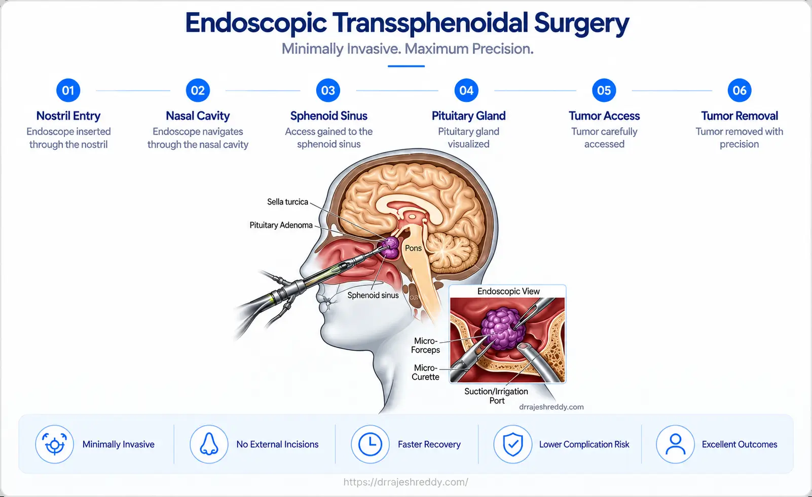
The endoscopic transsphenoidal approach to the pituitary gland is one of the great success stories of modern minimally invasive neurosurgery. It exploits a natural anatomical corridor — the nostrils → nasal cavity → sphenoid sinus → sella turcica — to access the pituitary gland without any external incision or brain retraction.
Step-by-Step: What Happens During the Procedure
- The patient is placed supine with the head slightly extended. General anaesthesia is administered.
- Neuronavigation is set up using pre-operative MRI data, providing real-time 3D GPS guidance throughout the procedure.
- A high-definition 4mm endoscope is introduced through one nostril. The nasal mucosa is gently displaced rather than incised where possible.
- The sphenoid sinus — an air-filled cavity directly in front of the sella turcica — is entered. The bony face of the sella is identified and a small opening is made.
- The dura (the protective membrane over the pituitary) is opened carefully, exposing the pituitary gland and the tumor within it.
- The tumor is removed using fine curettes, suction, and ring curettes, working within the pituitary fossa and — where the tumor extends upward — gently following it into the suprasellar space as it descends after intrasellar removal.
- The sella is then closed using a multilayer reconstruction — typically with a fat graft and a small piece of fascia harvested through a tiny 1–2 cm incision on the outer aspect of the thigh (a quick, separate part of the same procedure, leaving a small scar that heals well). The fat and fascia are used to seal the portal of entry in the sphenoid sinus and the floor of the sella, supplemented with a dural sealant and, where needed, a vascularised nasoseptal flap — to prevent CSF leak.
- The nasal passage is inspected and, if a septal flap was raised, repositioned. No external sutures. Nasal sponges (soft absorbable or non-absorbable packs) are placed in the nostrils at the end of the procedure and remain for 24–48 hours. During this period patients breathe through the mouth — the nursing team will ensure mouth care and adequate hydration. The sponges are removed bedside without anaesthesia before discharge.
What Makes Dr. Rajesh Reddy's Endoscopic Approach Different
- High-definition 4K endoscopy — providing panoramic, magnified visualisation of the pituitary fossa
- Neuronavigation integration — real-time 3D position confirmation relative to carotid arteries and optic chiasm
- Angled endoscopes (30° and 45°) — allowing the surgeon to look around corners in lateral recesses
- Intraoperative fluoroscopy — real-time imaging confirmation during complex cases
- Vascularised nasoseptal flap reconstruction — provides robust skull base reconstruction and dramatically reduces CSF leak risk
Endoscopic vs. Traditional Open Surgery: A Direct Comparison
| Feature | Endoscopic Transsphenoidal Surgery | Traditional Open Craniotomy |
|---|---|---|
| Access route | Through nostrils and sphenoid sinus — no skin incision | Open skull surgery with scalp incision and bone removal |
| Brain retraction | None — natural corridor used | Significant brain retraction required |
| Incision | None visible externally | Scalp incision; small bone flap removed |
| Visualisation | High-definition endoscope; panoramic view | Limited field through microscope; blind spots |
| Hospital stay | 2–3 days typically | 5–7+ days |
| Return to activity | 1–2 weeks | 4–6 weeks |
| Pain / discomfort | Mild nasal congestion; manageable headache | Significant post-operative pain; wound discomfort |
| Tumour removal | Excellent for intrasellar and suprasellar tumours | Preferred for large tumours with lateral extension |
| Risk to vision | Immediate decompression of optic chiasm | Delayed access compared to transsphenoidal approach |
| Recurrence rate | Comparable — depends on completeness of resection | Comparable — depends on completeness of resection |
Special Cases: Surgery for Cushing's Disease & Acromegaly
A Special Case: Surgery for Cushing's Disease
Cushing's disease — caused by an ACTH-secreting pituitary microadenoma — is among the most surgically challenging pituitary conditions. The responsible microadenoma is often very small (3–5 mm) and may not be visible on MRI. In these cases, surgical cure depends entirely on surgeon experience, quality IPSS lateralisation data, and meticulous intraoperative exploration.
Biochemical cure in Cushing's disease is defined as a morning serum cortisol of less than 50–138 nmol/L in the immediate post-operative period. Remission rates after endoscopic transsphenoidal surgery in experienced centres are 70–90% for microadenomas and 50–65% for macroadenomas.
Surgery for Acromegaly: Goals and Outcomes
Acromegaly — caused by a GH-secreting somatotroph adenoma — is treated primarily with surgery. The goals are:
- Achieve biochemical remission — normalisation of IGF-1 and suppression of GH to < 1 ng/mL on glucose tolerance test
- Decompress the optic chiasm if visual field defects are present
- Reduce tumor bulk to improve response to medical therapy (somatostatin analogues) if complete resection is not achieved
Remission rates after surgery for acromegaly are 85–90% for microadenomas and 50–70% for macroadenomas without cavernous sinus invasion.
Recovery After Endoscopic Pituitary Surgery
Day 0–1 (Surgery & Immediate Post-Op)
- Surgery typically takes 1.5–3 hours depending on tumor size and complexity.
- Patient wakes in the recovery room and is transferred to a neurosurgical ward (HDU monitoring for the first night).
- Mild nasal congestion and a sensation of fullness in the nose are the most common complaints.
- Headache is usually mild and well-controlled with simple analgesics.
- If a lumbar drain has been placed to reduce the risk of cerebrospinal fluid (CSF) leak, it is typically maintained for several days while drainage is carefully monitored.
- Sodium and fluid balance monitored closely for the first 48–72 hours — diabetes insipidus, if it occurs, typically presents in this window.
Days 2–3 (Discharge Planning)
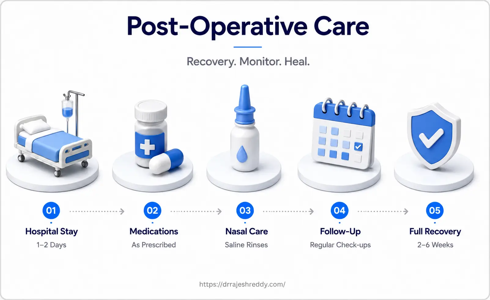
- Nasal packs (absorbable or sponge packs, if used) are usually removed on post-operative Day 2, resulting in improved nasal breathing and comfort.
- Most patients are fit for discharge on day 2 or 3.
- Hormonal blood tests repeated to assess immediate post-operative pituitary function.
- Nasal saline irrigation started to aid mucosal healing and prevent crusting.
- Clear instructions given on symptoms that require urgent return: clear fluid dripping from the nose (possible CSF leak), new severe headache, sudden vision change, or high fever.
Day 5 (If Lumbar Drain Was Placed)
- The lumbar drain, if inserted during surgery, is typically removed around Day 5 after confirming there is no evidence of a CSF leak and the skull base repair is healing satisfactorily.
Weeks 2–6
- Avoid nose blowing, heavy lifting, and strenuous activity for 4 weeks to protect the skull base reconstruction.
- Return to desk-based work typically possible within 1–2 weeks.
- Post-operative MRI at 6–12 weeks to assess resection extent.
- Endocrinology follow-up at 6 weeks for hormonal reassessment.
Long-Term Follow-Up
- Regular MRI surveillance: annually for 5 years, then every 2 years.
- Annual hormonal testing to monitor pituitary axis recovery.
- Ophthalmology review — most patients with pre-op bitemporal hemianopia show significant improvement within weeks.
Why Choose Dr. Rajesh Reddy for Pituitary Tumor Surgery in Hyderabad?
Pituitary surgery sits at the intersection of neurosurgery, endocrinology, and skull base surgery — and requires a surgeon who is fluent in all three domains. Here is what sets Dr. Rajesh Reddy apart:
- 4K High-Definition Endoscopy with angled scopes & neuronavigation
- FINR (Zurich) Endovascular Training enabling him to perform Inferior Petrosal Sinus Sampling (IPSS) in-house for Cushing's disease
- Visiting Scholar, Barrow Neurological Institute (Phoenix, Arizona) — a global reference centre for skull base and pituitary surgery
- Visiting Scholar — Okayama University, Japan; Klinikum Stuttgart, Germany
- Over 5,000 neurosurgical procedures across a 15+ year career
- Multidisciplinary coordination with endocrinology, ophthalmology, and radiation oncology