
Introduction
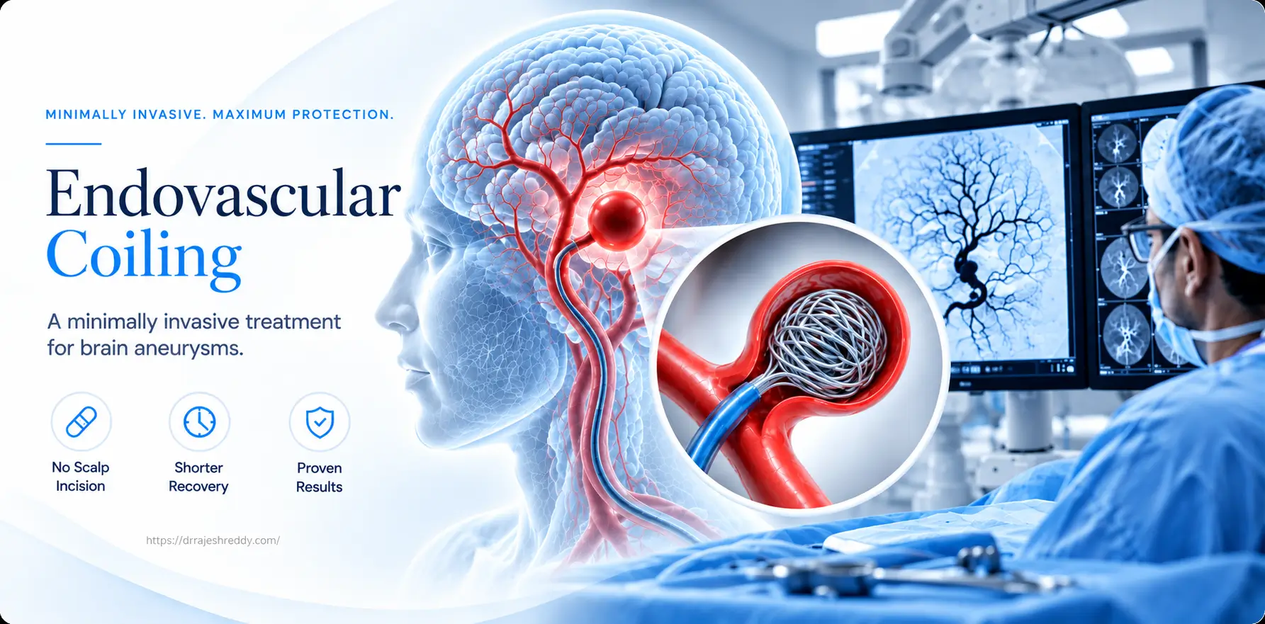
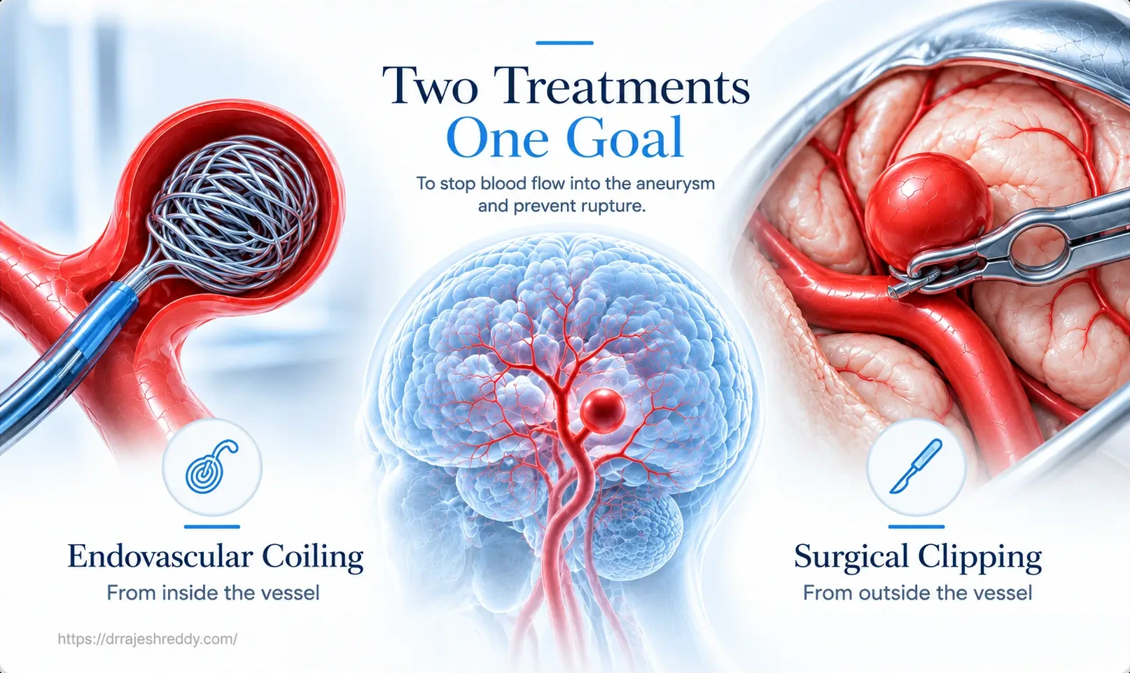
Brain aneurysm surgery once meant one thing: open skull surgery, a craniotomy under general anaesthesia, careful dissection to the base of the brain, and application of a titanium clip across the aneurysm neck. That open approach remains important and effective for specific aneurysm types — but for the majority of brain aneurysms encountered in clinical practice today, a fundamentally different route exists. Endovascular coiling reaches the aneurysm from inside the bloodstream — through a small puncture in the groin — without a single incision in the scalp, without removing any bone, and without touching the brain itself.
Endovascular coiling has transformed the treatment of brain aneurysms. For patients with ruptured aneurysms causing subarachnoid haemorrhage, the landmark ISAT trial demonstrated that coiling produces significantly better neurological outcomes than clipping for aneurysms suitable for both techniques. For patients with incidentally discovered unruptured aneurysms, coiling offers definitive treatment with a hospital stay of 1–3 days and return to normal activity within 1–2 weeks.
Dr. Rajesh Reddy Sannareddy is one of the few neurosurgeons in Hyderabad with a dedicated Fellowship in Interventional Neuroradiology (FINR) from the University Hospital, Zurich — a centre performing over 400 endovascular cerebrovascular procedures annually. He is trained in the full spectrum of endovascular aneurysm treatment: standard coiling, balloon-assisted coiling, stent-assisted coiling and Flow Diversion [Pipeline, Silk, Surpass, FRED, Derivo, p64, Contour, WEB]. His visiting scholar experience at Barrow Neurological Institute, Phoenix, Okayama University, Japan, and Klinikum Stuttgart, Germany further shaped his endovascular practice with exposure to high-volume, internationally recognised cerebrovascular programmes.
What Is Endovascular Coiling?
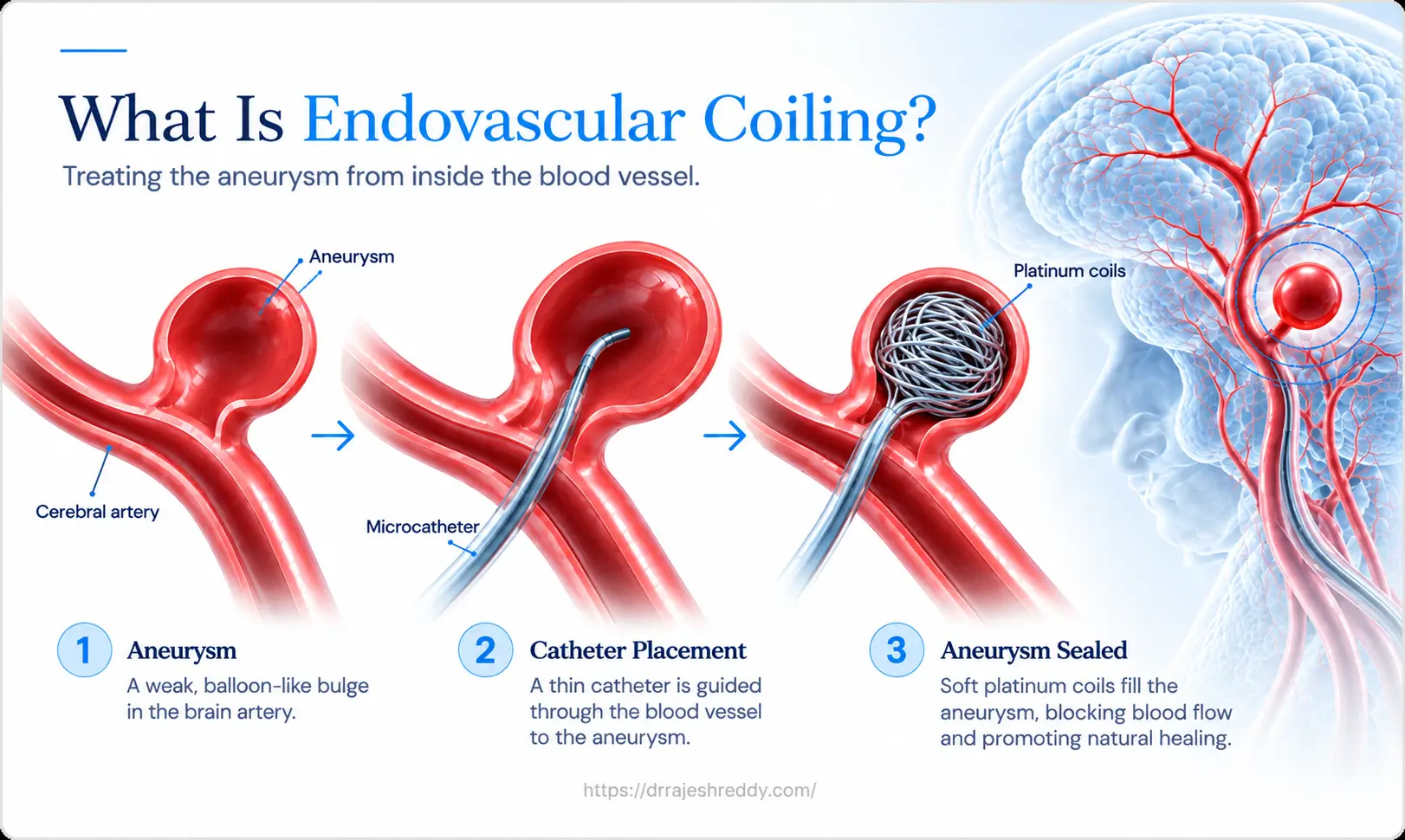
Endovascular coiling — formally called Guglielmi Detachable Coil (GDC) embolisation, after its inventor Dr. Guido Guglielmi — is a minimally invasive procedure that treats brain aneurysms by filling the aneurysm sac with soft platinum coils from within the bloodstream. The coils disrupt blood flow inside the aneurysm, promoting clot formation that progressively seals it from the parent artery circulation.
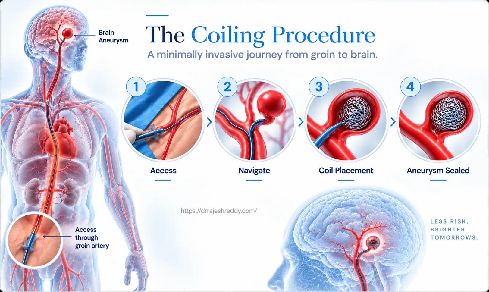
The procedure is performed in a specialised neurointerventional suite — a hybrid operating and imaging room equipped with biplane digital subtraction angiography (DSA), advanced 3D imaging, and fluoroscopic guidance. The entire procedure is performed through a small puncture in the femoral artery in the groin — the same access point used for a cardiac catheterisation. There is no incision in the scalp, no removal of skull bone, and no manipulation of brain tissue.
The Endovascular Coiling Procedure: Step by Step
Step 1: Pre-Procedure Preparation
Before the procedure, patients undergo a dedicated Digital Subtraction Angiography (DSA) with 3D rotational imaging to precisely characterise the aneurysm — its exact size, neck width, dome shape, relationship to parent and branch arteries, and the optimal working projection for treatment. This roadmap is essential for safe and complete coil delivery.
For elective (unruptured) aneurysm coiling, patients requiring stent-assisted coiling or flow diversion are pre-loaded with dual antiplatelet therapy (aspirin 150 mg plus clopidogrel 75 mg or Ticagrelor 180 mg/day) for 5–7 days before the procedure to prevent stent thrombosis. For emergency SAH cases, antiplatelet therapy is initiated on the day of procedure if stent assisted coiling or Flow diversion therapy is preferred over microsurgical clipping.
Step 2: Femoral Access and Guide Catheter Placement
Under general anaesthesia, the right (or occasionally left) femoral artery is accessed via a small puncture using a modified Seldinger technique. A short introducer sheath (typically 6 French, approximately 2 mm diameter) is placed in the femoral artery. A guide catheter — a flexible tube approximately 2 mm in outer diameter — is then navigated under fluoroscopic guidance from the femoral artery, through the iliac arteries and aorta, and into the internal carotid or vertebral artery supplying the aneurysm. This process takes 10–20 minutes in experienced hands.
Step 3: Microcatheter Navigation into the Aneurysm
Through the guide catheter, a microcatheter — a highly flexible, steerable tube approximately 1.5 mm in outer diameter — is navigated using a microwire guide into the target aneurysm sac. This requires precise, real-time fluoroscopic guidance and is the most technically demanding part of the procedure. The microcatheter tip must be positioned optimally within the aneurysm sac — not too deep (risking perforation) and not too shallow (risking coil herniation into the parent artery). Correct positioning is confirmed under fluoroscopic road map guidance.
Step 4: Coil Delivery and Packing
With the microcatheter in position, platinum coils are delivered one by one into the aneurysm sac. Each coil is detachable and is released only after confirming that it is correctly positioned and not herniated into the parent artery. If positioning is unsatisfactory, the coil can be retrieved and repositioned before detachment.
The first coil, called the framing coil, is the largest — it forms a basket within the aneurysm, conforming to its shape and defining the working space. Subsequent coils (filling and finishing coils) are progressively smaller and softer, packing the interior of the basket until no further coils can be introduced safely. The goal is maximum packing density — typically > 25–30% of the aneurysm volume — to minimise recanalisation risk.
Step 5: Final Angiogram and Closure
After coiling is complete, a final DSA run confirms the degree of aneurysm occlusion, patency of the parent artery, and absence of thromboembolic complications. Occlusion is classified using the Raymond-Roy scale: complete occlusion (no contrast entering the sac), neck remnant (coils at the neck level with no dome filling), or residual aneurysm (contrast entering the dome). The aim is always complete or near-complete occlusion.
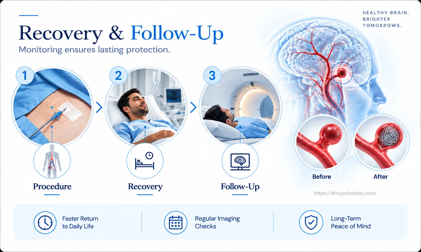
The femoral access site is closed using a vascular closure device or manual compression. The patient wakes from anaesthesia and is transferred to the neurosurgical ward or ICU depending on clinical status. The groin puncture site requires pressure dressing and 4–6 hours of bed rest to prevent haematoma.
Types of Coils Used in Aneurysm Embolisation
Coil technology has advanced significantly since the introduction of the original bare platinum Guglielmi Detachable Coil. Coil type is selected according to the aneurysm's size, shape, neck anatomy and packing requirements.
| Coil Type | When Used | Key Characteristic |
|---|---|---|
| Bare Platinum Framing Coil | First coil deployed — establishes the scaffold | Large diameter, forms a basket within the aneurysm; defines the working space for filling coils |
| Bare Platinum Filling Coil | Subsequent coils after framing | Progressively packs the sac within the basket; smaller diameters are used as packing density increases |
| Hydrocoil (Bioactive) | Wide-neck or large aneurysms where tight packing is critical | Polymer coating expands on contact with blood; achieves higher packing density and promotes organised thrombus |
| 3D / Complex-shaped Coil | Irregular or lobulated aneurysms | Pre-shaped 3D configuration adapts to complex aneurysm geometries for better coverage |
| Finishing / Soft Coil | Final coil to complete packing | Very soft, high compliance — fills residual spaces without displacing the frame |
Advanced Endovascular Techniques for Challenging Aneurysms
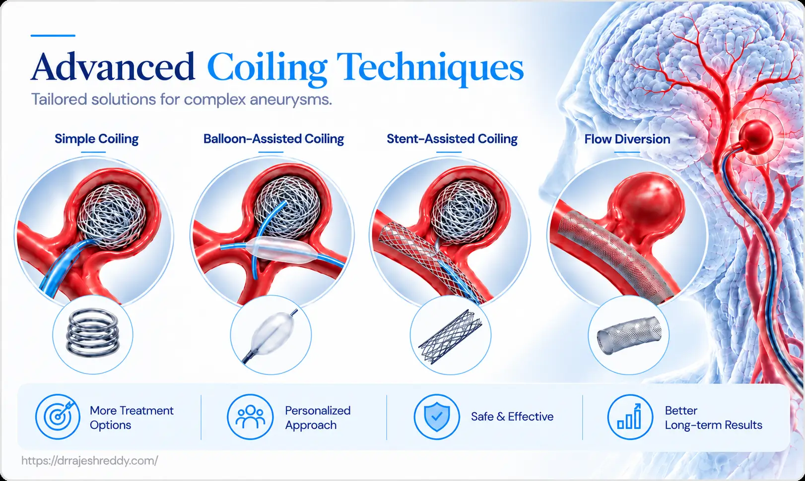
Standard coiling is ideal for narrow-neck saccular aneurysms. When anatomy is more complex — wide neck, bifurcation location, large size — advanced techniques are required:
| Technique | How It Works | Best For | Antiplatelet Needed? |
|---|---|---|---|
| Simple Coiling | Microcatheter in sac; coils deployed until the sac is packed | Narrow-neck saccular aneurysms (dome:neck ≥ 2) | No |
| Balloon-Assisted Coiling (BAC) | Remodelling balloon inflated across neck during coil delivery; prevents herniation | Wide-neck aneurysms where simple coiling risks coil prolapse | No (short-term aspirin only) |
| Stent-Assisted Coiling (SAC) | Laser cut or Braided stent deployed across neck first; coils packed through stent struts | Wide-neck aneurysms; fusosaccular aneurysms; coils alone insufficient | Yes — dual antiplatelet (aspirin + clopidogrel or Ticagrelor) 3–6 months |
| Intrasaccular Device | Nitinol mesh sphere deployed within sac; disrupts inflow without stent | Wide-neck bifurcation aneurysms (MCA, basilar tip, AComm) | Aspirin alone — no clopidogrel required |
| Flow Diversion(Pipeline/ Silk, Surpass, Derivo, p64, FRED) | Dense mesh stent across neck; redirects flow; aneurysm thromboses over months | Large/giant, fusiform, wide-neck ICA aneurysms; failed coiling | Yes — dual antiplatelet 3–6 months, single antiplatelet thereafter |
Balloon-Assisted Coiling (BAC) — The Remodelling Technique
BAC uses a compliant balloon catheter positioned across the aneurysm neck alongside the microcatheter. During coil delivery, the balloon is briefly inflated to cover the neck — preventing coils from prolapsing into the parent artery. Between coils, the balloon is deflated to restore blood flow. This technique allows tighter packing of wide-neck aneurysms without the need for a permanent stent implant and without requiring dual antiplatelet therapy.
Stent-Assisted Coiling (SAC)
When balloon assistance is insufficient, a low-profile braided or laser-cut microstent is deployed across the aneurysm neck before coiling. The stent provides a permanent scaffold — its struts prevent coil herniation into the parent artery and alter the haemodynamic environment at the aneurysm neck, promoting progressive thrombosis. The stent remains permanently in the parent artery. Dual antiplatelet therapy (aspirin + clopidogrel or Ticagrelor) is required for 3–6 months to prevent stent thrombosis, after which aspirin alone is continued indefinitely.
The WEB Device — Intrasaccular Flow Disruption
The WEB (Woven Endo Bridge) device is a self-expanding nitinol mesh sphere deployed entirely within the aneurysm sac through a single microcatheter. Unlike stent-assisted coiling — where the stent is deployed in the parent artery — the WEB sits completely within the aneurysm and requires no stent in the circulation. This eliminates the need for clopidogrel; aspirin alone is sufficient. The WEB is particularly well-suited to wide-neck bifurcation aneurysms at the MCA trifurcation, basilar tip, and anterior communicating artery — locations where stent placement would require multiple stents or complex navigation. Contour, Artisse, Seal are the other Intrasaccular flow disrupters.
Risks of Endovascular Coiling: An Honest Assessment
Endovascular coiling is a safe procedure when performed in expert hands, with complication rates that compare favourably to open surgical alternatives. Patients deserve a clear and honest understanding of the specific risks involved.
Thromboembolic Complications
The most common serious complication of endovascular coiling is thrombus (clot) formation on the coils or catheters, which can embolise to distal cerebral arteries causing ischaemic stroke. The risk is approximately 3–5% overall, with the majority being minor or clinically silent. Heparin anticoagulation during the procedure significantly reduces this risk. For procedures requiring stents, pre-procedural dual antiplatelet therapy is the primary preventive measure.
Aneurysm Perforation
Perforation of the aneurysm wall during coil delivery — caused by the guidewire or coil perforating the thin aneurysm dome — can cause intraprocedural haemorrhage. The risk is approximately 1–2%. If perforation occurs, the immediate management is to rapidly complete coiling to seal the perforation from within, while simultaneously managing the rise in intracranial pressure. An experienced operator can manage most perforations without catastrophic outcome.
Coil Herniation
A coil loop prolapsing into the parent artery during delivery can cause thrombosis or distal embolisation. This is managed by retrieving the offending coil with a micrograsper, repositioning, or — if deployed — inflating a balloon to push it back into the sac or deploying a stent across the neck. Balloon and stent-assisted techniques specifically address this risk for wide-neck aneurysms.
Groin Haematoma
A haematoma at the femoral access site occurs in approximately 2–5% of cases, usually resolving with compression and bed rest. Rarely, it requires surgical drainage. Vascular closure devices have significantly reduced this complication compared to manual compression alone.
Recanalisation
As discussed above, approximately 15–20% of coiled aneurysms show recanalisation at 5 years — requiring surveillance imaging and possible retreatment. This is not a complication of the initial procedure but a known limitation of coiling biology that mandates long-term follow-up.
Recovery After Endovascular Coiling: Elective vs. Emergency
Recovery differs substantially depending on whether coiling was performed electively for an unruptured aneurysm or as an emergency for a ruptured aneurysm causing SAH:
| Phase | Elective (Unruptured) | Emergency (Ruptured / SAH) |
|---|---|---|
| Procedure | 1–3 hours under GA; groin puncture; no craniotomy | 1–3 hours; same procedure but patient is acutely unwell; may have concurrent vasospasm management |
| Day 0–1 | Recovery room; neuro obs; groin site checked; ambulate within 4–6 hrs | ICU admission; hourly neuro obs; vasospasm monitoring; nimodipine started |
| Day 2–3 | Discharge home; mild groin bruising resolves in 1–2 weeks; gentle activity | Remains in ICU or HDU; vasospasm monitoring continues; EVD if hydrocephalus |
| Week 1–2 | Return to desk work; no heavy lifting or strenuous activity | Still hospitalised in most SAH cases; vasospasm peak risk period (days 4–14) |
| Week 4–6 | Return to full activity including driving (after medical clearance) | Discharge possible for good-grade SAH if no complications; rehabilitation begins |
| 6 months | Follow-up MRA, CTA or DSA to confirm aneurysm occlusion | Follow-up DSA mandatory; neuropsychological and physical rehabilitation continues |
| 18 months | Second follow-up MRA/CTA/DSA; annual thereafter if stable | Second DSA; cognitive recovery assessment; return-to-work evaluation |
Antiplatelet Medication After Coiling
Patients treated with stent-assisted coiling or flow diversion require dual antiplatelet therapy (aspirin + clopidogrel or Ticagrelor) for 3–6 months, after which aspirin is continued indefinitely. Stopping antiplatelet medication early — without medical advice — risks stent thrombosis, which can cause stroke. Patients must not stop any antiplatelet medication without first consulting Dr. Rajesh Reddy.
Activity Restrictions
- Groin site: avoid heavy lifting, strenuous exercise, and hot baths for 5–7 days while the puncture heals.
- Driving: typically permitted after 24–48 hours for simple coiling; deferred if antiplatelet medication affects driving safety; discuss with Dr. Rajesh Reddy
- Return to desk work: typically 2–5 days after elective coiling
- Vigorous exercise / contact sports: 2–4 weeks after elective coiling
- Sexual activity: generally safe from day 3–5 after elective coiling
Follow-Up After Coiling: Why Surveillance Is Non-Negotiable
Unlike surgical clipping — where a correctly placed clip provides an immediately verifiable, durable mechanical seal — coiling depends on progressive thrombosis of the aneurysm sac. The coil mass can compact over time, potentially allowing recanalisation. Follow-up imaging is therefore mandatory, not optional.
6-Month DSA, MRA or CTA:
The first post-procedure surveillance imaging. Confirms whether complete occlusion has been maintained or whether recanalisation has occurred. DSA remains the gold standard; high-resolution MRA at 3T or CTA is an acceptable alternative for stable, small aneurysms.
18-Month DSA, MRA or CTA:
Second surveillance. The majority of clinically significant recanalisation occurs within the first 18 months. A stable result at 18 months significantly reduces the probability of late recanalisation.
Annual MRA or CTA thereafter:
For coiled aneurysms that are stable at 18 months, annual or biennial MRA surveillance is recommended indefinitely — particularly for large aneurysms, those treated with stent assistance, and younger patients who have many decades of follow-up ahead.
Why Choose Dr. Rajesh Reddy for Endovascular Coiling in Hyderabad?
Endovascular aneurysm treatment is one of the most technically demanding procedures in all of medicine — requiring fluency with catheter navigation through tortuous cerebrovascular anatomy, mastery of coil selection and delivery, and the judgement to manage intraoperative complications rapidly. Volume and training matter enormously. Dr. Rajesh Reddy's endovascular credentials are among the strongest available in Hyderabad:
Endovascular Expertise — Dr. Rajesh Reddy
- FINR — Fellowship in Interventional Neuroradiology, University Hospital, Zurich (one of Europe's highest-volume cerebrovascular endovascular programmes)
- Visiting Scholar — Barrow Neurological Institute, Phoenix, Arizona; Okayama University, Japan; Klinikum Stuttgart, Germany
- Double board certified neurosurgeon — understands the surgical anatomy underpinning every endovascular decision
- Trained in: GDC coiling, hydrocoil, balloon-assisted coiling, stent-assisted coiling, Flow diversion, endovascular vasospasm management (angioplasty, intra-arterial vasodilators)
- Can perform Inferior Petrosal Sinus Sampling (IPSS) — an endovascular diagnostic procedure for Cushing's disease — integrated within his neurosurgical practice
- Several complex neurosurgical and endovascular procedures across a 15+ year career
- Performs both endovascular coiling and microsurgical clipping — treatment recommendation is based on aneurysm anatomy, not institutional preference
- Clinical acumen and judgement gained through years of training with leading global experts
Frequently Asked Questions
Consult Dr. Rajesh Reddy for Endovascular Coiling in Hyderabad
Whether you have been told you have a brain aneurysm and want to understand whether coiling is the right treatment for you — or you need emergency evaluation — Dr. Rajesh Reddy Sannareddy offers comprehensive endovascular consultations in Hyderabad. Book via the Dr. Reddy's Neuro Care app on Google Play.
Related Articles
Continue reading to explore brain aneurysm treatment, symptoms, flow diversion and recovery:
Brain Aneurysm Symptoms: When Is It an Emergency?
Thunderclap headache, warning signs and when immediate emergency care is needed.
Read GuideRuptured vs. Unruptured Brain Aneurysm: Key Differences
Symptoms, emergency management and treatment considerations.
Read GuideAneurysm Coiling vs. Surgical Clipping
Understand the differences between endovascular coiling and microsurgical clipping.
Read GuideFlow Diversion Treatment for Brain Aneurysms
Flow diversion for selected complex, wide-necked and giant aneurysms.
Read GuideSubarachnoid Hemorrhage (SAH): Causes and Treatment
Causes, emergency management, vasospasm prevention and critical care.
Read GuideLife After Brain Aneurysm Surgery: Recovery Guide
Recovery timeline, returning to work, driving and long-term follow-up.
Read GuideArteriovenous Malformation (AVM) Treatment Hyderabad
Endovascular embolisation, radiosurgery and microsurgical treatment for AVMs.
Read GuideBrain Aneurysm Treatment in Hyderabad
Visit the main pillar page for brain aneurysm treatment in Hyderabad.
View Pillar Page