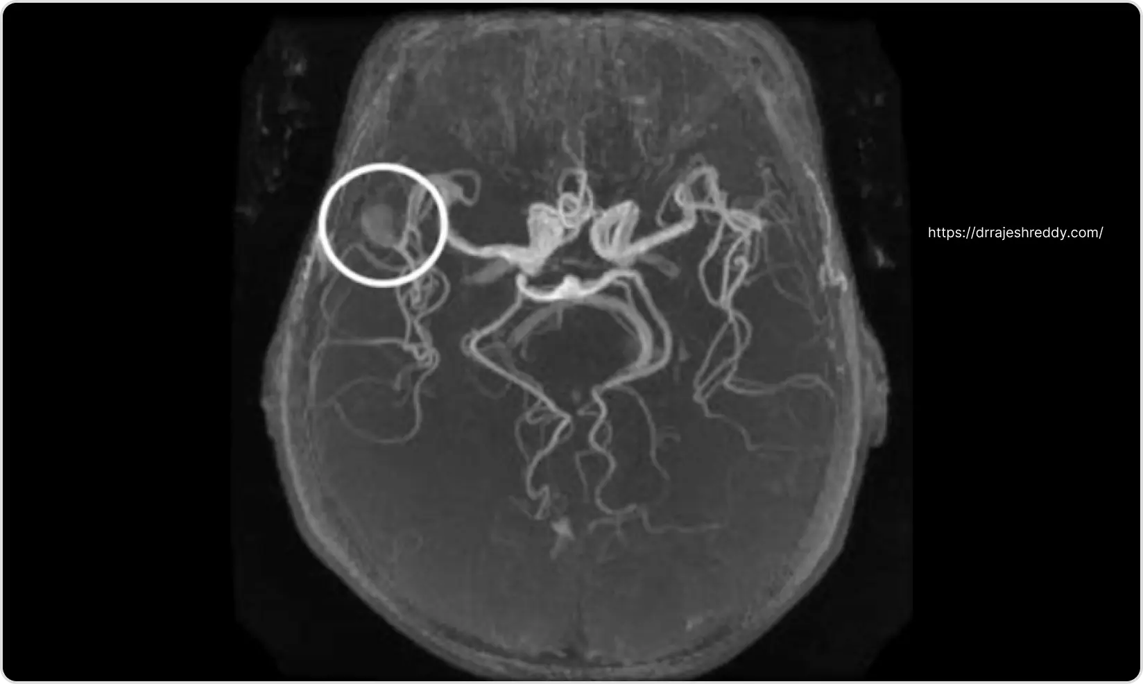
Introduction
Not every headache is a brain aneurysm. But some are — and when they are, the difference between acting immediately and waiting until morning can be the difference between a full recovery and permanent disability, or death. The challenge is that the most dangerous symptom of a brain aneurysm — a sudden, catastrophic headache — is also one of the most common symptoms in the general population. Knowing what distinguishes a true aneurysmal emergency from an ordinary severe headache is knowledge that could save your life or the life of someone you love.
This page explains the full spectrum of brain aneurysm symptoms — from subtle warning signs that develop over days or weeks, to the neurological emergencies that demand immediate action. It is written for patients, families, and anyone who wants to understand what a brain aneurysm actually feels like, what specific symptoms require emergency care, and which early warning signs should prompt urgent evaluation before disaster strikes.
Dr. Rajesh Reddy Sannareddy, Senior Consultant Endovascular Neurosurgeon in Hyderabad, manages brain aneurysms at every stage of their presentation — from incidental discovery to acute subarachnoid hemorrhage (SAH). His training in both endovascular and microsurgical cerebrovascular treatment, including a Fellowship in Interventional Neuroradiology (FINR) from the University Hospital, Zurich, means that patients reach the right treatment without delay.
Brain Aneurysm Symptoms: From Silent to Life-Threatening
Brain aneurysm symptoms span an enormous range — from no symptoms at all, to subtle warning signs, to sudden catastrophic events. The table below maps the full symptom spectrum with the action each level requires:
| Symptom Category | Symptoms | Action Required |
|---|---|---|
| EMERGENCY — Act immediately | Thunderclap headache (worst headache of life); sudden loss of consciousness; sudden severe vomiting with headache; neck stiffness with headache; seizure with headache; sudden vision loss or double vision with headache | Call emergency services NOW — suspected ruptured aneurysm / SAH |
| URGENT — Within 24–48 hours | New drooping eyelid + dilated pupil (third nerve palsy); sudden new severe headache different from usual; new double vision without headache; sudden onset visual field loss | Go to emergency department or call Dr. Rajesh Reddy's clinic immediately — possible symptomatic unruptured aneurysm |
| Soon — Within days | Persistent new headache behind one eye; recurrent headaches different from usual pattern; visual field changes noticed over days to weeks | Book urgent neurosurgical consultation; MRI and MRA required |
| Routine — Scheduled | Incidentally found aneurysm on MRI with no symptoms; family history of aneurysm; headaches with known migraine history but now changing in character | Elective neurosurgical consultation for risk assessment and treatment planning |
The Silent Majority: Most Aneurysms Cause No Symptoms
The majority of brain aneurysms — particularly small ones under 7 mm — produce no symptoms whatsoever. They are discovered purely by chance: on an MRI done for headaches, a CT after a minor head injury, or during screening in a patient with a family history of aneurysm. Patients are often astonished to learn that a potentially dangerous lesion has been present for years without causing any sensation.
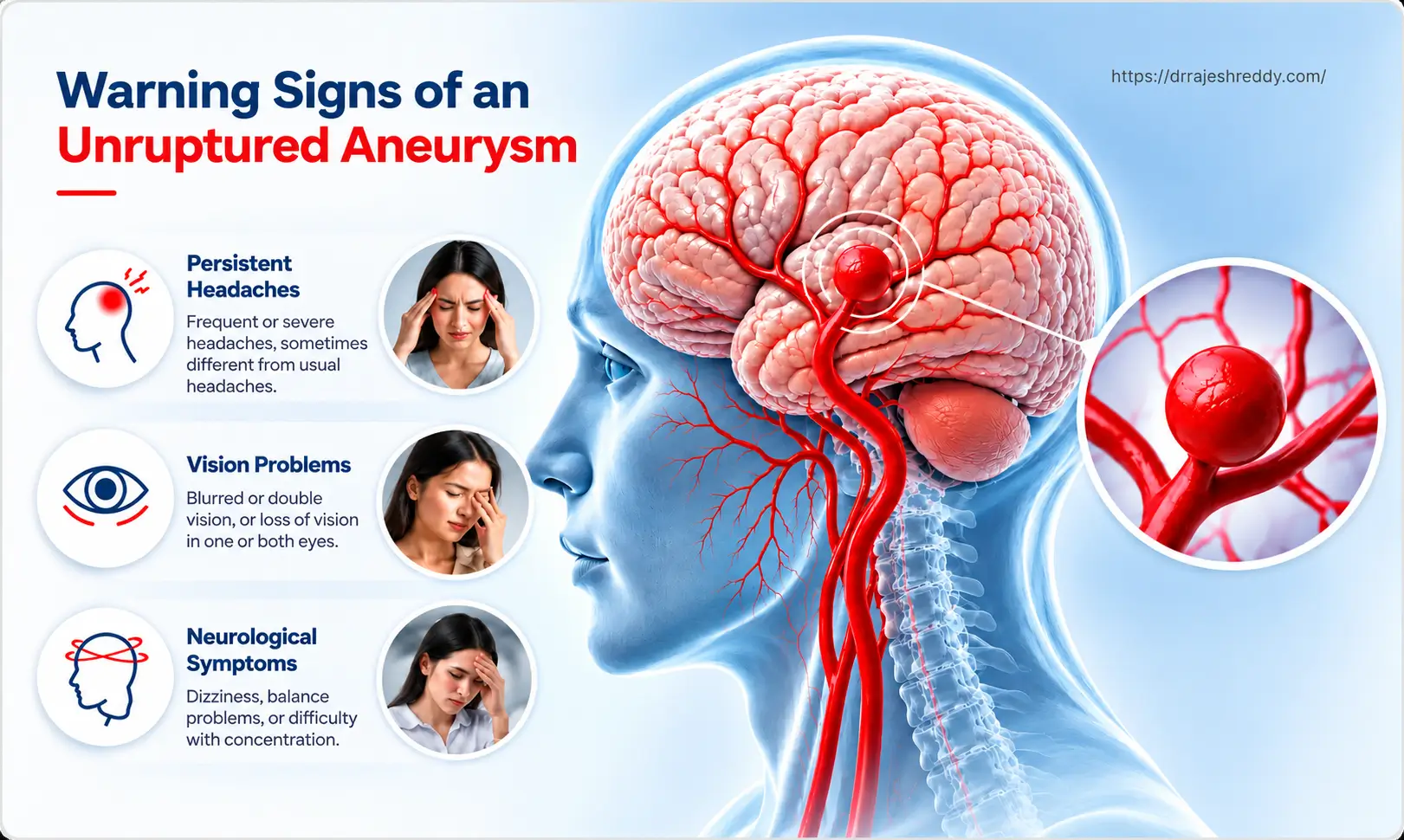
This silence is both reassuring and deceptive. Reassuring because it means the aneurysm has not ruptured and the brain is functioning normally. Deceptive because the absence of symptoms does not mean the absence of rupture risk — and some aneurysms rupture without any preceding warning whatsoever.
An incidentally discovered brain aneurysm is not an emergency — but it does require expert evaluation. Dr. Rajesh Reddy will assess the size, location, shape, and your individual risk factors to determine whether observation, endovascular treatment, or surgical clipping is the right approach. Doing nothing without an expert opinion is not the same as choosing observation.
Symptoms of an Unruptured but Symptomatic Aneurysm
A minority of unruptured aneurysms do cause symptoms — typically through mass effect (the aneurysm pressing on adjacent structures) rather than through bleeding. These symptoms are important because they are warning signs that the aneurysm is growing or is at higher risk of rupture.
Third Nerve Palsy — The Most Important Warning Sign
The single most important warning symptom of an unruptured brain aneurysm is a sudden drooping eyelid (ptosis) combined with a dilated, non-reactive pupil on the same side. This is called a third cranial nerve palsy (CN III palsy) and it signals that an aneurysm — almost always at the posterior communicating artery (PComm) — is pressing on or compressing the oculomotor nerve.
CRITICAL WARNING
NEW DROOPING EYELID + DILATED PUPIL = NEUROSURGICAL EMERGENCY. This combination, even without headache, indicates possible aneurysm compression of the third cranial nerve. Go to emergency immediately.
A CN III palsy from aneurysm compression is a neurological emergency even if the patient has no headache. The aneurysm may rupture within hours or days. Any patient who develops a new drooping eyelid with a dilated pupil must be taken to an emergency department immediately for CT angiography.
Other Mass Effect Symptoms of Unruptured Aneurysms
- Progressive visual loss: A large aneurysm at the ophthalmic artery or internal carotid artery can compress the optic nerve, causing gradual or sudden visual loss in one eye. A large anterior communicating or cavernous sinus aneurysm may cause bitemporal visual field loss by compressing the optic chiasm.
- Facial pain or numbness: A cavernous sinus or basilar artery aneurysm may compress the trigeminal nerve (CN V), causing facial pain, numbness, or a pins-and-needles sensation across the cheek, jaw, or forehead.
- Double vision without ptosis: Involvement of CN IV or CN VI — which control eye movement — produces double vision without the drooping eyelid or pupil dilation seen in CN III palsy. Seen with cavernous sinus or posterior circulation aneurysms.
- Pulsatile tinnitus: A rhythmic whooshing sound in the ear, synchronous with the heartbeat, occasionally reported with large aneurysms close to the ear or cavernous sinus.
- Headache: A persistent, new headache — particularly one felt behind one eye — associated with a known or suspected aneurysm warrants urgent evaluation. Aneurysmal headaches are often described as dull and persistent, rather than throbbing or episodic.
Giant Aneurysms — A Distinct Presentation
Giant aneurysms (≥ 25 mm) that are partially thrombosed — where blood has clotted within part of the aneurysm sac — can present in an entirely different way from smaller aneurysms. The clot within the sac can shed small emboli into the brain circulation, causing Transient Ischaemic Attacks (TIAs, or mini-strokes) — brief, reversible episodes of weakness, speech difficulty, or visual disturbance lasting minutes to hours. The partially thrombosed sac may also irritate adjacent cortex, triggering new-onset seizures. In large aneurysms exerting significant mass effect on the adjacent brain tissue, patients may develop progressive weakness of one side of the body (hemiparesis) that worsens over weeks — closely mimicking a brain tumour presentation. These mass-effect and embolic presentations of giant aneurysms are important to recognise because the correct diagnosis requires urgent neurovascular imaging, not just a standard stroke or tumour workup.
The Sentinel Headache: The Warning That Is Most Often Missed
In the days, weeks, or occasionally months before a major aneurysm rupture, approximately 20–50% of patients experience what is called a sentinel headache — a sudden, severe headache caused by a minor leak or rapid expansion of the aneurysm wall that does not constitute a full rupture. Blood leaks into the subarachnoid space in a small quantity, causing sudden irritation of the meninges — but not the catastrophic flooding of SAH.
The sentinel headache is clinically indistinguishable from the headache of full SAH in terms of its onset speed and initial severity. However, because the blood leak is small, the patient may recover relatively quickly — within hours — and be tempted to attribute the headache to stress, dehydration, or a particularly severe migraine. This is the most dangerous mistake in cerebrovascular medicine.
Characteristics of a Sentinel Headache
- Sudden onset — reaching maximum severity within seconds, not minutes
- Severe — typically described as the worst or one of the worst headaches the patient has ever experienced
- Genuinely new — different in character, location, or severity from any previous headache
- May be associated with nausea, vomiting, or transient neck stiffness
- May be followed by a period of feeling 'not quite right' or unusually fatigued
- Resolves within hours or a day — which reassures the patient but does not indicate safety
CRITICAL CLINICAL INSIGHT
A sudden severe headache that resolves within hours is not necessarily safe. If it was the worst headache of your life, you must be investigated. A CT scan negative for blood does not exclude a small sentinel bleed — a lumbar puncture is required to look for xanthochromia (yellow CSF from blood breakdown). Studies show that sentinel headaches are misdiagnosed as migraine in up to 50% of cases, and these patients return days to weeks later with a catastrophic rupture.
What Happens If a Sentinel Headache Is Missed
If a sentinel headache is not investigated and the underlying aneurysm is not treated, the risk of major rupture (full SAH) in the following weeks is very high — estimated at 30–50% within 30 days. Major SAH carries a mortality of approximately 40–50%, and half of survivors have permanent neurological disability. The sentinel headache represents the last window of opportunity to prevent catastrophe.
The Thunderclap Headache: A Ruptured Aneurysm Until Proven Otherwise
Rupture of a brain aneurysm causes subarachnoid hemorrhage (SAH) — sudden, high-pressure bleeding into the subarachnoid space that bathes the brain. The hallmark is the thunderclap headache: an instantaneous, explosive headache that reaches absolute maximum severity within 1–2 seconds.
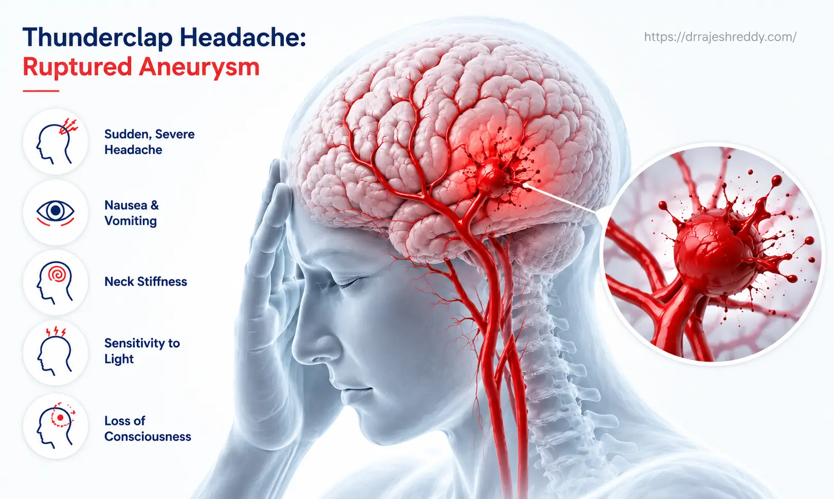
Patients who have experienced it describe it with remarkable consistency:
- "A gun went off inside my head"
- "Someone hit me on the back of the head with a cricket bat"
- "The worst pain I have ever felt in my life — like an explosion"
- "I was absolutely fine one moment and then could not see, hear, or move for the pain"
Associated Symptoms of Ruptured Aneurysm / SAH
- Immediate vomiting: Often projectile, occurring within seconds to minutes of the headache. The vomiting is caused by the sudden rise in intracranial pressure from the haemorrhage and the irritation of the meninges.
- Neck stiffness (meningism): Blood in the subarachnoid space irritates the meninges, causing painful resistance to neck flexion — typically developing within 3–6 hours of rupture. This is one of the classic signs of SAH on clinical examination.
- Photophobia and phonophobia: Extreme sensitivity to light and sound, also from meningeal irritation — similar to severe migraine, but in the context of a thunderclap headache.
- Loss of consciousness: Transient loss of consciousness at the time of rupture occurs in approximately 45% of SAH patients — caused by the sudden spike in intracranial pressure momentarily stopping cerebral perfusion. Prolonged unconsciousness indicates a more severe bleed.
- Seizure: Occurs in approximately 10–20% of SAH patients at or shortly after rupture. A seizure in the context of a thunderclap headache is a strong indicator of SAH.
- Focal neurological deficits: Weakness on one side, speech difficulty, or visual loss — indicating that the haemorrhage or associated haematoma has affected specific brain regions.
- Sudden death: Approximately 10–15% of SAH patients die before reaching hospital — from the initial haemorrhage causing catastrophic brain injury or cardiac arrest. This underlines why every thunderclap headache is a medical emergency.
IMMEDIATE EMERGENCY ACTION
CALL EMERGENCY SERVICES IMMEDIATELY if anyone experiences: a sudden severe headache reaching maximum intensity within seconds — particularly if accompanied by vomiting, neck pain, loss of consciousness, or seizure. Do not drive to hospital. Do not wait to see if the headache improves. Every minute without treatment after aneurysm rupture increases the risk of rebleeding, vasospasm, and death.
Thunderclap Headache vs. Migraine: The Critical Differences
The most dangerous diagnostic error in emergency medicine is dismissing a thunderclap headache as migraine. The two conditions can feel similar — both involve severe head pain, nausea, and light sensitivity. But the differences are critical:
| Feature | Thunderclap / Aneurysm Headache | Migraine / Tension Headache |
|---|---|---|
| Onset speed | Instantaneous — maximal within seconds | Builds over minutes to hours |
| Severity at onset | 10/10 immediately — 'worst ever' | Builds gradually; rarely 10/10 at onset |
| Character | Explosive, 'gun going off', 'hammer blow' | Throbbing, pressure, band-like |
| Associated features | Neck stiffness, vomiting, photophobia, LOC | Nausea, photophobia, aura — no neck stiffness |
| Time of onset | Any time — often during exertion or straining | Often morning; may be triggered by stress, food |
| Previous headaches | Usually first of its kind — 'never had this before' | Often recurrent with similar pattern |
| Response to analgesics | Poor or no relief | Often relieved |
| Red flag? | YES — emergency; CT scan immediately | Only if new features or changing pattern |
The single most important distinguishing feature of a thunderclap headache is its onset speed: instantaneous, maximal from the very first second. Migraine builds over minutes to hours. If a patient says the headache 'hit like a thunderbolt' or was 'instant and at its worst immediately,' this is an aneurysmal headache until proven otherwise — regardless of any previous migraine history.
Location-Specific Warning Symptoms: Aneurysm by Artery
The specific warning symptoms produced by an unruptured aneurysm depend on which artery it arises from and which surrounding structures it compresses. The table below maps aneurysm location to its characteristic warning symptoms:
| Aneurysm Location | Specific Warning Symptoms | Why These Symptoms Occur |
|---|---|---|
| Posterior Communicating Artery (PComm) | Drooping eyelid (ptosis) + dilated, fixed pupil on same side; double vision | PComm aneurysm compresses the third cranial nerve (CN III) — a neurological emergency even without headache |
| Internal Carotid Artery (ICA) / Ophthalmic | Progressive visual loss in one eye; visual field defect | Compression of optic nerve or ophthalmic artery reducing blood supply to the eye |
| Middle Cerebral Artery (MCA) | Headache; contralateral weakness or speech difficulty if large or leaking | MCA aneurysms can cause mass effect on the frontal/temporal lobe or leak blood into the Sylvian fissure |
| Anterior Communicating Artery (AComm) | Headache; memory disturbance; personality change if large | AComm aneurysms lie between the frontal lobes; large ones compress the hypothalamus and basal forebrain |
| Basilar Tip | Headache; drowsiness; double vision; facial numbness | Basilar tip aneurysms lie near the brainstem and third cranial nerve; compression causes pontine/midbrain symptoms |
| Posterior Inferior Cerebellar Artery (PICA) | Severe occipital headache; neck stiffness; balance disturbance | PICA aneurysms are in the posterior fossa; rupture causes posterior SAH with marked meningism |
| Giant Aneurysm (any location, > 25 mm) | Persistent headache; progressive focal neurological deficit; visual symptoms | Mass effect from the aneurysm sac itself, independent of rupture |
What to Do If You Suspect a Brain Aneurysm Symptom
Emergency — Thunderclap Headache or Loss of Consciousness
- Call emergency services (108 or nearest emergency number) immediately — do not drive
- Lay the patient on their side if they are vomiting — to prevent aspiration
- Stay with the patient and monitor their level of consciousness
- Do not give aspirin or ibuprofen — these may worsen bleeding
- At hospital: insist on an urgent CT brain scan — any delay in imaging increases risk
Urgent — New Drooping Eyelid with Dilated Pupil
- Go to the nearest emergency department immediately — do not wait for an outpatient appointment
- Tell the treating doctor: "I have a new drooping eyelid and dilated pupil — please rule out posterior communicating artery aneurysm"
- CT angiography or MR angiography must be performed urgently to visualise the cerebral arteries
Urgent — Sentinel Headache That Has Resolved
- Go to an emergency department on the same day — even if the headache has improved
- Request a CT brain scan — if negative, ask specifically about lumbar puncture to test for xanthochromia
- Do not accept a diagnosis of migraine without neuroimaging if the headache was sudden-onset and unlike your usual headaches
Elective — Incidentally Discovered Aneurysm
- Book a neurosurgical consultation with Dr. Rajesh Reddy for a comprehensive risk assessment
- Bring all imaging (MRI, MRA, CT angiogram reports and digital films)
- Avoid heavy exertion, straining, or activities that significantly raise blood pressure until the assessment is complete
- Do not stop any prescribed blood pressure or other medications without medical advice
How a Brain Aneurysm Is Investigated After Symptoms
CT Brain (Non-Contrast) — First Line in Emergency
CT brain is the first investigation in any patient presenting with a thunderclap headache. It is 98% sensitive for subarachnoid hemorrhage within the first 12 hours — blood appears as a white (hyperdense) area in the basal cisterns, sylvian fissures, or around the brainstem. A negative CT in the first 6 hours makes SAH very unlikely, but does not exclude it entirely.
Lumbar Puncture — When CT Is Negative
If CT is negative but the headache was genuinely sudden-onset and severe, a lumbar puncture is essential. CSF is examined for xanthochromia — a yellow discolouration caused by the breakdown of red blood cells in the subarachnoid space. Xanthochromia persists for up to 2 weeks after SAH, even when CT has normalised. A positive LP in the context of a thunderclap headache is diagnostic of SAH.
CT Angiography (CTA) — Identifying the Aneurysm
Once SAH is confirmed (or suspected based on symptoms and CT), CT angiography of the cerebral vessels is performed urgently to identify the responsible aneurysm — its location, size, shape, and relationship to surrounding vessels. Modern multi-slice CT scanners provide near-angiographic resolution and are the standard first-line vascular imaging in the emergency setting.
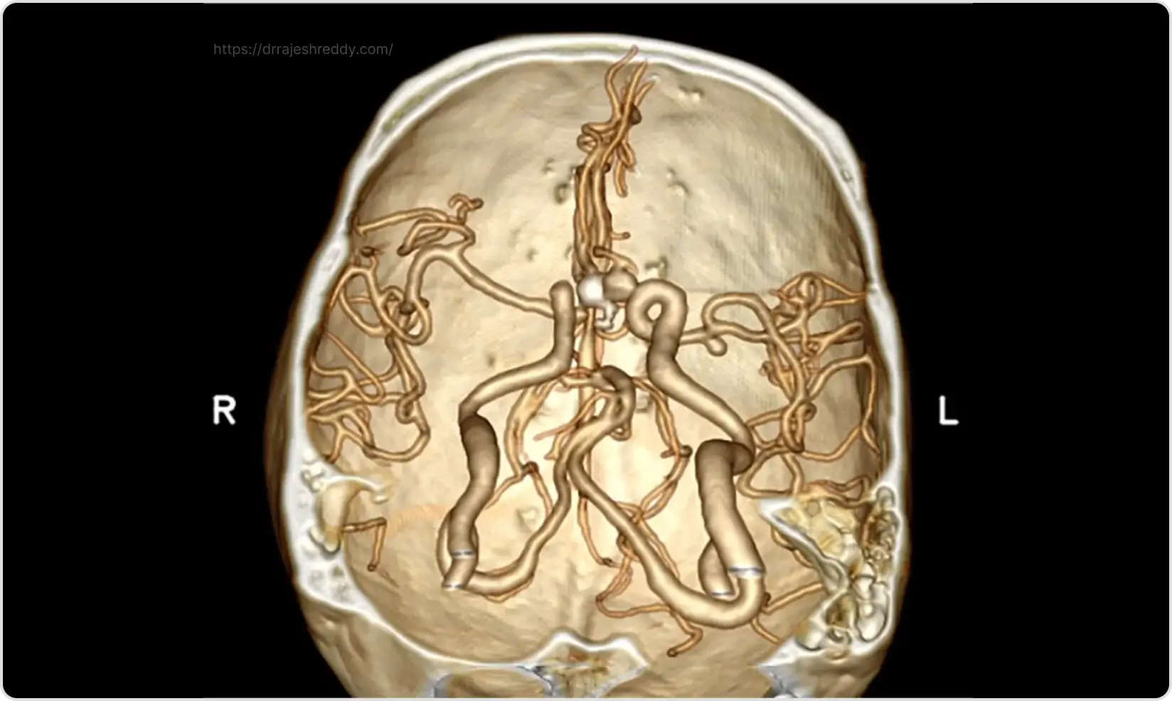
Digital Subtraction Angiography (DSA) — The Gold Standard
DSA provides the highest resolution imaging of cerebral vessels and is mandatory before any endovascular treatment (coiling, flow diversion). It allows real-time visualisation of blood flow, 3D rotational imaging of the aneurysm, and precise measurement of the aneurysm neck and dome — essential for planning the most appropriate treatment. Dr. Rajesh Reddy performs DSA as part of his integrated diagnostic and treatment pathway.
.webp)
MRI Brain and MR Angiography (MRA) — For Non-Emergency Cases
For patients with an incidentally discovered aneurysm, or for follow-up imaging after treatment, MRI and MRA provide excellent non-invasive assessment without radiation. High-resolution 3T MRI with dedicated MRA sequences can detect aneurysms as small as 2–3 mm.