

Introduction
The moment a brain aneurysm is successfully treated — whether by endovascular coiling, surgical clipping, or flow diversion — is a significant milestone. The immediate threat of rupture or re-bleeding has been addressed. But for most patients, particularly those who have survived subarachnoid hemorrhage (SAH), the journey is far from over. Recovery from brain aneurysm surgery is a process that unfolds over months — shaped by the type of treatment received, the severity of any bleeding that occurred, and the individual patient's baseline health and resilience.
This guide is written for patients and families navigating recovery from brain aneurysm treatment — whether that was an elective procedure for an unruptured aneurysm, or emergency treatment after SAH. It covers what to expect at each stage, what is normal, what is not, when to seek help, and how to approach the practical decisions — driving, returning to work, exercise, medications — that patients face in the weeks and months after treatment.
Dr. Rajesh Reddy Sannareddy, Senior Consultant Endovascular Neurosurgeon in Hyderabad, follows every aneurysm patient through the entire arc of their treatment and recovery — from the procedure itself through to long-term neurovascular surveillance. His team is available through the Dr. Reddy's Neuro Care app on Google Play for post-operative queries, follow-up appointments, and emergency guidance.
Recovery at a Glance: Coiling vs. Clipping vs. SAH
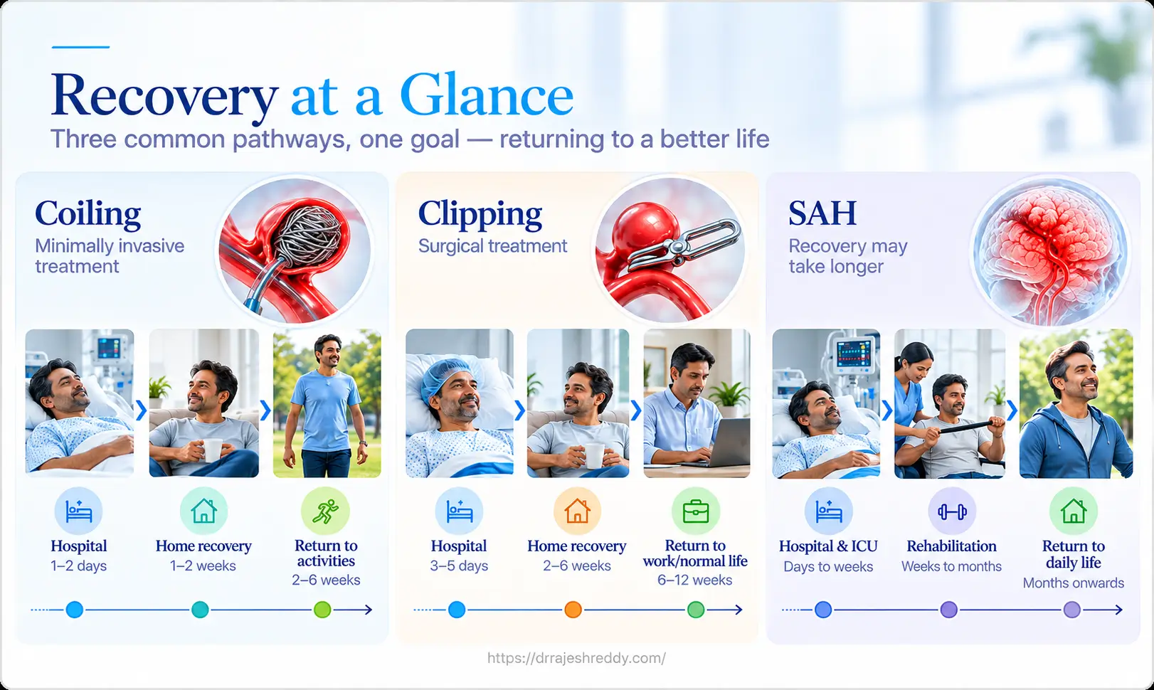
Recovery timelines differ substantially depending on what treatment was performed and whether the aneurysm had ruptured:
| Phase | After Coiling (Elective) | After Clipping (Elective) | After SAH (Any Treatment) |
|---|---|---|---|
| Hospital stay | 1–3 days | 5–7 days | 10-15 days (vasospasm monitoring) |
| ICU / HDU monitoring | Overnight only | 1–2 days | 5-7 days average |
| Groin / wound pain | Mild (groin site only) | Moderate (scalp wound) | Determined by procedure |
| Return to light activity | 3–5 days | 2–3 weeks | 6–12 weeks (varies with grade) |
| Return to driving | 1–2 weeks (no seizure) | 4–6 weeks | 6–12 months (seizure risk) |
| Return to desk work | 1–2 weeks | 4–6 weeks | 3–6 months |
| Return to physical work | 2–4 weeks | 6–10 weeks | 6–12 months |
| Antiplatelet medication | Dual for 6 months (stent/flow diversion). Thereafter Aspirin – duration is upto discretion of treating doctor | Not required after clipping | Same as procedure performed |
| Follow-up imaging | DSA/MRA/CTA at 6 months, 18 months | MRA/CTA at 6 months | DSA/CTA at 6 months mandatory |
Recovery after SAH is primarily determined by the severity of the initial bleed (WFNS grade) and the complications that followed — not by the aneurysm treatment technique itself. A patient who had coiling for a ruptured aneurysm but suffered severe vasospasm will have a much longer recovery than a patient who had surgical clipping for an unruptured aneurysm.
Week-by-Week Recovery After Elective Aneurysm Treatment (Unruptured)
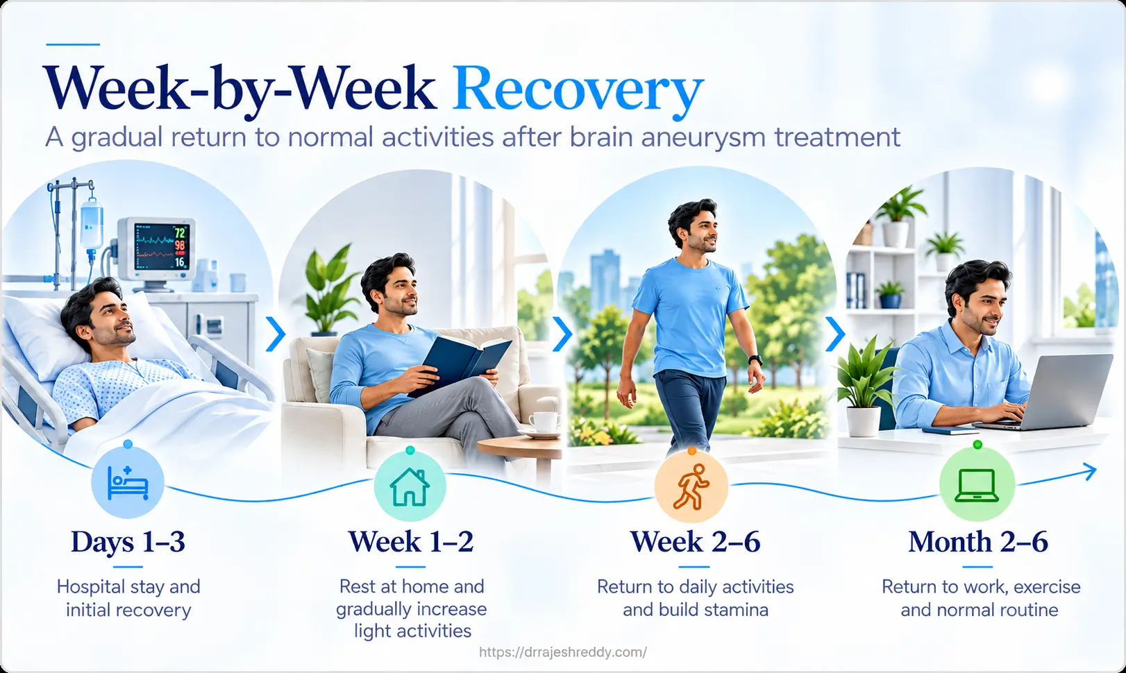
For patients who had elective treatment of an unruptured aneurysm — the most straightforward recovery scenario — here is what to expect at each stage:
Days 1–3: Procedure and Discharge
After Coiling
- Waking from anaesthesia in the recovery room; neurological check performed immediately
- Groin puncture site monitored — pressure dressing in place; 4–6 hours bed rest to prevent haematoma
- Mild groin bruising and tenderness is normal and typically resolves within 1–2 weeks
- Headache after coiling is common — usually mild and manageable with paracetamol
- Discharge day 1–2 for simple coiling; day 2–3 for stent-assisted coiling or flow diversion
- Antiplatelet medication: aspirin + clopidogrel or Ticagrelor (stent/flow diversion) — do not miss doses
After Clipping
- Recover in neurosurgical ICU for 24–48 hours; neurological observations every 2–4 hours
- Scalp wound with sutures or staples — wound checked daily; removed at 10–14 days
- Headache after craniotomy is more significant than after coiling — managed with regular analgesics
- Facial swelling around the eye on the side of the craniotomy — peaks at 48–72 hours and resolves over 1–2 weeks
- Anti-seizure medication commenced and continued for atleast 3–6 months
- Early physiotherapy — walking with support begins day 1–2
Week 1–2: Early Home Recovery
Whether home after 2 days (coiling) or 7 days (clipping), the first two weeks at home are about rest and gradual recovery. The brain and body are healing, and fatigue is the most universal — and most underestimated — early symptom.
What Is Normal in Week 1–2
- Fatigue — often profound; activities that were effortless before may feel exhausting
- Mild headache — improving progressively; paracetamol is usually sufficient
- Scalp numbness or tingling around the clipping wound — normal nerve regeneration
- Mild mood changes — feeling emotional, anxious, or flat is a normal response to the experience
- Poor appetite — common in the first week; small frequent meals help
- Sleep disturbance — difficulty sleeping or excessive sleepiness; both are temporary
Activity Restrictions — Week 1–2
- No driving — minimum 4 weeks for clipping; 1–2 weeks for coiling (in the absence of neurological deficit or seizure)
- No heavy lifting (> 5 kg) — avoid straining, which raises blood pressure and intracranial pressure
- No alcohol — particularly while on anti-epileptic or antiplatelet medications
- No strenuous exercise — light walks are encouraged and beneficial
- No swimming — until wound is fully healed (minimum 4–6 weeks)
- Constipation management — straining at stool causes dangerous spikes in intracranial pressure; use stool softeners from day 1 if needed; maintain adequate hydration
Important Note: Managing constipation actively is essential for all brain aneurysm patients in the recovery period. Straining at stool causes sudden surges in intracranial pressure that can be dangerous. Stool softeners (lactulose or sodium docusate) should be used from the first day at home if bowel movements are not occurring easily.
Week 2–6: Progressive Recovery
By week 2–3, most patients begin to feel meaningfully better — more energy, clearer thinking, reducing headaches. This is when gradual reintegration into normal activity begins.
Physical Recovery Milestones
- Week 2–3: Short walks (15–30 minutes), light household activity, reading and screen time in limited amounts
- Week 3–4: Increasing walking distance; light cooking; social activity; short car trips as a passenger
- Week 4–6 (coiling): Return to desk work; driving after medical clearance; light exercise (walking, gentle swimming after wound healed)
- Week 6–10 (clipping): Return to desk work; driving after 6-week neurosurgical review; suture removal at 10–14 days; physiotherapy if any motor deficit
6-Week Neurosurgical Review
A clinic review — in person or via video consultation — is scheduled at approximately 6 weeks. This appointment covers:
- Neurological assessment — any ongoing deficits, headache status, functional recovery
- Anti-epileptic medication review — tapering plan if no seizures have occurred
- Antiplatelet medication review — plan for clopidogrel or Ticagrelor cessation at 6 months (stent/flow diversion)
- Driving fitness — formal clearance for driving based on neurological status and seizure risk
- Return-to-work discussion — what activities are safe, what modifications may be needed
- Follow-up imaging schedule confirmed — DSA or MRA/ CTA at 6 months
Month 2–6: Functional Reintegration
For elective patients, month 2–6 marks a return to most normal activities. For SAH survivors, this period may still be early in recovery — particularly for those with higher-grade bleeds or vasospasm-related deficits.
Return to Work
- Desk-based / office work: typically possible from week 4–6 (coiling) or week 6–8 (clipping), starting with part-time hours and building up over 2–4 weeks.
- Professional roles (doctors, lawyers, teachers): typically week 6–10; cognitive demands should be considered — early return may require reduced caseload.
- Manual or physically demanding roles: 3–6 months; formal occupational health assessment recommended.
- SAH survivors: 3–6 months for good-grade; 6–12 months for moderate-grade; some poor-grade patients require permanent work modification.
Driving After Brain Aneurysm Treatment
Driving regulations after brain aneurysm treatment depend on the type of treatment and whether seizures have occurred:
- Coiling (unruptured, no seizure): driving typically from 1–2 weeks; confirm with Dr. Rajesh Reddy at follow-up
- Clipping (unruptured, no seizure): driving from 4–6 weeks after neurosurgical clearance
- Any patient who has had a seizure (at rupture or post-operative): typically 6–12 months seizure-free before driving is permitted — discuss with Dr. Rajesh Reddy and the transport authority
- SAH survivors (no seizure): driving typically from 3–6 months; return to driving requires formal neurosurgical clearance
Exercise and Physical Activity
- Light aerobic exercise (walking, gentle cycling) — from week 2–3 for coiling; week 4–6 for clipping
- Swimming — from week 4–6 once all wounds are fully healed
- Gym / weight training — from month 3 for coiling; month 3–4 for clipping; avoid breath-holding, heavy squats, or explosive lifting
- Contact sports — discuss with Dr. Rajesh Reddy; generally avoided for 6–12 months after any aneurysm treatment
- Sexual activity — generally safe from week 2–3 for coiling; week 3–4 for clipping
Recovery After SAH: A Different Journey
Recovery after subarachnoid hemorrhage is a fundamentally different experience from recovery after elective aneurysm treatment. The aneurysm treatment itself is only one part of what the brain has endured — the haemorrhage, the ICP spike, the blood-brain barrier disruption, and the complications (vasospasm, hydrocephalus) all contribute to a recovery that unfolds over months to years.
Physical Recovery After SAH
- Total hospital stay: 3–6 weeks for good-grade SAH (WFNS I–II) without major complications
- Rehabilitation: physiotherapy (motor), occupational therapy (daily function), speech and language therapy (language and swallowing) initiated in hospital and continued as outpatient
- Fatigue: the most persistent physical symptom; typically present for 6–12 months; pace activity carefully
- Headaches: often present for weeks to months after SAH; typically tension-type in character; managed with regular analgesics and headache diary
- VP shunt (15–20% of patients): typically inserted 3–6 weeks after SAH; dramatic improvement in walking, cognition, and quality of life usually follows within days
Post-SAH Syndrome: The Invisible Recovery
Even patients who make a full physical recovery after SAH frequently experience what is now recognised as post-SAH syndrome — a constellation of cognitive and psychological symptoms that are not visible on imaging but profoundly affect quality of life. Families often recognise the change before the patient does.
- Fatigue: A profound, qualitatively different tiredness from anything experienced before SAH. Unlike normal tiredness, it is not always relieved by sleep and can be triggered by cognitive effort as much as physical activity. It typically improves progressively over 12–24 months but may be permanent in severe SAH.
- Memory and concentration: Difficulty forming new memories, word-finding problems, slowed information processing, and difficulty multitasking. These are collectively described as 'brain fog' and are a recognised sequela of SAH — not a sign of ongoing damage, but of a brain that is still healing.
- Headaches: Persistent — often daily in the first months — and can be severe enough to interfere with return to work. The mechanism is multifactorial: vasomotor changes, medication effects, and psychological contributors all play a role.
- Depression and anxiety: Affect approximately 40–50% of SAH survivors. The combination of a frightening experience, changed life circumstances, ongoing physical symptoms, and the neurobiological effects of SAH on mood regulation creates a high risk of affective disorder. Proactive psychological support — counselling, psychiatry if needed — is not optional; it is part of good SAH care.
- Personality and behavioural changes: Subtle shifts in emotional regulation, patience, impulsivity, or social judgement may be noticed by family members. These typically improve over time but may require psychological or neuropsychological support.
Medications After Brain Aneurysm Treatment
Understanding the medications prescribed after brain aneurysm treatment — what each is for and how long to take it — helps patients stay adherent and avoid dangerous interruptions:
- Aspirin (75–150 mg daily): After stent-assisted coiling or flow diversion — continued for at least 3–5 years (decision of treating doctor) as single antiplatelet after clopidogrel is stopped. Never stop without consulting Dr. Rajesh Reddy.
- Clopidogrel (75 mg daily) or Ticagrelor (90 mg twice daily): After stent-assisted coiling or extrasaccular flow diversion — dual therapy with aspirin for 6 months, then stopped. Stopping clopidogrel/ Ticagrelor early risks in-stent thrombosis and stroke.
- Anti-epileptic medication: After surgical clipping (and in some SAH patients) — typically continued for 3–6 months. Tapered under medical supervision; never stopped suddenly.
- Nimodipine (60 mg every 4 hours): After SAH — the only proven medical prophylaxis against vasospasm-related delayed cerebral ischaemia. Continued for the full 21-day course from the day of SAH regardless of how well the patient feels.
- Stool softeners (lactulose or sodium docusate): For all post-operative patients — preventing straining at stool and the associated dangerous surges in intracranial pressure. Continue until bowel habits are normal and easy.
- Analgesics (paracetamol): For post-operative headache — safe and effective. Avoid ibuprofen and aspirin-containing analgesics in the first weeks (unless aspirin is prescribed as antiplatelet therapy).
Follow-Up Imaging: Why It Is Non-Negotiable
The aneurysm treatment does not end in the operating or angiography suite. Post-treatment surveillance imaging is an essential part of aneurysm management — particularly after endovascular coiling, where recanalisation can occur over time.
After Coiling or Flow Diversion
- 6 months: DSA (preferred) or high-resolution 3T MRA/ CTAngiogram — confirms aneurysm occlusion, absence of recanalisation, in-stent patency
- 18 months: DSA or MRA /CT angiogram— second surveillance point; most clinically significant recanalisation detected here
- Annual MRA thereafter: for large aneurysms, stented cases, and flow diversion — long-term surveillance to confirm sustained occlusion and sac shrinkage
- If recanalisation detected: retreatment with further coiling, flow diversion, or — rarely — surgical clipping is recommended before the aneurysm returns to its pre-treatment anatomy
After Surgical Clipping
- 6 months: MRA / CT angiogram— confirms clip position and aneurysm occlusion; assesses parent vessel patency
- 5 years: MRA / CT angiogram— checks for de novo aneurysm formation (patients with one aneurysm have 15–20% chance of another)
- Annual thereafter if stable: for patients with additional unruptured aneurysms being observed
WARNING: Skipping follow-up imaging is not safe. Approximately 15–20% of coiled aneurysms show recanalisation at 5 years — most can be treated with a further straightforward procedure if caught early. An undetected recanalised aneurysm that re-ruptures is a catastrophe that follow-up imaging prevents.
Warning Signs: When to Seek Urgent Help
Most aspects of recovery from brain aneurysm treatment are predictable and manageable at home. The following symptoms, however, require urgent medical attention:
| Symptom | What It May Indicate | Action |
|---|---|---|
| New sudden severe headache — different from usual post-op headache | Re-bleeding from aneurysm (coiling recanalisation); new aneurysm rupture | Go to emergency department immediately |
| New or worsening weakness, speech difficulty, or vision change within 3 weeks of aneurysm treatment | Stroke from vasospasm (SAH patients); thromboembolic event after coiling | Emergency — call 108 / go to ED immediately |
| First seizure or prolonged seizure | Cortical irritation; cerebral oedema; recurrent haemorrhage | Emergency — call 108; do not drive |
| Fever + headache + neck stiffness | Meningitis — post-operative or chemical from blood products | Urgent — same-day ED attendance |
| Clear fluid dripping from nose (pituitary/skull base patients) | CSF leak | Urgent — same-day clinic or ED attendance |
| Rapidly increasing confusion or drowsiness | Hydrocephalus (VP shunt malfunction or new shunt need) | Urgent — ED attendance; CT brain required |
| Groin haematoma after coiling — expanding painful swelling in groin | Femoral artery complication | Urgent — same-day vascular/surgical review |
| Wound redness, discharge, swelling after clipping | Wound infection or CSF leak | Urgent clinic review within 24 hours |
Long-Term Life After Brain Aneurysm Treatment

Most patients who undergo successful treatment of a brain aneurysm — whether ruptured or unruptured — go on to live full, active lives. The key to long-term wellbeing is a combination of ongoing surveillance, risk factor control, and realistic expectations about the recovery timeline.
Risk Factor Control for Life
- Blood pressure: target < 130/80 mmHg; medication if needed; home BP monitoring encouraged
- Smoking: cessation mandatory — smoking significantly increases the risk of recurrence and new aneurysm formation
- Constipation: manage actively long-term — straining at stool remains a risk for both treated and any remaining untreated aneurysms
- Alcohol: moderate — heavy alcohol use is associated with increased cerebrovascular risk
- Exercise: regular moderate aerobic exercise is beneficial and recommended after the initial recovery period
- Stress management: chronic psychological stress elevates blood pressure; mindfulness, relaxation techniques, and psychological support are part of long-term aneurysm health
Multiple Aneurysms & Family Screening
Patients with one brain aneurysm have a 15–20% chance of harbouring one or more additional aneurysms. Any untreated additional aneurysms detected at the time of initial treatment are monitored with regular MRA — their risk assessment and treatment planning follows the same evidence-based framework used for the index aneurysm. Dr. Rajesh Reddy will discuss the surveillance and management plan for any additional aneurysms at your follow-up consultation.
First-degree relatives of a patient with a confirmed brain aneurysm have 3–7 times the population risk of harbouring an aneurysm themselves. Screening with MRA — non-invasive, no radiation — is recommended for relatives with two or more affected first-degree relatives, or for those with one affected relative combined with other risk factors (hypertension, smoking, Polycystic Kidney Disease, Ehler Danlos or Marfan Syndrome). Please discuss family screening with Dr. Rajesh Reddy at your follow-up appointment.