Introduction & What Is a Glioma?
A glioma diagnosis carries a weight unlike almost any other. These are tumors that arise from within the brain's own tissue — from the glial cells that support and sustain neurons — and their treatment demands a level of surgical precision, intraoperative technology, and multidisciplinary coordination that few neurosurgical conditions require. For patients and families navigating a glioma diagnosis in Hyderabad, the questions are urgent: Who should perform the surgery? What approach is best for my tumor? What can realistically be achieved, and at what risk?
This page provides a comprehensive, honest guide to glioma surgery — what gliomas are, how they are graded, what surgery involves for different glioma types, the advanced technologies Dr. Rajesh Reddy Sannareddy uses to maximise safe resection, and what multimodal treatment looks like after surgery.
Dr. Rajesh Reddy is a Senior Consultant Neurosurgeon in Hyderabad with over 5,000 neurosurgical procedures to his credit, international fellowship training in Zurich, and visiting scholar experience at Barrow Neurological Institute (Phoenix, Arizona) — one of the world's leading centres for brain tumor surgery. His expertise in microsurgical, awake craniotomy, and fluorescence-guided glioma resection places him among the most experienced glioma surgeons in the region.
What Is a Glioma?
Gliomas are primary brain tumors — tumors that arise from within the brain itself rather than spreading from elsewhere in the body. They originate from glial cells, which are the brain's non-neuronal supportive cells. Glial cells provide structural support, nourishment, and insulation to neurons, and include several subtypes: astrocytes, oligodendrocytes, and ependymal cells. Tumors arising from each of these cell types carry different names, behaviours, and prognoses.
Gliomas account for approximately 80% of all malignant primary brain tumors and are among the most challenging tumors in all of oncology — not because surgery cannot remove them, but because their tendency to infiltrate surrounding normal brain tissue means that complete eradication is rarely possible. The goal of glioma surgery is maximum safe resection — removing as much tumor as possible while preserving the neurological function that determines a patient's quality of life.
How Are Gliomas Classified?
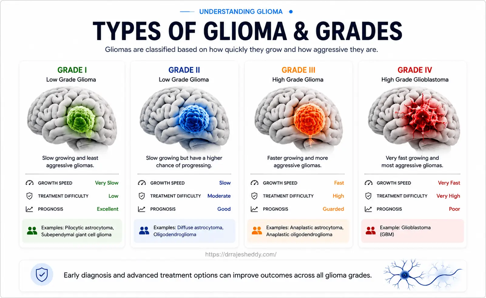
Gliomas are classified by the World Health Organisation (WHO) according to two parameters: cell type of origin and grade (I–IV). The 2021 WHO update added molecular markers — particularly IDH mutation status — as a mandatory part of glioma classification, because molecular profile predicts behaviour and treatment response more accurately than histology alone.
- IDH-mutant gliomas — grow more slowly, respond better to chemotherapy, and are associated with better survival. Found predominantly in younger adults.
- IDH-wildtype gliomas — more aggressive, less chemosensitive, and associated with shorter survival even at lower histological grades. GBM is almost always IDH-wildtype.
- 1p/19q codeletion — a molecular hallmark of oligodendrogliomas, associated with the best prognosis among diffuse gliomas and high sensitivity to PCV chemotherapy.
- MGMT promoter methylation — predicts response to temozolomide chemotherapy in GBM. Methylated tumors respond significantly better to alkylating chemotherapy.
Types of Glioma: Grades, Molecular Profile, and Prognosis
The table below summarises the key glioma subtypes treated by Dr. Rajesh Reddy, along with their WHO grade, IDH status, typical presentation, and prognosis:
| Glioma Type | WHO Grade | IDH Status | Typical Presentation | Prognosis |
|---|---|---|---|---|
| Pilocytic Astrocytoma | I | Wildtype | Children / young adults; cerebellum or optic pathway | Excellent — often curable with surgery alone |
| Diffuse Astrocytoma | II | Usually mutant | Young adults; seizures; frontal or temporal lobe | Good — median survival 10+ years with IDH mutation |
| Oligodendroglioma | II–III | Mutant + 1p/19q | Adults; seizures; frontal lobe predominance | Very good — most chemosensitive glioma subtype |
| Anaplastic Astrocytoma | III | Variable | Adults; progressive deficits; any lobe | Intermediate — surgery + radiation + chemotherapy |
| Anaplastic Oligodendroglioma | III | Mutant + 1p/19q | Adults; rapid symptom progression | Relatively favourable with PCV or temozolomide |
| Glioblastoma (GBM) | IV | Usually wildtype | Adults 50–70; rapid onset; any lobe | Poor — median survival ~14–16 months with treatment |
| IDH-mutant GBM | IV | Mutant | Younger adults; often transformed from LGG | Better than IDH-wildtype GBM; more chemosensitive |
A glioma's grade and molecular profile — not just its appearance on MRI — determine the treatment plan and prognosis. This is why surgical resection for tissue diagnosis and molecular profiling is critical even when complete removal is not the immediate goal.
Glioma Symptoms: What to Look For
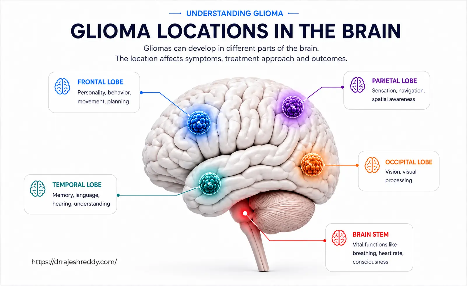
Glioma symptoms depend on the tumor's grade, size, and location. High-grade gliomas typically cause symptoms that develop rapidly over days to weeks; low-grade gliomas often evolve over months to years and may present with only a single symptom for a prolonged period.
Common Glioma Symptoms
- New-onset seizures — particularly in younger adults; often the first symptom of a low-grade glioma
- Progressive headaches — worsening over days to weeks, worst in the morning
- Focal neurological deficits — progressive weakness, numbness, or coordination problems on one side of the body
- Language difficulties — word-finding problems, slurred speech, or comprehension difficulties
- Cognitive and personality changes — memory problems, impaired judgement, apathy, or behavioural change
- Visual disturbances — field defects, blurring, or double vision
- Nausea and vomiting — from raised intracranial pressure as the tumor grows
Important Red Flag:
New-onset seizures in an adult who has never had epilepsy should always prompt an urgent MRI of the brain. Low-grade gliomas commonly present this way — and early identification gives patients the widest range of surgical and treatment options.
Diagnosing a Glioma: The Workup Before Surgery
Before surgery is recommended, Dr. Rajesh Reddy conducts a thorough pre-operative evaluation to characterise the tumor precisely and plan the safest, most effective surgical approach.
MRI Brain with Contrast — The Essential First Step
MRI is the gold standard for glioma diagnosis and surgical planning. Gliomas have characteristic MRI appearances: low-grade gliomas typically appear as non-enhancing lesions with T2/FLAIR signal change, while high-grade gliomas show ring-like contrast enhancement around a necrotic core. The MRI defines the tumor's size, location, relationship to eloquent brain areas, and degree of surrounding oedema (swelling).
Functional MRI (fMRI)
For gliomas near language or motor areas, functional MRI maps the precise location of these eloquent regions relative to the tumor. This information is essential for planning whether an awake craniotomy is needed and for defining safe surgical corridors. fMRI data is loaded into the neuronavigation system and used in real time during surgery.
Diffusion Tensor Imaging (DTI) and Tractography
DTI maps the brain's white matter fibre tracts — including the corticospinal tract (motor), arcuate fasciculus (language), and optic radiations (vision). By overlaying DTI tractography on the surgical plan, Dr. Rajesh Reddy identifies which critical pathways run through or adjacent to the glioma, and plans resection boundaries accordingly. This is a key component of connectomics-informed surgical planning — protecting not just individual eloquent areas but the networks that connect them.
MR Spectroscopy
MR spectroscopy analyses the biochemical profile of the tumor and surrounding tissue. It helps differentiate high-grade from low-grade gliomas, identify the most metabolically active (and therefore most malignant) region of the tumor to target for biopsy, and distinguish tumor recurrence from radiation-induced changes after treatment.
Stereotactic Biopsy
When a glioma is located in a region that does not permit safe resection — such as the thalamus, basal ganglia, or dominant hemisphere language areas where the risk-benefit of surgery is unfavourable — a stereotactic biopsy is performed. Using 3D image guidance, a small needle is introduced through a burr hole to obtain a tissue sample for histopathological and molecular analysis. This is a minimally invasive procedure performed under local anaesthesia with sedation in most cases.
Glioma Surgery: Techniques and Technology
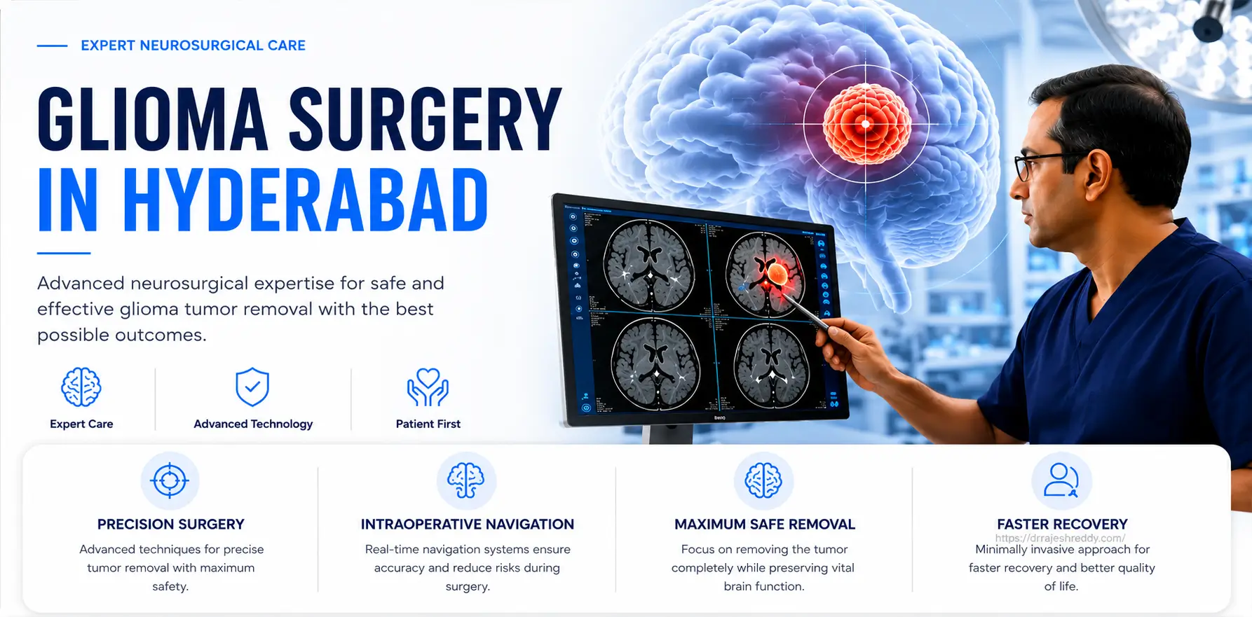
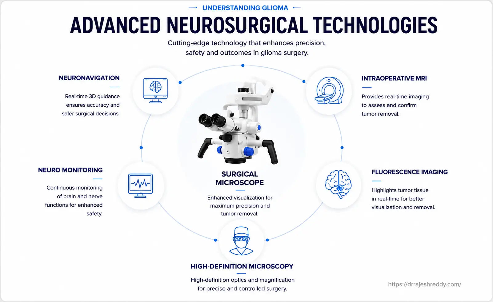
The surgical management of gliomas has been transformed over the past two decades by a convergence of advanced imaging, intraoperative technology, and neurosurgical technique. Dr. Rajesh Reddy employs a comprehensive toolkit for glioma surgery, choosing and combining techniques based on each patient's tumor characteristics and functional anatomy.
| Technique | When Used for Glioma | Key Benefit |
|---|---|---|
| Microsurgical Craniotomy | Most glioma resections — any grade, any lobe | High-magnification precision; optimal tumour control |
| Awake Craniotomy with Brain Mapping | Gliomas in or near eloquent cortex (speech, motor, language) | Real-time function preservation during maximum resection |
| 5-ALA / Sodium Fluorescein Guided Resection | High-grade gliomas (GBM, Grade III) | Visually distinguishes tumour from normal brain intraoperatively |
| Neuronavigation (StealthStation / BrainLab) | All glioma cases — used with other techniques | Sub-millimetre GPS accuracy for tumour localisation |
| Diffusion Tensor Imaging (DTI) Integration | Gliomas near white matter tracts (language, motor) | Maps critical fibre pathways to protect during resection |
| Intraoperative Neurophysiological Monitoring | All cases near motor or sensory pathways | Real-time detection of nerve stress during resection |
| Intraoperative MRI (iMRI) | Select complex high-grade glioma cases | Confirms extent of resection before closing; guides further removal |
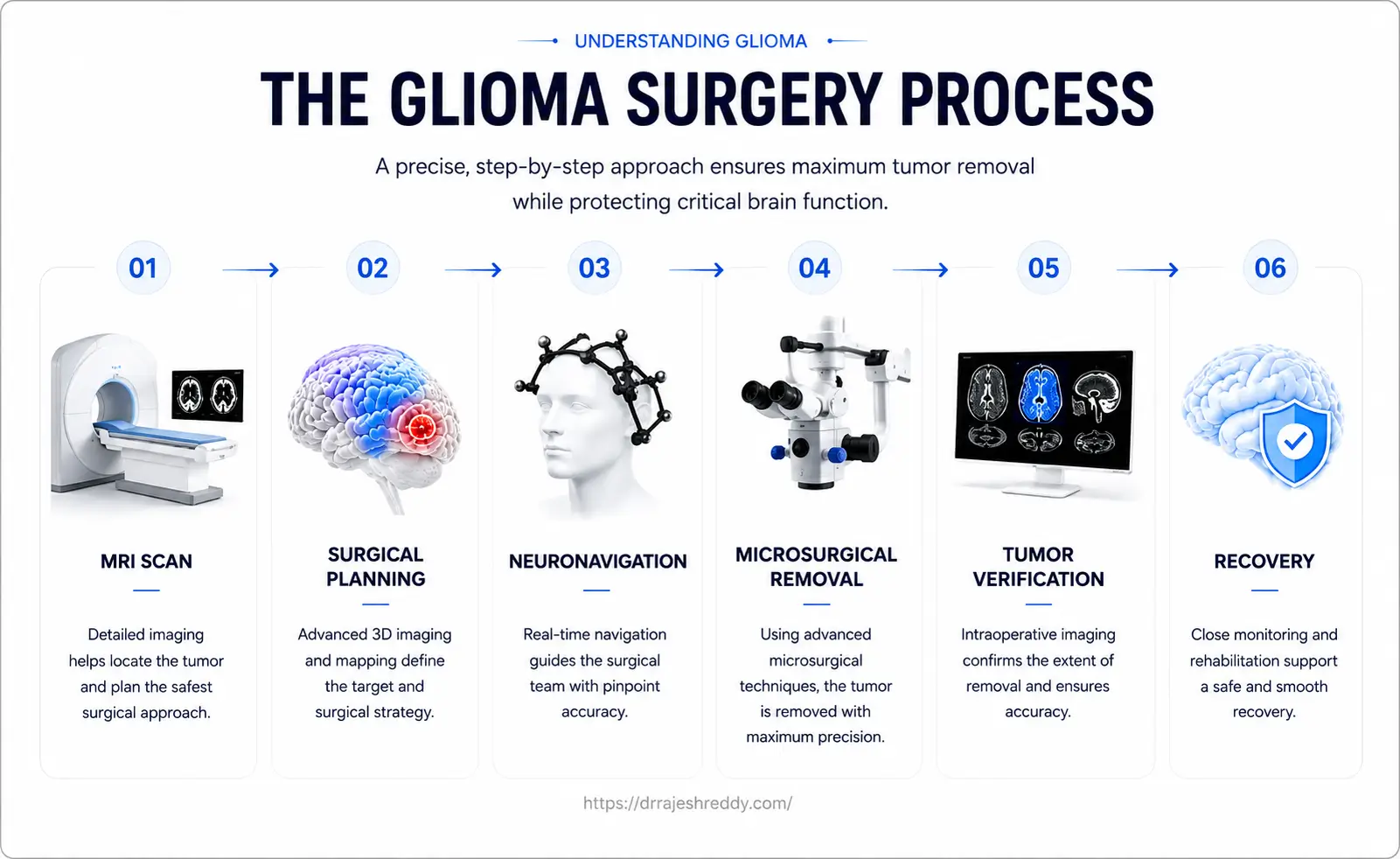
Maximum Safe Resection: Why Extent of Resection Matters
The extent of resection (EOR) in glioma surgery is one of the most important determinants of outcomes — both survival and quality of life. For GBM, studies consistently show that gross total resection (complete removal of all contrast-enhancing tumor) is associated with longer overall survival and longer time to recurrence compared to subtotal resection.
For low-grade gliomas, increasing EOR reduces the risk of malignant transformation and improves seizure control — even when the tumor is not immediately life-threatening. The challenge is achieving maximum EOR without causing permanent neurological deficits that would impair the patient's quality of life — which is precisely where the combination of awake craniotomy, neuronavigation, fluorescence guidance, and intraoperative monitoring becomes decisive.
Awake Craniotomy for Gliomas in Eloquent Areas
When a glioma involves or is adjacent to the motor cortex, Broca's area (speech production), Wernicke's area (speech comprehension), or primary sensory cortex, awake craniotomy is the surgical technique of choice. The patient is kept awake and interactive during the tumor resection phase while the surgical team monitors neurological function in real time.
Dr. Rajesh Reddy uses direct cortical and subcortical stimulation mapping during awake craniotomy to identify the precise boundaries of functional tissue. As resection proceeds, the patient is asked to speak, move their limbs, name objects, or count — depending on which functions are at risk. If stimulation at a particular point causes a temporary speech arrest or motor twitch, that point is marked and avoided. This technique allows glioma resection right up to the boundary of eloquent cortex without crossing it.
5-ALA and Sodium Fluorescein: Seeing Glioma Cells in Real Time
5-ALA (5-aminolevulinic acid) is an oral agent taken by the patient 3–4 hours before surgery. It is selectively metabolised by high-grade glioma cells into a fluorescent compound (protoporphyrin IX) that glows pink-red under violet-blue light in the operating microscope. Normal brain tissue does not fluoresce. This visual contrast allows the surgeon to identify and remove glioma tissue that would be invisible under standard white-light illumination — significantly improving the completeness of GBM resection.
Sodium Fluorescein — More Commonly Used in India: Sodium Fluorescein is an intravenous dye injected at the time of surgery that accumulates in areas of blood-brain barrier disruption — where high-grade glioma is present. Under a specific yellow-fluorescence filter (the YELLOW 560 filter) in the operating microscope, the glioma tissue glows bright yellow against the non-fluorescing normal brain tissue, allowing precise real-time identification of tumor margins during resection. Sodium Fluorescein is more commonly used than 5-ALA in India due to its wider availability, lower cost, and ease of administration — and is the primary fluorescence agent used by Dr. Rajesh Reddy in high-grade glioma surgery. Both agents can be used depending on the tumor and clinical context.
Connectomics and Surgical Planning
Connectomics maps the brain's neural networks — not just where individual functional areas are located, but how they communicate through white matter pathways. By integrating connectomic data with pre-surgical fMRI and DTI, Dr. Rajesh Reddy plans surgical approaches that protect critical neural network connections alongside maximising tumor removal, giving patients the best chance of preserving complex cognitive and motor abilities after glioma surgery.
Stereotactic Biopsy and Frameless Stereotaxy
When a glioma is located in a region that does not permit safe resection — such as the thalamus, basal ganglia, brainstem, or dominant hemisphere language areas where the risk-benefit of open surgery is unfavourable — a stereotactic biopsy is performed to obtain a tissue sample for histopathological and molecular diagnosis. This is a minimally invasive procedure performed under local anaesthesia with sedation.
Frameless Stereotaxy: Unlike traditional frame-based stereotactic biopsy — which requires a rigid metal frame bolted to the patient's skull — frameless stereotaxy uses neuronavigation (a surface-registration-based 3D GPS system) to guide the biopsy needle to the precise target within the brain without any frame. The patient's pre-operative MRI is loaded into the neuronavigation system and registered to the patient's head position in the operating room using surface landmarks or fiducial markers. The surgeon then uses a tracked biopsy needle holder to navigate to the target in real time, with the neuronavigation screen confirming instrument position at every step. Frameless stereotaxy is more patient-friendly, faster to set up, and allows the surgeon to adjust the biopsy trajectory intraoperatively if needed — making it the preferred approach in experienced centres.
Beyond Surgery: Multimodal Treatment for Gliomas
Surgery is the first and most important step in glioma management — but it is rarely the last. The treatment protocol after surgery depends on the tumor grade, molecular profile, and extent of resection:
| Glioma Grade | Surgery | Radiation | Chemotherapy |
|---|---|---|---|
| Grade I (Pilocytic) | Complete resection — often curative | Not routinely required | Not routinely required |
| Grade II (LGG) | Maximum safe resection | After surgery if high-risk features present | Temozolomide or PCV in high-risk cases |
| Grade III (Anaplastic) | Maximum safe resection | Post-operative radiotherapy (standard) | Temozolomide (MGMT methylated) or PCV (1p/19q deleted) |
| Grade IV (GBM) | Maximum safe resection + 5-ALA | 6-week concurrent chemoradiation (Stupp protocol) | Concurrent + adjuvant temozolomide; TTFields device |
The Stupp Protocol for GBM
The standard of care for newly diagnosed GBM following maximum safe resection is the Stupp protocol: 6 weeks of concurrent radiotherapy (60 Gy in 30 fractions) with daily oral temozolomide chemotherapy, followed by 6 cycles of adjuvant temozolomide. In patients with MGMT promoter methylation, response rates are significantly better, and some patients achieve long-term disease control well beyond the median survival figures.
Tumour Treating Fields (TTFields)
TTFields (Optune device) use alternating electric fields delivered via scalp electrodes to disrupt glioma cell division. They are used concurrently with adjuvant temozolomide in GBM and have been shown in Phase III trials to improve both progression-free and overall survival when added to standard chemotherapy. The decision to use TTFields is made in the tumour board meeting by Dr. Rajesh Reddy alongside radiation and medical oncology specialists.
Treatment of Recurrent Glioma
Gliomas — particularly GBM — almost invariably recur despite optimal primary treatment. Options at recurrence include re-operation (if the tumor is resectable and the patient's condition allows), repeat stereotactic radiosurgery, bevacizumab (anti-angiogenic therapy), clinical trial enrolment, and best supportive care. Each decision is made individually in a tumour board setting, weighing expected benefit against treatment burden and the patient's wishes.
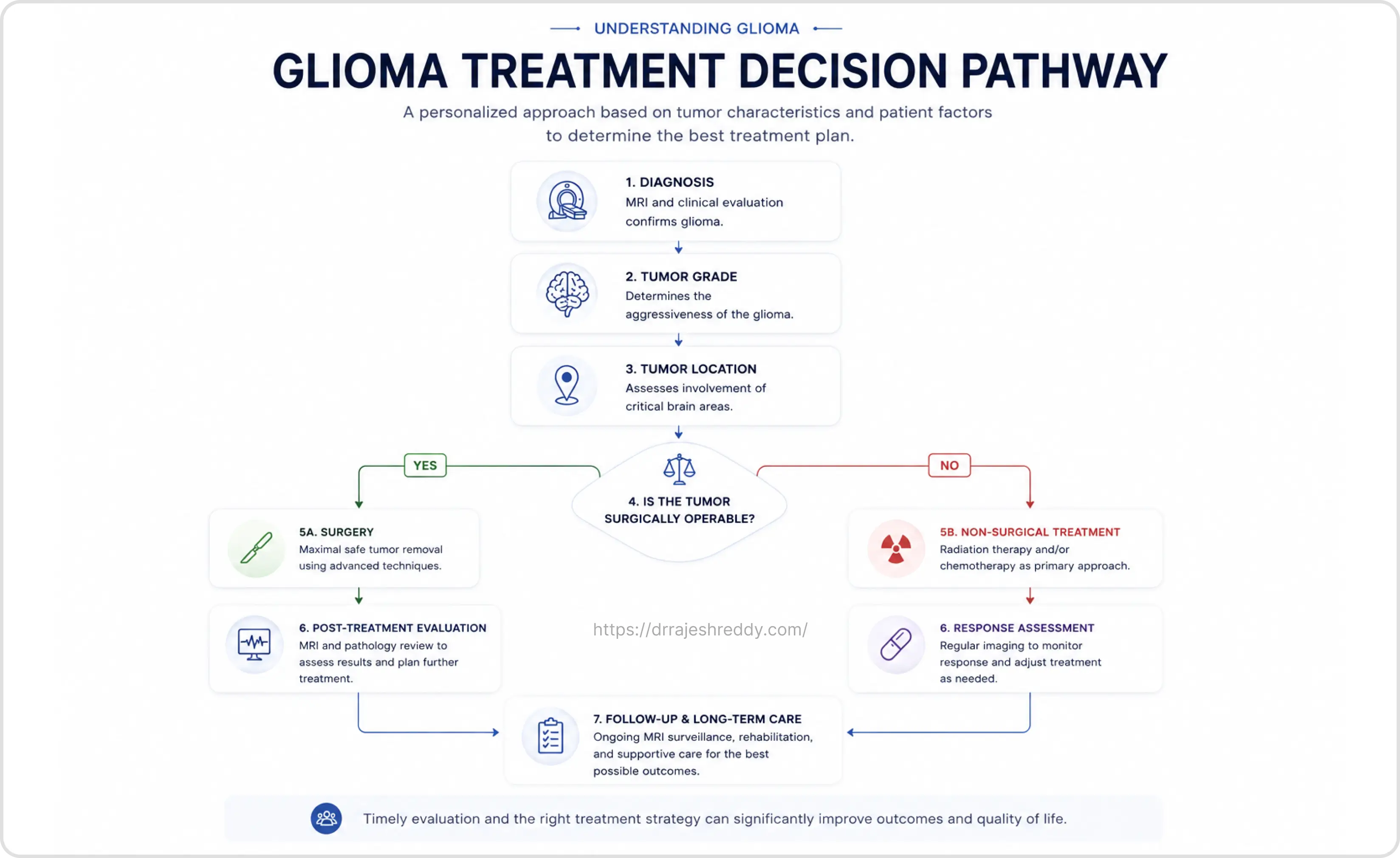
Why Choose Dr. Rajesh Reddy for Glioma Surgery in Hyderabad?
Glioma surgery — particularly for tumors in eloquent locations — is one of the most technically demanding procedures in neurosurgery. The difference between a surgeon who can remove a glioma and one who can remove it safely while preserving function is significant. Here is what sets Dr. Rajesh Reddy apart:
Expertise & Credentials
- MCh Neurosurgery (Exam Topper), NIMS Hyderabad; DNB Neurosurgery
- FINR — Fellow in Interventional Neuroradiology, University Hospital, Zurich
- Visiting Scholar — Barrow Neurological Institute, Phoenix, Arizona (a global centre of excellence for brain tumor surgery)
- Visiting Scholar — Okayama University, Japan; Klinikum Stuttgart, Germany
- Over 5,000 neurosurgical procedures; extensive glioma surgery experience across all grades
- Expertise in awake craniotomy, 5-ALA / Sodium Fluorescein guided resection, neuronavigation, DTI-integrated surgical planning, and intraoperative neurophysiological monitoring
- Clinical acumen and judgement gained through years of training with leading experts globally
Multidisciplinary Coordination:
Dr. Rajesh Reddy works closely with medical oncology and radiation oncology teams to ensure that every glioma patient receives a coordinated, tumour board-reviewed treatment plan from the outset — not just a surgical consultation in isolation.
Recovery After Glioma Surgery
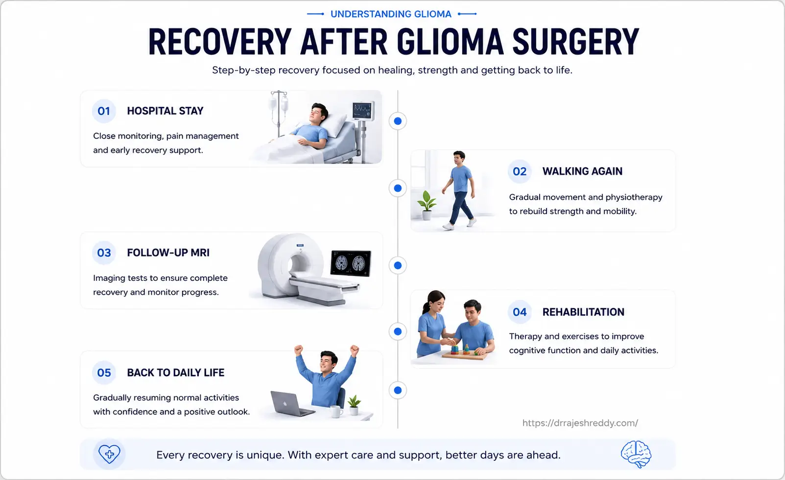
Recovery after glioma surgery varies depending on the tumor grade, location, extent of resection, and any post-operative neurological changes. Here is what patients can typically expect:
Immediate Post-Operative Period (Days 1–5)
- Monitoring in a dedicated neurosurgical ICU for the first 24–48 hours
- Neurological assessment every few hours to detect and respond to any early complications
- Steroids (dexamethasone) to control brain swelling — typically tapered over 1–2 weeks
- Anti-seizure medication continued or initiated as appropriate
- Early mobilisation — most patients are sitting up within 24 hours and walking within 48 hours
Weeks 2–6
- Return home typically after 5–7 days for uncomplicated cases
- Fatigue is the most common complaint — rest is important but progressive activity is encouraged
- Wound healing monitored; sutures or staples removed at 10–14 days
- Post-operative contrast MRI performed at 24–72 hours and again at 3–4 weeks to assess resection extent and plan adjuvant treatment — earlier re-imaging is essential for gliomas as treatment planning cannot be delayed
- Oncology referral and treatment planning meeting initiated during this period for malignant gliomas
3 Months and Beyond
- Malignant glioma patients continue with radiation and chemotherapy; regular MRI every 2–3 months
- Low-grade glioma patients typically return to normal activities within 6–8 weeks
- Neuropsychological rehabilitation if cognitive or language deficits persist
- Seizure management reviewed and anti-epileptic medications adjusted based on post-operative seizure control