
Introduction
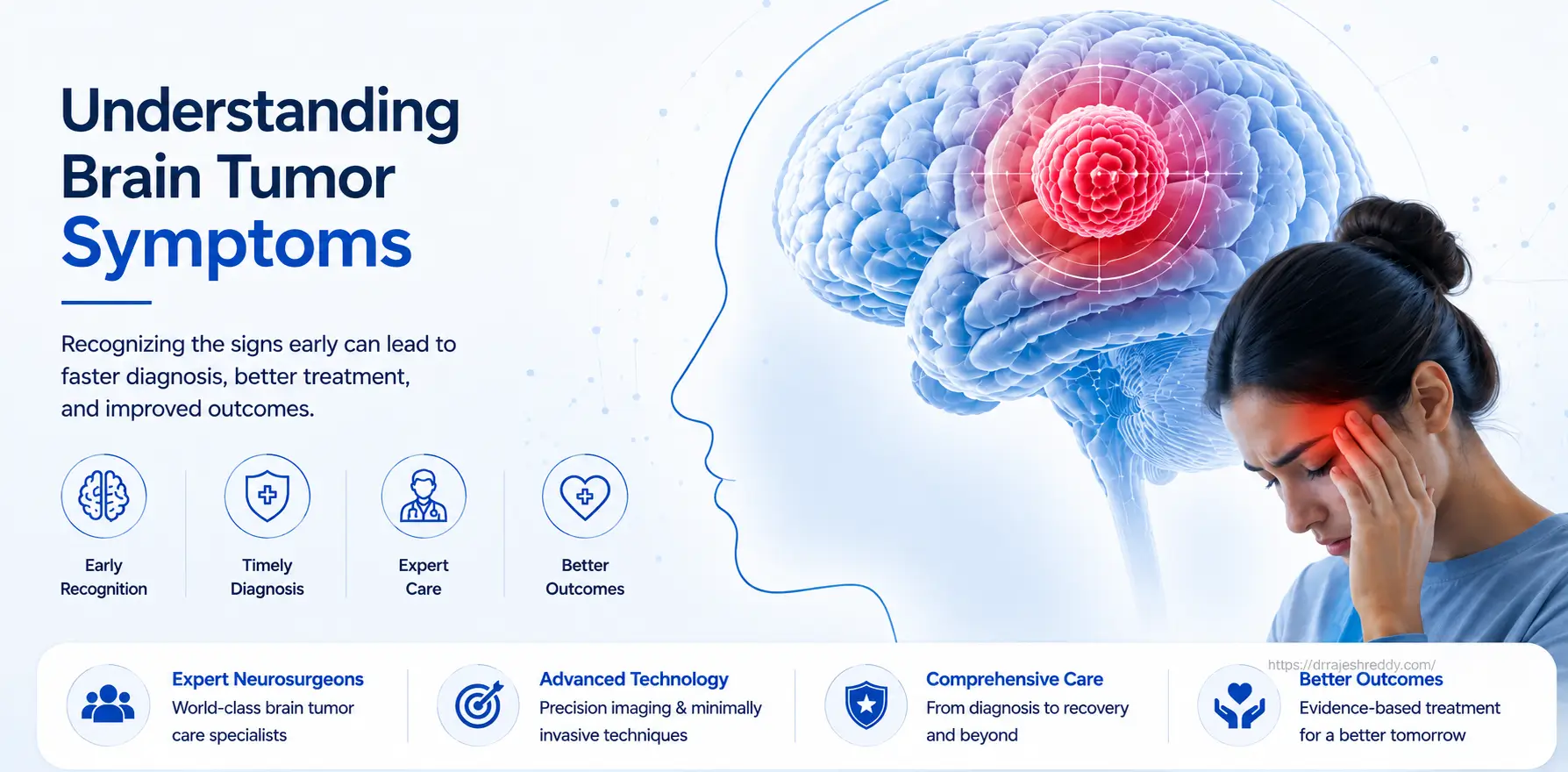
Brain tumor symptoms are among the most varied and easily misattributed of any medical condition. A persistent headache is dismissed as stress. A new seizure is explained away as sleep deprivation. A gradual change in personality goes unnoticed for months until a family member raises the alarm. The challenge with brain tumor symptoms is not that they are invisible — it is that they mimic so many other, far more common conditions.
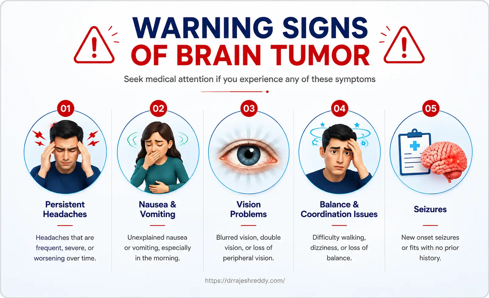
This guide is written to help patients, families, and caregivers understand what brain tumor symptoms actually look like — why they occur, how they differ by tumor location, which symptoms demand urgent attention, and when it is time to see a neurosurgeon. It is not written to cause alarm. The vast majority of people who experience headaches or memory problems do not have a brain tumor. But for those who do, early recognition and timely diagnosis make a significant difference in outcomes.
Dr. Rajesh Reddy Sannareddy, Senior Consultant Neurosurgeon in Hyderabad, has evaluated thousands of patients presenting with neurological symptoms. This guide reflects the clinical patterns he encounters in practice and the questions patients most frequently ask at their first consultation.
Why Does a Brain Tumor Cause Symptoms?
Brain tumor symptoms arise through two distinct mechanisms. Understanding these mechanisms helps explain why the same tumor type can cause very different symptoms in different patients — and why some tumors cause severe symptoms while others are discovered incidentally with no symptoms at all.
Mechanism 1: Raised Intracranial Pressure (ICP)
The skull is a rigid, closed box. The brain, blood, and cerebrospinal fluid (CSF) inside it exist in a precise pressure equilibrium. When a tumor grows, it adds volume to this fixed space. The brain initially compensates — compressing CSF spaces and venous blood — but once these compensatory mechanisms are exhausted, intracranial pressure rises.
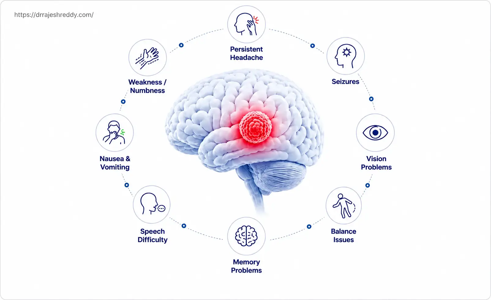
Raised ICP produces a characteristic set of symptoms regardless of where the tumor is located:
- Headache — typically progressive, worse in the morning or when lying flat
- Nausea and vomiting — particularly on waking or with sudden changes in head position
- Visual disturbances — blurring of vision, or episodes of graying-out of vision (visual obscurations)
- Papilloedema — swelling of the optic disc visible on fundoscopy, caused by pressure transmitted along the optic nerve sheath
- Altered consciousness — drowsiness, confusion, or reduced responsiveness in severe cases
Mechanism 2: Focal (Location-Specific) Effects
Every region of the brain has specific functions. A tumor growing in or adjacent to a functional area disrupts those functions directly — either by invading the tissue, compressing it, or irritating it. This produces focal neurological symptoms that point specifically to the tumor's location in the brain. A motor cortex tumor causes weakness; a visual cortex tumor causes vision loss; a language area tumor causes speech difficulty.
The interplay between these two mechanisms — raised ICP producing general symptoms and focal effects producing location-specific symptoms — is what makes brain tumor presentations so variable from patient to patient.
Brain Tumor Symptoms by Location
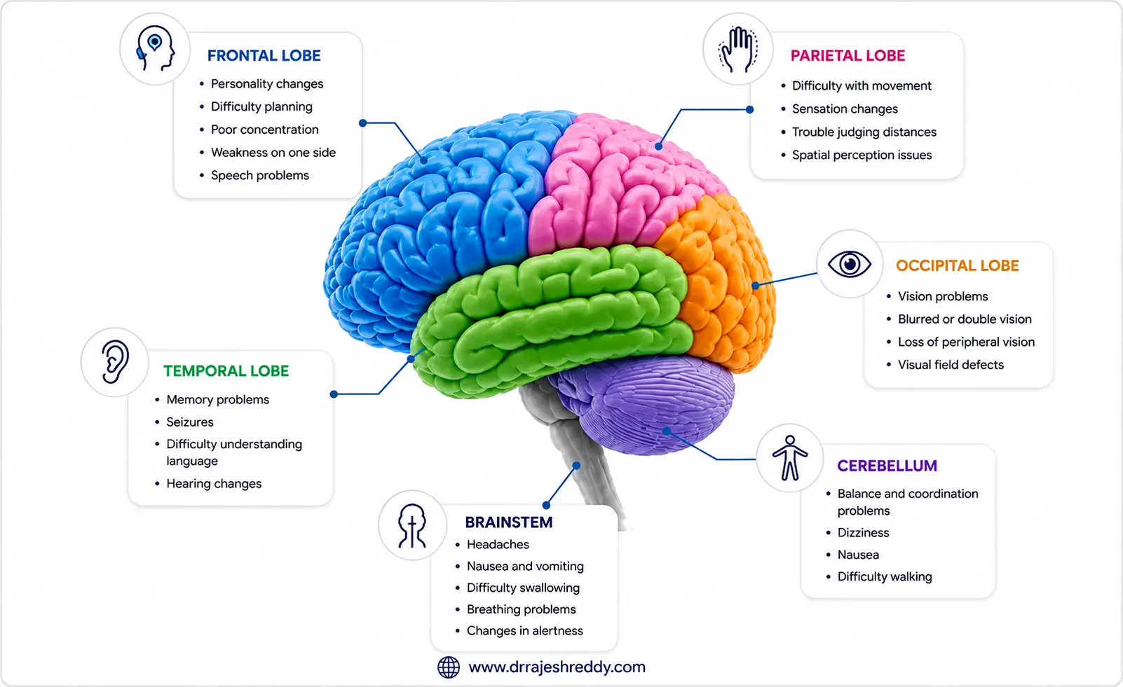
The table below maps common brain tumor symptoms to the region of the brain where the tumor is located and explains why each symptom occurs:
| Tumor Location | Typical Symptoms | Why It Happens |
|---|---|---|
| Frontal Lobe | Personality & behaviour changes, poor planning, weakness in opposite arm/leg, speech difficulty (dominant side) | Frontal lobe governs executive function, personality, and motor control of the opposite side of the body |
| Temporal Lobe | Memory problems, language comprehension issues, new-onset seizures, unusual smells or sounds (aura) | Temporal lobe handles memory, language comprehension, and is the most common site for seizure onset |
| Parietal Lobe | Numbness / tingling on one side, difficulty reading or writing, spatial disorientation | Parietal lobe processes sensory information and spatial awareness |
| Occipital Lobe | Visual field loss, blurred vision, visual hallucinations, difficulty recognising objects | Occipital lobe is the brain's visual processing centre |
| Cerebellum | Loss of balance and coordination, unsteady gait, difficulty with fine movements, slurred speech | Cerebellum controls balance, coordination, and smooth muscle movements |
| Brainstem | Double vision, facial weakness or numbness, difficulty swallowing, severe coordination problems | Brainstem houses cranial nerve nuclei and controls vital functions including breathing and heart rate |
| Pituitary Gland | Hormonal disturbances, visual field defects (bitemporal hemianopia), headache behind the eyes | Pituitary regulates hormones; enlarging tumors compress the optic chiasm directly above |
| Ventricles | Headache, nausea, drowsiness, sudden deterioration (hydrocephalus) | Ventricular tumors block CSF flow, raising intracranial pressure rapidly |
Brain Tumor Headaches: How to Tell Them Apart
Headache is the symptom most people associate with brain tumors — and one of the most frequently Googled. The reality is nuanced: headache is a symptom in only about 50% of brain tumor patients at diagnosis, and most headaches are not caused by brain tumors. However, certain headache characteristics are genuine red flags that warrant investigation.
Characteristics of a Brain Tumor Headache
- Progressive — worsening in frequency and intensity over days to weeks, rather than following the episodic pattern of migraine or tension headache
- Morning predominance — often worst on waking, because lying flat overnight allows ICP to rise
- Position-dependent — worsens when bending forward, coughing, sneezing, or straining (Valsalva manoeuvre)
- Associated with nausea or vomiting — particularly in the morning
- Accompanied by neurological symptoms — weakness, vision changes, speech difficulty, or seizures alongside the headache
- Not relieved by standard painkillers — over-the-counter analgesics provide partial or no relief
- New in onset — a headache pattern that is genuinely new for the patient, particularly in middle age or older adulthood
The comparison table below contrasts brain tumor headaches with the more common tension and migraine headaches:
| Feature | Brain Tumor Headache | Tension / Migraine Headache |
|---|---|---|
| Onset pattern | Gradually worsening over days to weeks | Often episodic; known pattern for the patient |
| Worst time of day | Morning; may wake patient from sleep | Usually improves with rest; variable timing |
| Position effect | Worsens when lying flat or bending forward | Typically not position-dependent |
| Associated symptoms | Nausea, vomiting, neurological signs (weakness, vision changes) | May have nausea/photophobia; no focal neurology |
| Response to painkillers | Partial or no relief with standard analgesics | Often relieved by painkillers or sleep |
| Family history | No specific association | Often positive family history of migraine |
| Red flag? | Yes — warrants MRI evaluation | Only if pattern changes or new features appear |
A headache that is new, progressively worsening, worst in the morning, or accompanied by any neurological symptom — weakness, vision change, speech difficulty, memory problems — warrants an MRI of the brain. Do not assume a new headache pattern is benign without investigation.
Seizures: A Key Red Flag
New-onset seizures in an adult who has no previous history of epilepsy are one of the most important red flags for an underlying brain tumor. Seizures occur when a tumor irritates the surrounding brain tissue, creating abnormal electrical activity that spreads through the cortex.
Types of Seizures in Brain Tumor Patients
Focal (partial) seizures: Affect a specific part of the body corresponding to the tumor's location — for example, rhythmic twitching of one hand (frontal lobe), a rising sensation in the abdomen or déjà vu (temporal lobe), or visual flashes (occipital lobe). The patient may remain conscious.
Focal seizures with secondary generalisation: Begin as a focal seizure but spread to involve the whole brain, producing a generalised tonic-clonic (convulsive) seizure with loss of consciousness.
Generalised seizures: Involve the whole brain from the outset — less common as a first presentation of brain tumor than focal seizures.
What to Do After a First Seizure
Any first seizure in an adult — even a mild one — requires urgent medical evaluation. The standard workup includes blood tests, an EEG, and crucially, an MRI of the brain with contrast. Do not wait to see if 'it happens again.' A single unexplained seizure in a previously healthy adult is sufficient reason for neuroimaging.
Low-grade gliomas in particular often present with seizures as their only symptom for months or years before the tumor is discovered. Early detection through post-seizure MRI gives patients the widest range of treatment options.
Cognitive Changes and Personality Shifts
Changes in thinking, memory, and personality are among the most distressing — and most frequently overlooked — symptoms of brain tumors. They are often attributed to stress, depression, ageing, or burnout before a neurological cause is considered. Family members frequently notice these changes before the patient does.
Cognitive Symptoms to Watch For
- Memory problems — difficulty forming new memories, forgetting recent conversations or appointments
- Concentration difficulties — inability to focus, losing track mid-sentence or mid-task
- Word-finding problems — struggling to retrieve words during speech, even familiar ones
- Slowed thinking — processing information more slowly than usual, taking longer to respond
- Disorientation — confusion about time, place, or recent events
Personality and Behavioural Changes
- Apathy and loss of motivation — previously engaged individuals becoming withdrawn and disinterested
- Disinhibition — saying or doing things that are socially inappropriate without apparent awareness
- Increased irritability or aggression — low frustration tolerance, uncharacteristic outbursts
- Depression or anxiety — new-onset mood disturbances without a clear psychological trigger
- Impaired judgement — making poor decisions, difficulty assessing risk
These symptoms are particularly associated with tumors of the frontal lobe — the brain's centre of executive function, personality regulation, and social behaviour. Frontal lobe tumors can grow to a significant size before causing the headaches and neurological deficits that typically prompt a scan, making cognitive and personality changes the earliest and sometimes only warning signs.
Vision Changes and Brain Tumors
The brain devotes a substantial portion of its territory to visual processing, and the visual pathway runs from the retina all the way to the occipital lobe at the back of the skull. Tumors anywhere along this pathway can cause visual symptoms.
Types of Visual Disturbance
Bitemporal hemianopia: Loss of the outer (temporal) half of the visual field in both eyes. This is the classic sign of a pituitary tumor pressing on the optic chiasm — the point where the optic nerves cross. Patients often describe it as difficulty seeing objects to the side, or walking into door frames.
Homonymous hemianopia: Loss of the same half of the visual field in both eyes — for example, loss of the right visual field in both the right and left eye. This results from tumors affecting the optic radiation or occipital lobe.
Vision loss in one eye: Can occur when a tumor — such as a Meningioma — directly compresses one optic nerve or compromises its blood supply, depriving it of nutrition. This may present as progressive blurring or complete loss of vision in that eye and should be investigated urgently.
Diplopia (double vision): Two images of a single object. Common with brainstem tumors or tumors affecting the cranial nerves that control eye movement (CN III, IV, VI).
Papilloedema: Not a visual symptom the patient necessarily notices, but a sign the doctor sees on examining the back of the eye — swelling of the optic disc caused by raised intracranial pressure. When severe, it can cause visual obscurations (momentary greying out of vision) and eventually permanent visual loss if ICP is not relieved.
Visual hallucinations: Seeing shapes, colours, or images that are not present — associated with occipital lobe tumors.
Weakness, Numbness, and Speech Problems
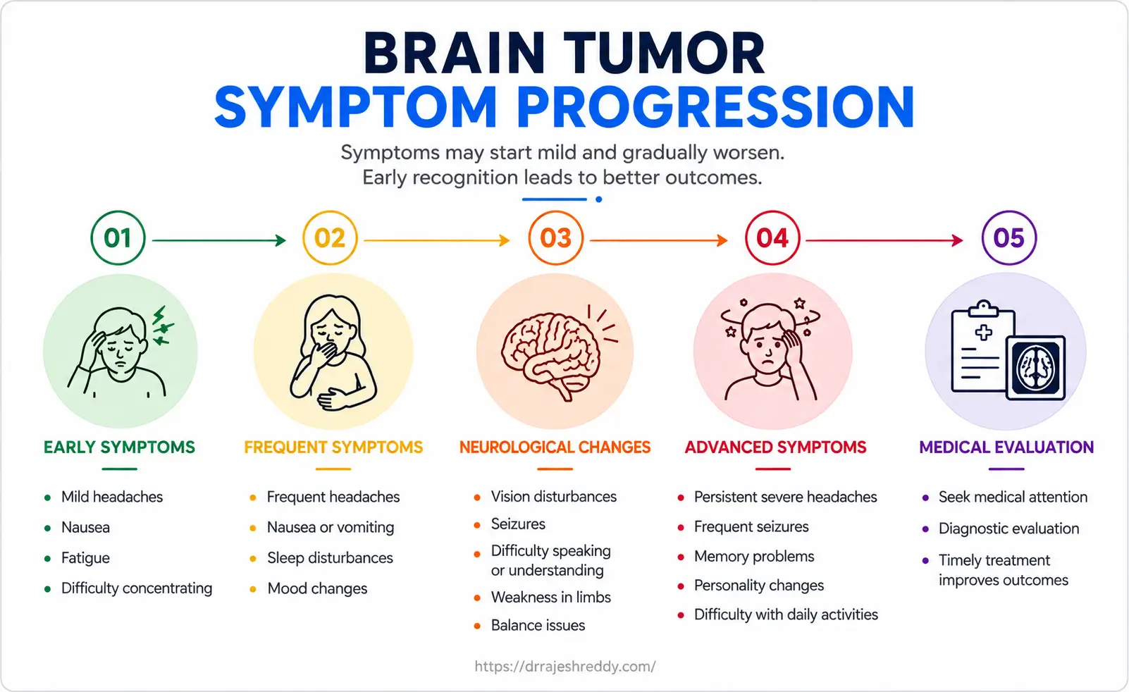
Focal neurological deficits — weakness, sensory loss, or speech difficulty — are among the most diagnostically useful symptoms of brain tumors because they point directly to the tumor's location.
Motor Weakness
Progressive weakness on one side of the body — hemiparesis — develops when a tumor involves the motor cortex (located in the posterior frontal lobe) or the motor pathways running through the internal capsule. The weakness typically affects the arm and leg on the opposite side to the tumor (contralateral weakness), because the motor pathway crosses before descending to the spinal cord.
Patients describe it as clumsiness, dropping objects, difficulty climbing stairs, or dragging one foot. It is gradual and progressive — unlike the sudden onset of stroke — though tumors that bleed (haemorrhage into a tumor) can cause sudden onset deficits that mimic stroke.
Speech and Language Difficulties
Speech problems from brain tumors take two distinct forms, depending on which language area is affected:
- Broca's aphasia (expressive aphasia): The patient understands what is being said but has difficulty forming words or sentences. Speech is laboured, telegraphic, and frustrating. Associated with tumors in Broca's area in the inferior frontal gyrus of the dominant hemisphere.
- Wernicke's aphasia (receptive aphasia): The patient speaks fluently but the words are jumbled or nonsensical, and they have difficulty understanding speech. Associated with tumors in Wernicke's area in the posterior temporal lobe.
Language difficulties are among the most distressing symptoms for patients and families. They are also among the most important to identify before surgery — because planning an awake craniotomy requires knowing exactly which language areas are at risk, so they can be mapped and protected during tumor resection.
Sensory Symptoms
Numbness, tingling, or altered sensation on one side of the body — contralateral to the tumor — indicate involvement of the sensory cortex in the parietal lobe or the sensory pathways. Some patients describe a feeling that one hand or foot 'doesn't belong to them' or that sensations feel different on the two sides of the body.
Cranial Nerve Dysfunction: An Overlooked Group of Symptoms
Brain tumors — particularly those at the skull base, in the posterior fossa, or along the brainstem — can affect the cranial nerves, which control many essential functions of the head, face, and throat. Cranial nerve dysfunction produces a specific and recognisable set of symptoms that patients and families should be aware of:
- Drooping of one eyelid (ptosis) — caused by compression or involvement of the third cranial nerve (CN III); may also be accompanied by a dilated, unreactive pupil on the same side
- Double vision (diplopia) — from involvement of CN III, IV, or VI, which control eye movement; the patient sees two images of a single object
- Facial asymmetry or weakness — caused by facial nerve (CN VII) involvement; one side of the face droops, the patient cannot close one eye fully, and the smile appears uneven
- Numbness over the face — caused by trigeminal nerve (CN V) compression; may feel like a dental anaesthesia that does not wear off, or persistent tingling across one cheek, jaw, or forehead
- Loss of hearing or tinnitus — from involvement of the vestibulocochlear nerve (CN VIII); a classic presentation of vestibular schwannoma (acoustic neuroma)
- Swallowing difficulty (dysphagia) — from glossopharyngeal (CN IX) or vagus nerve (CN X) involvement; patients may cough or choke on food or liquids
- Nasal regurgitation of feeds — food or liquid returning through the nose during or after swallowing, indicating soft palate weakness from CN IX or X dysfunction
- Change in voice quality — hoarseness or a nasal quality to the voice, caused by vagus nerve (CN X) involvement affecting the vocal cords or soft palate
- Tongue weakness — causing difficulty with chewing, forming words clearly, or moving food within the mouth; caused by hypoglossal nerve (CN XII) involvement; patients may inadvertently bite the tongue due to impaired tongue muscle control
- Shoulder drooping or weakness in raising the arm — from accessory nerve (CN XI) involvement; patients cannot shrug one shoulder or turn their head against resistance
These cranial nerve symptoms are particularly associated with skull base tumors (meningiomas, schwannomas, chordomas, craniopharyngiomas), posterior fossa tumors, and brainstem gliomas. Their presence should prompt urgent neurological evaluation and dedicated MRI of the brain and skull base.
Hormonal Symptoms: Pituitary Tumor Presentation
Pituitary tumors deserve special mention because their symptoms are often hormonal rather than neurological — leading patients to consult gynaecologists, endocrinologists, or general physicians before a neurosurgeon is ever involved.
- Prolactinoma: irregular or absent periods, milk discharge from the breasts (galactorrhoea), infertility in women; reduced libido, erectile dysfunction, and infertility in men
- Cushing's disease (ACTH-secreting): weight gain concentrated around the abdomen and face (moon face), purple stretch marks (striae), easy bruising, high blood pressure, diabetes, and mood disturbances
- Acromegaly (GH-secreting): gradual enlargement of the hands, feet, and jaw; coarsening of facial features; excessive sweating; joint pain; carpal tunnel syndrome
- Non-functioning macroadenoma: bitemporal visual field loss, headache behind the eyes, fatigue from pituitary insufficiency
Pituitary tumors can be present for years before diagnosis. If you have been treated for infertility, irregular periods, or unexplained hormonal abnormalities without a clear cause, ask your doctor whether a pituitary MRI has been performed.
Emergency Symptoms: When to Go to Hospital Immediately
Most brain tumor symptoms develop gradually over weeks to months. However, certain situations demand immediate emergency care — they indicate either a rapidly expanding tumor, hemorrhage into a tumor, or acute hydrocephalus.
SEEK EMERGENCY CARE IMMEDIATELY IF YOU EXPERIENCE:
- A sudden, severe headache described as 'the worst headache of my life'
- Sudden loss of speech, vision, or ability to move one side of the body
- A first seizure or a prolonged seizure
- Sudden loss of consciousness or unresponsiveness
- Sudden extreme confusion or agitation
Hemorrhage into a brain tumor — where the tumor's abnormal blood vessels rupture — can cause sudden, stroke-like symptoms. This is a neurosurgical emergency. Similarly, a colloid cyst or ventricular tumor that suddenly blocks CSF circulation can cause acute hydrocephalus with rapid deterioration. Both require immediate CT imaging and neurosurgical assessment.
When Should You See a Neurosurgeon?
You should seek a neurosurgical consultation — which will typically begin with an MRI of the brain — if you experience any of the following:
- New-onset seizures at any age with no prior epilepsy history
- A headache pattern that is genuinely new, progressively worsening, or worst in the morning
- Any focal neurological symptom: progressive weakness, numbness, vision change, or speech difficulty
- Unexplained personality or cognitive changes noticed by yourself or family members
- Hormonal symptoms suggesting a pituitary tumor (menstrual irregularity, visual field loss, acromegaly features)
- Cranial nerve symptoms: drooping eyelid, facial numbness, hearing loss, swallowing difficulty, change in voice
- An incidental finding on MRI — a lesion found when imaging was done for another reason
- A previously diagnosed benign lesion showing change on follow-up imaging
Expert Advice:
Dr. Rajesh Reddy Sannareddy sees patients presenting at all stages — from an incidental MRI finding with no symptoms, to patients with advanced neurological deficits who have been managing symptoms for months. Whatever stage you are at, early evaluation provides clarity, narrows uncertainty, and — where treatment is needed — maximises the options available to you.
Frequently Asked Questions
Concerned About Your Symptoms? Consult Dr. Rajesh Reddy
If you are experiencing symptoms that concern you — or if a family member has shown changes that worry you — do not wait. A neurosurgical consultation with Dr. Rajesh Reddy Sannareddy in Hyderabad will provide a thorough evaluation, the right investigations, and a clear answer. Book an in-person or video consultation via the Dr. Reddy's Neuro Care app on Google Play.
.webp)
Related Articles
Continue reading to understand more about specific brain tumor types, treatment options, and what to expect from surgery:
Types of Brain Tumors: A Complete Guide
Understand gliomas, meningiomas, pituitary adenomas, schwannomas, and metastatic tumors.
Read GuideGlioma Surgery in Hyderabad
Microsurgical & awake craniotomy techniques for high-grade and low-grade gliomas.
Read GuideMeningioma Treatment Without Open Surgery
Minimally invasive options, radiosurgery, and observation strategies for meningiomas.
Read GuidePituitary Tumor Surgery: Endoscopic Approach
Transsphenoidal endoscopic surgery through the nose with no visible facial scars.
Read GuideAwake Craniotomy: What to Expect
Understanding why awake brain surgery protects speech & motor functions during tumor removal.
Read GuideBrain Tumor Surgery Recovery: Week-by-Week
Step-by-step healing timeline, hospital discharge, neuro-rehab, and returning to activities.
Read GuideMinimally Invasive Brain Tumor Removal
Advanced neuronavigation, keyhole surgery, and endoscopic resection techniques.
Read GuideBrain Tumor Removal Surgery (Pillar Page)
Visit our comprehensive overview pillar page for brain tumor treatment in Hyderabad.
View Pillar Page