Introduction
A brain aneurysm is a silent threat — a weakness in the wall of a brain artery that can sit undetected for years, causing no symptoms, until the moment it ruptures. When rupture occurs, it is one of the most catastrophic neurological emergencies medicine faces: a sudden, explosive haemorrhage into the space surrounding the brain, carrying a mortality risk of up to 30–50% in the first month, and leaving survivors with a race against time to prevent re-bleeding, vasospasm, and the devastating neurological consequences that follow.
Yet not every brain aneurysm ruptures — and not every detected aneurysm needs emergency treatment. The management of brain aneurysms, both ruptured and unruptured, has been transformed over the past two decades by the development of endovascular (minimally invasive, catheter-based) techniques that can seal an aneurysm from the inside without opening the skull. Today, the majority of brain aneurysms can be treated through a small puncture in the groin — without craniotomy, without brain retraction, and with recovery measured in days rather than weeks.
Dr. Rajesh Reddy Sannareddy is a Senior Consultant Brain, Spine and Endovascular Neurosurgeon in Hyderabad — one of a small group of specialists in India who can offer the complete spectrum of brain aneurysm treatment: endovascular coiling, flow diversion, intrasaccular devices, and microsurgical clipping, under one roof. His Fellowship in Interventional Neuroradiology (FINR) at the University Hospital, Zurich — one of the world's foremost centres for complex neurovascular interventions — and his visiting scholar experience at Barrow Neurological Institute, Phoenix (a global reference centre for cerebrovascular surgery) place him among the most comprehensively trained neurovascular specialists in the region.
What Is a Brain Aneurysm?
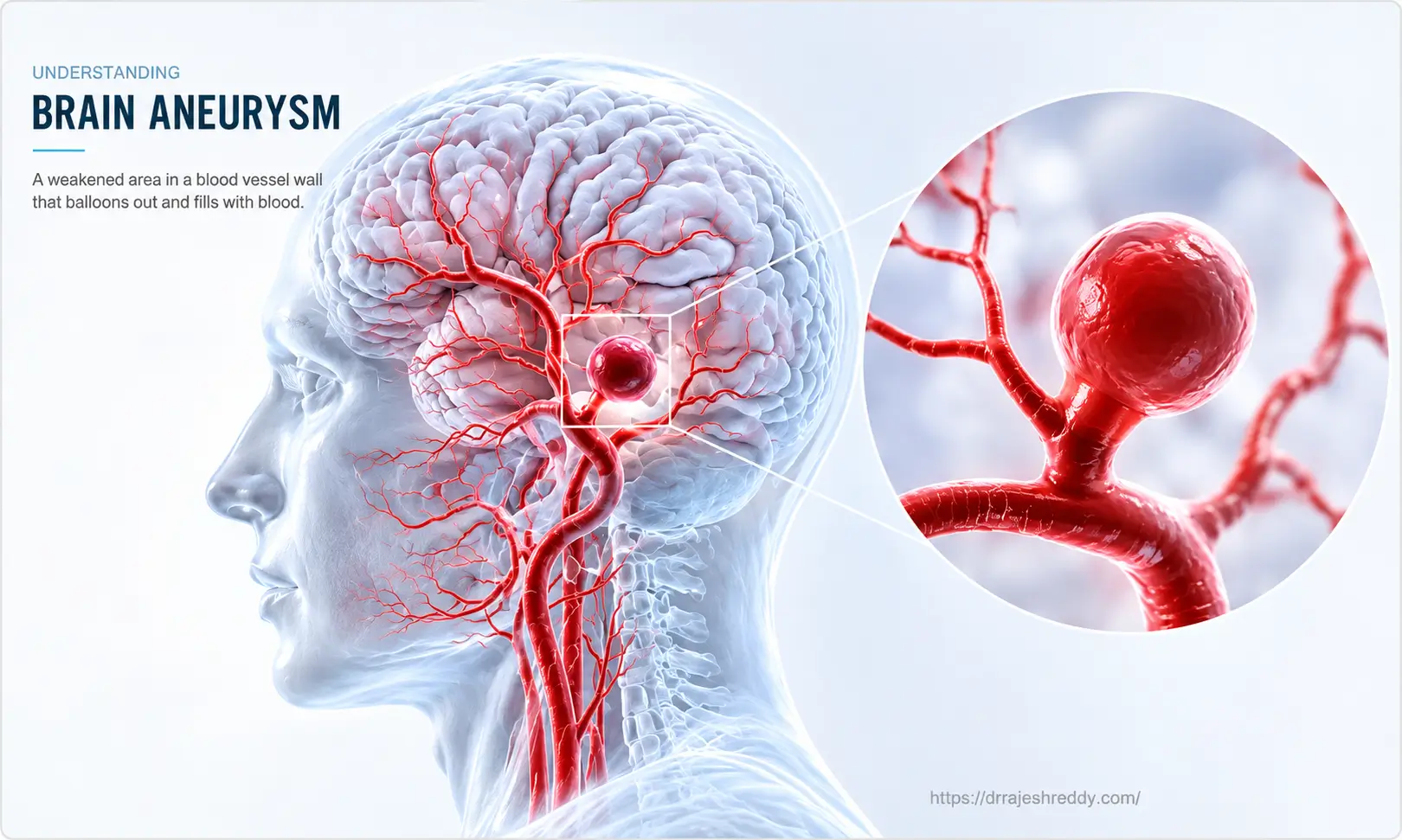
A brain aneurysm (also called an intracranial or cerebral aneurysm) is a localised ballooning or bulging of a brain artery caused by a weakness in the arterial wall. As blood pulses through the artery, the weakened segment expands under pressure — much like a weak spot in a bicycle tyre bulging outward. The aneurysm may remain stable for years, or it may grow and eventually rupture, releasing blood into the subarachnoid space (the fluid-filled area between the brain and its protective membranes).
Brain aneurysms are more common than most people realise — population studies estimate a prevalence of approximately 2–5% of the adult population, meaning millions of people worldwide carry an unruptured aneurysm without knowing it. The challenge is that the vast majority cause no symptoms until — or unless — they rupture.
Where Do Brain Aneurysms Form?
Aneurysms preferentially develop at the branching points of cerebral arteries, where haemodynamic stress is highest. The most common locations include:
- Anterior communicating artery (ACoA) — most common single site (~30%)
- Posterior communicating artery (PCoA) — arises at the junction of the internal carotid and posterior communicating arteries (~25%)
- Middle cerebral artery (MCA) bifurcation — the most common location for aneurysms treated surgically (~20%)
- Internal carotid artery — including cavernous and paraclinoid segments (~10%)
- Basilar artery tip and posterior circulation — (~10%); these carry higher rupture risk
- Anterior cerebral / pericallosal artery — less common
Risk Factors for Brain Aneurysm Formation and Rupture

- Hypertension (high blood pressure) — the most significant modifiable risk factor
- Smoking — doubles the risk of aneurysm formation and substantially increases rupture risk
- Family history — first-degree relatives of aneurysm patients have 3–7x higher risk
- Polycystic kidney disease (ADPKD) — associated with intracranial aneurysms in ~10% of patients
- Female sex — women have a higher risk of rupture, particularly post-menopause
- Prior aneurysm — patients with one aneurysm have a 15–20% chance of harbouring additional aneurysms
- Connective tissue disorders — Ehlers-Danlos syndrome, Marfan syndrome
- Coarctation of the aorta — associated with increased cerebrovascular risk
Types of Brain Aneurysms
Understanding the type and classification of a brain aneurysm is essential for treatment planning. The table below summarises the major aneurysm categories, their characteristics, and treatment implications:
| Type | Size / Category | Characteristics | Treatment Implication |
|---|---|---|---|
| Saccular (Berry) | Small (<7mm), Medium (7–12mm), Large (13–24mm), Giant (≥25mm) | Most common type (~85%); round or lobulated outpouching at arterial bifurcation | All sizes considered for treatment; giant aneurysms most challenging |
| Fusiform | Variable | Entire vessel wall dilated; no defined neck | Endovascular flow diversion preferred; surgical clipping rarely possible |
| Dissecting | Variable | Tear within arterial wall causing false lumen | Often managed with flow diversion or parent vessel sacrifice |
| Traumatic / Mycotic | Variable | Caused by injury or infection; may not have typical dome-neck morphology | Underlying cause treated; surgical or endovascular repair of aneurysm |
| Ruptured (SAH) | Any size | Has bled — presents as subarachnoid haemorrhage (SAH) | Emergency treatment — coiling or clipping within 24–72 hours |
| Unruptured | Any size | Found incidentally or on family screening | Risk-benefit assessment; observation vs. elective treatment |
Brain Aneurysm Symptoms: Unruptured vs. Ruptured

Unruptured Brain Aneurysms: The Silent Majority
The majority of unruptured brain aneurysms produce no symptoms and are discovered incidentally — on an MRI or CT scan performed for an unrelated reason such as headache investigation, a head injury, or routine screening in a patient with a family history. This incidental discovery, while initially alarming, is actually the best possible scenario — it allows a measured, unhurried evaluation of the aneurysm's risk and an informed treatment decision.
However, some large unruptured aneurysms — particularly those compressing adjacent structures — can produce symptoms:
- Posterior communicating artery aneurysm pressing on the third cranial nerve → sudden onset drooping eyelid (ptosis) and double vision — a neurosurgical emergency requiring immediate evaluation
- Large internal carotid artery aneurysm → visual field loss from optic nerve or chiasm compression
- Giant aneurysms → headache, focal neurological deficits from mass effect on adjacent brain
- Cavernous sinus aneurysm → facial numbness, double vision, or eye movement problems
Warning Sign: A sudden, painful drooping of one eyelid with double vision — even without headache — should prompt immediate neurovascular evaluation. This combination of signs is classic for a posterior communicating artery aneurysm compressing the third cranial nerve and may precede rupture by hours to days.
Ruptured Brain Aneurysm: The Thunderclap Headache
When a brain aneurysm ruptures, it causes subarachnoid haemorrhage (SAH) — sudden bleeding into the subarachnoid space around the brain. The hallmark symptom is a thunderclap headache: a sudden, explosive headache of maximal severity at onset, often described as 'the worst headache of my life' or 'like a thunderbolt to the head.'
Other symptoms of ruptured aneurysm (SAH) include:
- Sudden onset neck stiffness (meningism) — from blood irritating the meninges
- Photophobia — severe sensitivity to light
- Nausea and vomiting — often projectile
- Loss of consciousness — occurs in approximately 50% of patients at ictus
- Focal neurological deficits — depending on the aneurysm location and extent of bleeding
- Seizures — occur in approximately 10% of SAH patients
- Coma — in severe cases (WFNS Grade IV–V)
EMERGENCY: A sudden, severe headache of maximal intensity at onset — the 'thunderclap headache' — requires immediate emergency department attendance and urgent CT brain scan. Do not wait to see if it improves. Up to 50% of SAH patients describe a 'sentinel headache' in the days before major rupture — a warning sign that is too often dismissed. Every thunderclap headache must be treated as a ruptured aneurysm until proven otherwise.
Diagnosing a Brain Aneurysm: The Workup
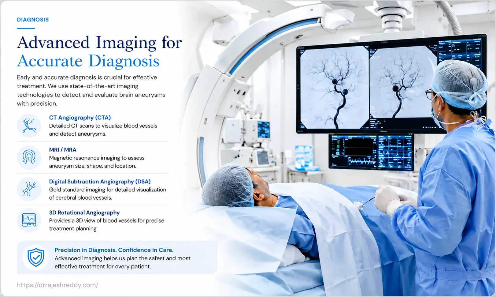
CT Brain (Non-Contrast) — The Emergency Investigation
In a patient presenting with thunderclap headache, a non-contrast CT brain scan is the first-line investigation. It detects subarachnoid blood in approximately 98% of SAH cases within 6 hours of symptom onset. The sensitivity decreases after 24 hours as blood is reabsorbed — which is why immediate investigation is essential.
CT Angiography (CTA)
If the CT brain confirms SAH — or if the clinical suspicion of unruptured aneurysm is high — CT angiography is performed immediately. CTA provides a rapid, non-invasive three-dimensional roadmap of the cerebral vasculature, identifying the aneurysm's location, size, shape, neck morphology, and relationship to surrounding vessels. Modern CTA has a sensitivity approaching 97–99% for aneurysms ≥ 3mm.
Lumbar Puncture (LP)
If the CT brain is normal but clinical suspicion of SAH remains high — particularly if the patient presents more than 6 hours after symptom onset — a lumbar puncture is performed to analyse the cerebrospinal fluid (CSF) for xanthochromia (yellow discolouration from haemoglobin breakdown products). A positive LP in the context of thunderclap headache confirms SAH even when CT is negative.
Digital Subtraction Angiography (DSA) — The Gold Standard
Digital subtraction angiography — conventional catheter angiography — remains the gold standard for definitive aneurysm characterisation. A catheter is introduced through the femoral artery and advanced under X-ray guidance to selectively inject contrast into each cerebral vessel. DSA provides the highest resolution imaging of aneurysm anatomy — including the neck width, dome-to-neck ratio, relationship to branch vessels, and aneurysm haemodynamics — which is essential for precise treatment planning.
In the setting of confirmed SAH, DSA is performed as a combined diagnostic and therapeutic procedure: the aneurysm is characterised and — when anatomy is suitable — treated with coiling or other endovascular devices in the same sitting.
MRI Brain and MR Angiography (MRA)
MRI is used for subacute evaluation, for detecting associated intracranial pathology, and for surveillance of treated aneurysms. MR angiography (time-of-flight MRA) is a non-invasive screening tool for unruptured aneurysms — particularly useful for family screening in relatives of aneurysm patients or for surveillance of known small aneurysms managed conservatively.
Subarachnoid Haemorrhage (SAH): Emergency Management
The management of ruptured brain aneurysm begins the moment the patient arrives at the emergency department and is a race to prevent re-bleeding — the most dangerous early complication of SAH, carrying a mortality of 50–80% when it occurs within the first 24 hours.
WFNS Grading: Assessing SAH Severity
The World Federation of Neurological Surgeons (WFNS) grading scale assesses clinical severity of SAH and guides prognosis and management decisions:
| WFNS Grade | GCS Score | Motor Deficit | Prognosis |
|---|---|---|---|
| Grade I | 15 | Absent | Excellent — >90% good outcome with prompt treatment |
| Grade II | 13–14 | Absent | Good — most patients recover well with treatment |
| Grade III | 13–14 | Present | Moderate — significant morbidity; recovery variable |
| Grade IV | 7–12 | Present or absent | Poor — high morbidity and mortality even with treatment |
| Grade V | 3–6 | Present or absent | Very poor — majority do not survive to discharge |
Immediate Management Priorities
- Airway protection — intubation for WFNS Grade IV–V patients and those with deteriorating consciousness
- Blood pressure control — target systolic BP < 160 mmHg to reduce re-bleeding risk while maintaining cerebral perfusion pressure
- Nimodipine — oral calcium channel blocker commenced immediately and continued for 21 days to reduce vasospasm risk
- Pain management — adequate analgesia reduces sympathetic activation and blood pressure spikes
- Seizure prophylaxis — anti-epileptic medication in the acute phase
- Hydrocephalus management — acute hydrocephalus from blood blocking CSF drainage treated with external ventricular drain (EVD) if present
- Aneurysm treatment — targeted within 24–72 hours of admission for good-grade SAH (WFNS I–III)
Vasospasm — The Delayed Killer
Cerebral vasospasm — narrowing of the cerebral arteries in response to the presence of subarachnoid blood — is the most feared delayed complication of SAH. It typically develops between days 4–14 after bleeding, causing delayed cerebral ischaemia (DCI) and potentially devastating stroke. Vasospasm affects approximately 30–70% of SAH patients to some degree.
Management of vasospasm includes: nimodipine (commenced on admission and continued for 21 days), induced hypertension (targeting higher blood pressure to drive blood through narrowed vessels), hypervolaemia, and — for refractory vasospasm — endovascular intervention with intra-arterial nimodipine infusion or balloon angioplasty of spastic segments. Dr. Rajesh Reddy's endovascular expertise makes him uniquely placed to perform both the initial aneurysm treatment and subsequent vasospasm interventions when required.
Brain Aneurysm Treatment Options: A Complete Overview
The goal of brain aneurysm treatment — whether the aneurysm has ruptured or not — is to exclude it from the circulation, eliminating the risk of (re-)bleeding. The choice between endovascular and surgical approaches depends on the aneurysm's anatomy, location, size, the patient's clinical grade, and the treating team's expertise. Dr. Rajesh Reddy's combined endovascular and microsurgical training allows him to offer the full spectrum:
| Treatment | Approach | Best Suited For | Key Advantage |
|---|---|---|---|
| Endovascular Coiling (GDC) | Catheter via groin → aneurysm filled with platinum coils | Most ruptured aneurysms; posterior circulation; elderly patients | No craniotomy; faster recovery; lower procedural mortality in good-grade SAH |
| Flow Diversion (Pipeline / Surpass) | Stent-like device in parent artery diverts blood away from aneurysm | Large/giant aneurysms; fusiform; wide-neck aneurysms | Treats entire aneurysm; high occlusion rates; single device |
| Intrasaccular Devices (WEB device) | Mesh device deployed inside aneurysm sac to disrupt flow | Wide-neck bifurcation aneurysms (MCA, basilar tip) | No stent in parent vessel; single device; avoids dual antiplatelet |
| Balloon-Assisted Coiling | Balloon inflated in parent artery during coiling to prevent coil herniation | Wide-neck aneurysms where simple coiling is insufficient | Allows coiling of aneurysms that would otherwise require stent |
| Stent-Assisted Coiling | Stent placed in parent artery; coils packed through stent mesh | Wide-neck aneurysms; bifurcation aneurysms with unfavourable anatomy | Allows dense coiling of complex aneurysms; scaffold for coil retention |
| Microsurgical Clipping | Craniotomy; titanium clip placed across aneurysm neck | Middle cerebral artery aneurysms; large MCA; giant aneurysms; failed coiling | Immediate, permanent obliteration; no antiplatelet medication required |
| Parent Vessel Occlusion / Surgical Bypass with Aneurysm Trapping | Deliberate occlusion of the parent vessel feeding the aneurysm — combined with a surgical bypass graft to maintain distal blood flow. | Giant or fusiform aneurysms not amenable to coiling, flow diversion, or clipping; complex skull base aneurysms; recurrent aneurysms after failed prior treatment | Eliminates aneurysm permanently by removing it from the circulation entirely; bypass preserves distal perfusion |
Endovascular Coiling: Treating a Brain Aneurysm Without Opening the Skull
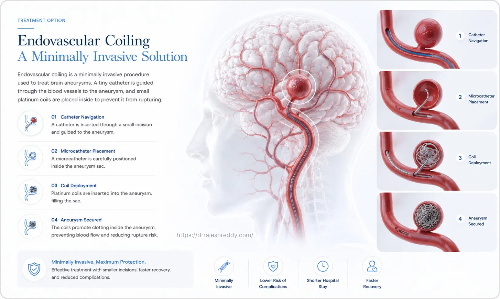
Endovascular coiling has become the preferred treatment for the majority of ruptured brain aneurysms following the landmark ISAT trial (International Subarachnoid Aneurysm Trial, 2002), which demonstrated superior outcomes for coiling compared to surgical clipping in patients suitable for both techniques.
How Coiling Works
Under general anaesthesia, a catheter is introduced through a small puncture in the femoral artery (groin) and navigated under X-ray fluoroscopy guidance up through the aorta, the carotid or vertebral arteries, and into the cerebral vasculature. A microcatheter is then advanced into the aneurysm sac, and soft platinum coils — thinner than a human hair — are deployed to fill the aneurysm. The coils cause thrombosis (clotting) within the aneurysm, which progressively seals it from the circulation.
For aneurysms with a wide neck — where simple coiling risks coil herniation into the parent artery — additional techniques are used: balloon-assisted coiling (temporarily inflating a balloon across the aneurysm neck during coil deployment) or stent-assisted coiling (placing a stent in the parent artery to provide a scaffold that holds the coils within the aneurysm).
Flow Diversion: Extrasaccular Flow Diverters — Pipeline, Surpass, SILK, p64, FRED, Derivo
Flow diversion represents the most significant advance in endovascular aneurysm treatment of the past decade. Rather than filling the aneurysm with coils, a flow diverter is a densely woven, stent-like device deployed in the parent artery across the aneurysm neck. The device redirects blood flow away from the aneurysm, causing progressive thrombosis and eventual complete occlusion — while simultaneously providing a scaffold for neo-endothelialisation across the aneurysm neck.
Flow diversion achieves complete aneurysm occlusion in approximately 75–85% of cases at 6 months and up to 95% at 2 years. It has become the treatment of choice for large and giant aneurysms, fusiform aneurysms, and wide-neck aneurysms that are not amenable to simple coiling. The patient requires dual antiplatelet medication (aspirin + clopidogrel) for 3–6 months after device deployment to prevent device thrombosis.
Intrasaccular Devices: WEB, Contour, Artisse
The Woven EndoBridge (WEB) device is a single-layer or double-layer mesh device deployed entirely within the aneurysm sac, disrupting flow at the aneurysm neck. It is particularly suited to wide-neck bifurcation aneurysms — such as MCA bifurcation and basilar tip aneurysms — where it avoids the need for a stent in the parent vessel and the associated requirement for dual antiplatelet therapy. The WEB device can be deployed in a single session and does not require a stent, simplifying the procedure and the post-operative medication regimen. Other intrasaccular devices now available include the Contour and Artisse devices — each designed for specific aneurysm neck geometries, offering expanding options for wide-neck bifurcation aneurysms without the need for parent vessel stenting.
Microsurgical Clipping: When Surgery Is the Better Choice
Despite the widespread adoption of endovascular techniques, microsurgical clipping remains an essential treatment for a significant proportion of brain aneurysms — and in experienced hands, it offers a level of durability and completeness that endovascular treatment has not yet matched in all aneurysm types.
When Clipping Is Preferred
- Middle cerebral artery (MCA) aneurysms — the MCA's complex branch anatomy makes endovascular treatment more challenging; direct surgical access provides excellent visualisation and clip placement
- Aneurysms with a broad base or complex shape — where complete endovascular occlusion is difficult to achieve reliably
- Giant aneurysms — particularly those causing mass effect; surgical decompression and clip reconstruction may be required
- Failed or recanalised endovascular treatment — clipping provides a definitive solution when coiling has been incomplete
- Young patients — the long-term durability of clipping is particularly valuable in patients who would otherwise face decades of angiographic surveillance after coiling
- Aneurysms with associated haematoma — where the haematoma requires surgical evacuation in any case
The Clipping Procedure
Under general anaesthesia, a craniotomy is performed — typically a pterional (frontotemporal) craniotomy for anterior circulation aneurysms, or a retrosigmoid / suboccipital craniotomy for posterior fossa aneurysms. The Sylvian fissure is opened under the operating microscope, the aneurysm-bearing vessel is exposed, and the aneurysm neck is identified. A titanium clip is then placed across the aneurysm neck under direct microscopic visualisation, permanently excluding the aneurysm from the circulation while preserving blood flow in the parent and branch vessels.
Intraoperative fluorescence angiography (indocyanine green, ICG) is performed after clip placement to confirm complete aneurysm occlusion and patency of all adjacent vessels — providing immediate confirmation of the surgical result without the need for a separate post-operative angiogram.
Coiling vs. Clipping: A Direct Comparison
| Feature | Endovascular Coiling | Microsurgical Clipping |
|---|---|---|
| Access | Catheter via femoral artery — no craniotomy | Craniotomy under general anaesthesia |
| Hospital stay | 3–5 days (SAH recovery determines stay) | 5–7 days post-craniotomy |
| Recovery | Faster — 2–4 weeks | 4–8 weeks |
| Immediate aneurysm occlusion | Complete in ~85–90% immediately; may need repeat imaging | Near 100% immediate permanent obliteration |
| Long-term durability | ~15–20% recanalization rate; surveillance angiography needed | Excellent — durable; rarely recanalizes |
| Antiplatelet medication | Required for stent-assisted / flow diversion cases | Not required |
| Best for posterior circulation | Yes — basilar tip aneurysms particularly suited | Technically demanding; higher risk in posterior fossa |
| Best for MCA aneurysms | Less ideal — complex branch anatomy | Preferred — direct surgical access to MCA |
| Risk of re-bleeding (SAH) | Comparable in short term; slightly higher recanalization risk long term | Lower long-term re-bleeding risk after complete clipping |
| Decision maker | Multidisciplinary — neurosurgeon + interventional neuroradiologist | Multidisciplinary — neurosurgeon + interventional team |
The choice between coiling and clipping is not made by the patient's preference alone — it is a clinical decision made by Dr. Rajesh Reddy and the neurovascular team based on the specific aneurysm's anatomy, location, the patient's age and clinical condition, and the most likely treatment to achieve durable, complete occlusion. Many patients are suitable for either approach, and the team's combined expertise ensures the safest and most effective option is selected for each individual.
Managing Unruptured Brain Aneurysms: Treatment vs. Observation
The discovery of an unruptured brain aneurysm inevitably raises the question: does it need to be treated? The answer requires balancing the risk of aneurysm rupture (without treatment) against the risk of the treatment itself.
Annual Rupture Risk: Size and Location Matter
The annual risk of rupture for an unruptured intracranial aneurysm varies significantly by size and location. The PHASES score — incorporating Population (country), Hypertension, Age, Size, Earlier SAH, and Site (location) — is used to calculate an individualised annual rupture risk. As a general guide:
- Aneurysms < 7mm in low-risk locations: annual rupture risk approximately 0.1–0.5%
- Aneurysms 7–12mm: annual rupture risk approximately 0.5–1.5%
- Aneurysms > 12mm: annual rupture risk > 1.5–3% per year
- Posterior circulation (basilar tip, posterior communicating artery) aneurysms: significantly higher rupture risk at any size
- Prior SAH from another aneurysm: higher rupture risk for remaining unruptured aneurysms
Treatment Risk: Procedural Morbidity and Mortality
In experienced hands, the procedural morbidity and mortality for both endovascular and surgical treatment of unruptured aneurysms is low — approximately 1–3% for major morbidity and < 1% mortality for elective procedures in good-grade patients. However, this risk increases with patient age, medical comorbidities, aneurysm size, and complexity.
Dr. Rajesh Reddy uses the PHASES score and UIATS (Unruptured Intracranial Aneurysm Treatment Score) in combination with his clinical assessment to provide patients with an individualised, evidence-based recommendation on whether treatment or observation is more appropriate for their specific aneurysm.
When Observation Is Appropriate
- Small aneurysms (< 5–7mm) in low-risk locations in elderly patients or those with significant medical comorbidities
- Patient preference after thorough counselling on rupture and treatment risk
- Aneurysms showing stability on serial imaging over 2+ years
Risk Factor Modification — Essential in All Cases
Regardless of whether treatment or observation is chosen, all patients with a known brain aneurysm should:
- Control blood pressure rigorously — target < 130/80 mmHg
- Stop smoking — smoking substantially increases aneurysm growth and rupture risk
- Avoid cocaine and stimulant drugs — associated with acute aneurysm rupture
- Moderate caffeine and alcohol intake
- Attend all surveillance imaging appointments without delay
- Avoid straining and manage constipation actively — sudden rises in intracranial pressure from straining can trigger aneurysm rupture; stool softeners should be used routinely in all aneurysm patients
Why Choose Dr. Rajesh Reddy for Brain Aneurysm Treatment in Hyderabad?
Brain aneurysm treatment — particularly in the acute setting of SAH — demands a rare combination of endovascular expertise, microsurgical skill, and the clinical judgement to choose the right approach for each aneurysm and each patient. Dr. Rajesh Reddy is one of a small group of specialists in Hyderabad and India who can offer the complete spectrum of aneurysm treatment under one roof:
Dr. Rajesh Reddy — Neurovascular Expertise at a Glance
- FINR (Fellow in Interventional Neuroradiology) — University Hospital, Zurich: one of the world's foremost centres for complex neurovascular endovascular intervention
- MCh Neurosurgery (Exam Topper), NIMS Hyderabad; DNB Neurosurgery — dual expertise in endovascular and microsurgical aneurysm treatment
- Visiting Scholar — Barrow Neurological Institute, Phoenix, Arizona: a global reference centre for cerebrovascular surgery and complex aneurysm management
- Visiting Scholar — Okayama University, Japan; Klinikum Stuttgart, Germany
- Over 5,000 neurosurgical procedures — extensive aneurysm treatment experience across all modalities: coiling, flow diversion, WEB device, stent-assisted coiling, and microsurgical clipping
- Vasospasm intervention — endovascular management of delayed cerebral ischaemia including intra-arterial nimodipine and balloon angioplasty
- AVM treatment — combined surgical and endovascular management of arteriovenous malformations
- Clinical acumen and judgement gained through years of training with leading experts globally
Dr. Rajesh Reddy works within a multidisciplinary neurovascular team — neurosurgery, intensive care, neurology, and rehabilitation — ensuring that every SAH patient receives coordinated, protocol-driven care from the moment of arrival through to long-term follow-up.
Recovery After Brain Aneurysm Treatment
After Endovascular Coiling (Elective, Unruptured Aneurysm)
- Procedure duration: 1–3 hours under general anaesthesia
- Hospital stay: 2–3 days; groin puncture site monitored
- Return to normal activity: 1–2 weeks for most patients
- Driving: typically from 1–2 weeks after procedure (in the absence of neurological deficit)
- Follow-up angiography: DSA or MRA at 6 months, then at 18 months, then at 3–5 years to confirm sustained aneurysm occlusion
- Antiplatelet medication: no antiplatelet therapy is required after simple coiling. After stent-assisted coiling or extrasaccular flow diversion — two antiplatelet medications (aspirin + clopidogrel) for 6 months, followed by a single antiplatelet medication for at least 3–5 years (duration at the discretion of the treating doctor).
After SAH and Aneurysm Treatment (Ruptured Aneurysm)
Recovery from subarachnoid haemorrhage is determined primarily by the severity of the initial bleed (WFNS grade) and the occurrence of complications (re-bleeding, vasospasm, hydrocephalus) — not by the treatment technique itself.
- ICU stay: typically 14–21 days for vasospasm monitoring and management
- Total hospital stay: 3–6 weeks depending on complications
- Cognitive and functional recovery: months to 1–2 years; fatigue, headache, memory, and concentration problems are common
- Chronic hydrocephalus: develops in approximately 20% of SAH patients; managed with a ventriculoperitoneal (VP) shunt
- Psychological impact: depression and anxiety are common after SAH; psychological support is an essential part of recovery
- Return to work: highly variable — 50–70% of good-grade SAH survivors return to previous employment
After Microsurgical Clipping
- Hospital stay: 5–7 days post-craniotomy (longer after SAH)
- Return to normal activity: 4–8 weeks
- Post-operative angiography: typically at 3–6 months to confirm clip position and aneurysm occlusion
- Long-term durability: excellent — recurrence after complete clipping is rare (< 1%)
Frequently Asked Questions
Consult Dr. Rajesh Reddy for Brain Aneurysm Treatment in Hyderabad
Whether you have been found to have an unruptured brain aneurysm on imaging, have a family history that warrants screening, or are seeking a second opinion on a treatment recommendation, Dr. Rajesh Reddy Sannareddy provides comprehensive neurovascular consultations — with the full range of endovascular and surgical treatment options available under one roof. Book an in-person or video consultation via the Dr. Reddy's Neuro Care app on Google Play.
Join the Brain Aneurysm Self Help Group
Are you interested in volunteering to be a part of a Self Help Group to help patients and families with brain aneurysm navigate the decision-making phase and the journey thereafter? Reach out to us to know more about how you can make a difference.
Related Articles (Cluster Pages)
Explore these in-depth guides on specific aspects of brain aneurysm diagnosis, treatment, and recovery:
| Cluster Article | Suggested URL |
|---|---|
| Brain Aneurysm Symptoms: When Is It an Emergency? | /brain-aneurysm-symptoms |
| Ruptured vs. Unruptured Brain Aneurysm: Key Differences | /ruptured-unruptured-brain-aneurysm |
| Aneurysm Coiling vs. Surgical Clipping: Which Is Better? | /aneurysm-coiling-vs-clipping |
| Endovascular Coiling for Brain Aneurysm in Hyderabad | /endovascular-coiling-hyderabad |
| Flow Diversion Treatment for Brain Aneurysms | /flow-diversion-brain-aneurysm |
| Subarachnoid Hemorrhage (SAH): Causes and Treatment | /subarachnoid-hemorrhage-treatment |
| Life After Brain Aneurysm Surgery: Recovery Guide | /brain-aneurysm-surgery-recovery |
| Arteriovenous Malformation (AVM) Treatment Hyderabad | /avm-treatment-hyderabad |