Introduction
When a brain tumor is first suspected or diagnosed, one of the first questions most patients and families ask is: what kind of tumor is it? The answer matters enormously — different types of brain tumors behave in fundamentally different ways, require different treatments, and carry very different prognoses. A glioblastoma is not a meningioma. A pituitary adenoma is not a medulloblastoma. Understanding the distinctions is the foundation of understanding your diagnosis and the road ahead.
This guide provides a comprehensive, plain-language overview of the most common types of brain tumors — their origins, characteristics, symptoms, and how they are typically treated. It has been written to help patients and families prepare informed questions for their neurosurgeon and better understand what their MRI or biopsy report is telling them.
Dr. Rajesh Reddy Sannareddy, Senior Consultant Neurosurgeon in Hyderabad, treats the full spectrum of brain tumors — benign and malignant, primary and metastatic, simple and complex. This guide reflects the breadth of tumor types he encounters and manages in clinical practice.
How Are Brain Tumors Classified?
Brain tumors are classified along two primary axes: their origin (where they arise from) and their grade (how aggressive they are). Understanding both helps explain why two patients with 'brain tumors' may have entirely different experiences.
1. By Origin: Primary vs. Secondary
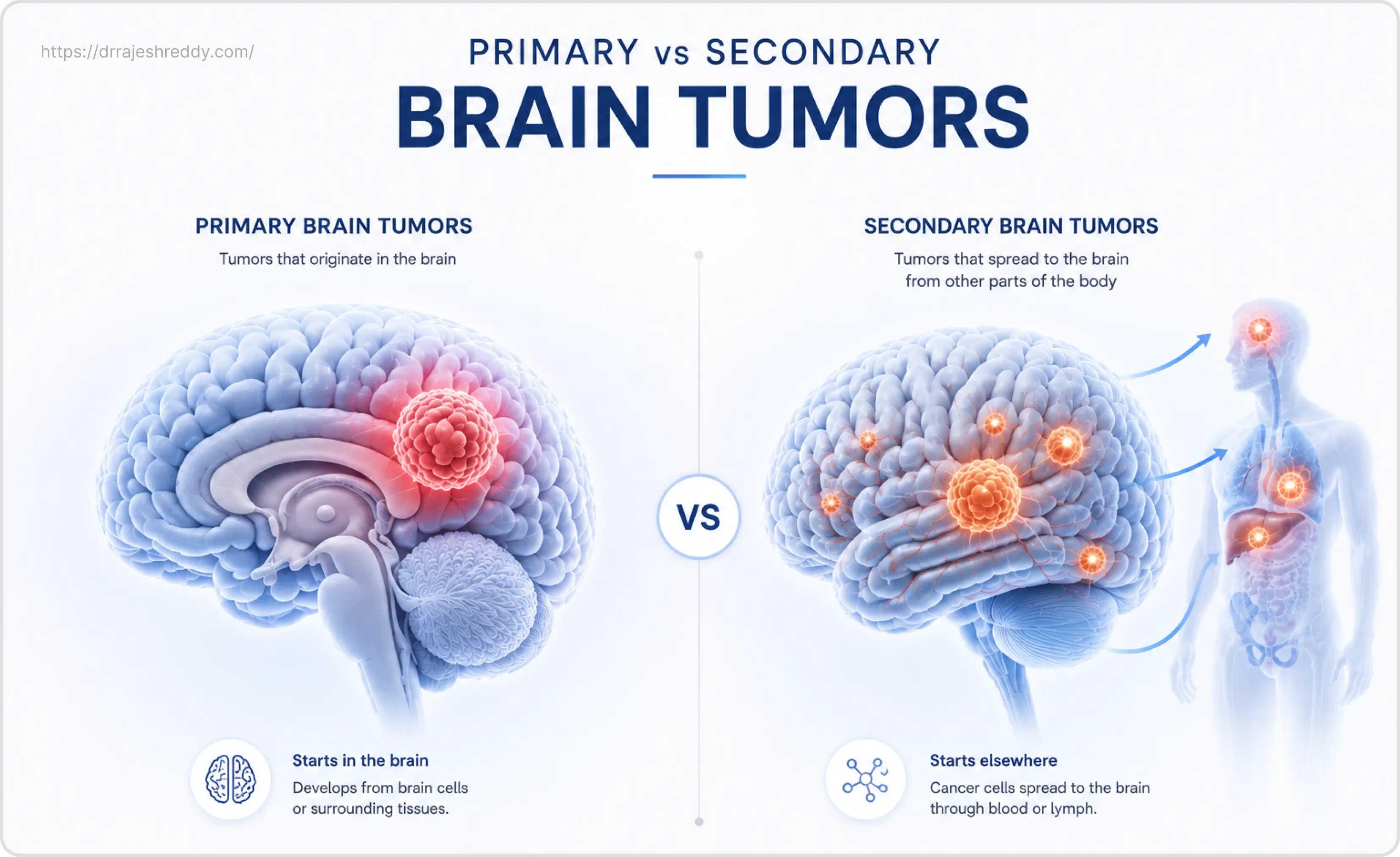
Primary brain tumors originate in the brain or its surrounding structures — the meninges, cranial nerves, pituitary gland, or the brain's own supporting cells (glial cells). They may be benign or malignant, but they begin in the brain itself.
Secondary (metastatic) brain tumors do not originate in the brain. They are cancer cells that have spread from a primary cancer elsewhere in the body — commonly the lungs, breast, kidney, colon, or skin (melanoma) — and lodged in the brain. Metastatic tumors are actually more common than primary brain tumors and require treatment of both the brain lesions and the original cancer.
2. By Grade: The WHO Classification
The World Health Organisation (WHO) classifies brain tumors on a grading scale from Grade I to Grade IV, based on how the tumor cells look under the microscope and their predicted behaviour:
- Grade I — Slow-growing, well-defined borders, least aggressive. Often curable with surgery alone. Example: pilocytic astrocytoma.
- Grade II — Relatively slow-growing but may infiltrate surrounding tissue and can recur or progress to a higher grade over time. Example: low-grade glioma.
- Grade III — Malignant. Actively reproducing abnormal cells, likely to invade nearby tissue. Example: anaplastic astrocytoma.
- Grade IV — Most aggressive. Rapid growth, extensive invasion, high recurrence. Example: glioblastoma multiforme (GBM).
Important Molecular Update:
The WHO classification was significantly updated in 2021 to incorporate molecular markers — such as IDH mutation status and MGMT promoter methylation — alongside histology. This means two tumors that look similar under the microscope may be classified differently (and treated differently) based on their molecular profile. Tissue biopsy and molecular profiling are now essential parts of brain tumor diagnosis.
Benign vs Malignant Brain Tumors
Non-Cancerous & Slow Growing
- Well-defined edges: Encapsulated and does not invade surrounding brain tissue.
- Slow growth: Grows gradually over years and causes symptoms over a longer period.
- Easier to treat: Often removed completely with surgery and has a lower recurrence rate.
Cancerous & Fast Growing
- Irregular edges: Invades and destroys surrounding healthy brain tissue.
- Fast growing: Grows rapidly and can cause severe, acute symptoms.
- Complex treatment: May require extensive surgery plus radiation and chemotherapy; higher risk of recurrence.
Quick Reference: Types of Brain Tumors at a Glance
The table below summarises the most commonly encountered brain tumor types, their origin, WHO grade, and key distinguishing characteristic:
| Tumor Type | Origin | Benign / Malignant | WHO Grade | Key Characteristic |
|---|---|---|---|---|
| Glioblastoma (GBM) | Glial cells | Malignant | IV | Most aggressive primary brain tumor; infiltrates surrounding tissue |
| Low-Grade Glioma | Glial cells | Slow-growing | I–II | Often presents with seizures in younger adults; may transform to high-grade |
| Anaplastic Glioma | Glial cells | Malignant | III | Intermediate grade; between LGG and GBM in aggressiveness |
| Meningioma | Meninges | Usually benign | I–III | Most common benign brain tumor; grows slowly over years |
| Pituitary Adenoma | Pituitary gland | Benign | — | May secrete hormones; causes visual field defects when large |
| Vestibular Schwannoma | Acoustic nerve (CN VIII) | Benign | I | Causes progressive hearing loss and tinnitus |
| Trigeminal Schwannoma | Trigeminal nerve (CN V) | Benign | I | Causes facial numbness, pain or weakness |
| Medulloblastoma | Cerebellum | Malignant | IV | Most common malignant brain tumor in children |
| Ependymoma | Ependymal cells (ventricles) | Variable | II–III | Blocks CSF flow; causes hydrocephalus |
| Craniopharyngioma | Skull base / sellar region | Benign | I | Causes hormonal and visual disturbances |
| Colloid Cyst | Third ventricle | Benign | — | Can cause acute hydrocephalus; requires timely surgery |
| Epidermoid Cyst | Skull base / cisterns | Benign | — | Slow-growing; arises from trapped skin cells during fetal development |
| Haemangioblastoma | Cerebellum / brainstem | Benign | I | Vascular tumor; associated with Von Hippel-Lindau syndrome |
| Metastatic Brain Tumor | Spreads from lungs, breast, kidney, melanoma | Malignant | — | More common than primary tumors; may be single or multiple |
Gliomas: The Most Common Primary Brain Tumors
Gliomas are tumors that arise from glial cells — the supportive cells of the brain that provide structure, insulation, and nourishment to neurons. They account for the majority of primary malignant brain tumors in adults.
Glioblastoma Multiforme (GBM) — Grade IV
GBM is the most aggressive and unfortunately the most common malignant primary brain tumor in adults. It grows rapidly, invades surrounding brain tissue extensively, and has a high recurrence rate even after optimal treatment. The median survival with standard treatment (surgery + radiation + temozolomide chemotherapy) is approximately 14–16 months, though outcomes vary considerably based on molecular markers such as MGMT promoter methylation and IDH mutation status.
Symptoms develop quickly — often over days to weeks — and include headaches, personality changes, cognitive decline, weakness on one side of the body, or seizures. Surgery is typically the first step: maximum safe resection using neuronavigation, fluorescence guidance (5-ALA or Sodium Fluorescein), and awake craniotomy where needed, followed by radiation and chemotherapy.
Low-Grade Glioma (LGG) — Grade I–II
Low-grade gliomas grow more slowly and typically affect younger adults. They often first present as new-onset seizures in an otherwise healthy person. While they are not immediately life-threatening, they carry the risk of malignant transformation to a higher-grade tumor over time — which is why active monitoring and timely surgery are recommended even when the tumor is not causing severe symptoms.
IDH mutation status is a key prognostic marker in LGG: IDH-mutant tumors have a significantly better prognosis than IDH-wildtype tumors. Awake craniotomy is commonly used for LGGs near eloquent cortex to allow maximum safe resection while preserving speech and motor function.
Pilocytic Astrocytoma: A typically slow-growing WHO Grade 1 glioma, more common in children and young adults. It generally has an excellent prognosis and can often be cured with complete surgical removal, with a much lower risk of malignant transformation compared with diffuse low-grade gliomas.
Anaplastic Glioma — Grade III
Anaplastic gliomas sit between low-grade gliomas and GBM in terms of aggressiveness. They include anaplastic astrocytoma, anaplastic oligodendroglioma, and anaplastic ependymoma. Treatment typically involves surgery, radiation, and chemotherapy — with specific regimens depending on the molecular profile of the tumor.
Meningiomas: The Most Common Benign Brain Tumor
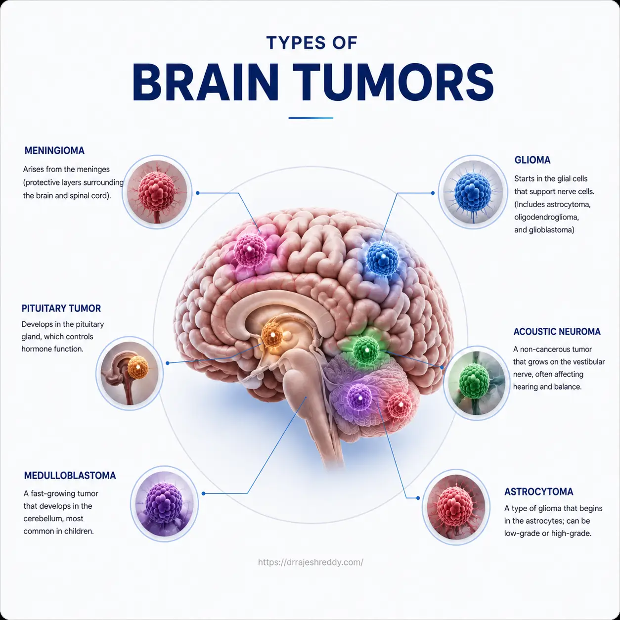
Meningiomas arise from the meninges — the three-layered protective membrane surrounding the brain and spinal cord. They are the most common primary brain tumor overall, accounting for approximately 37% of all primary brain tumors, and the vast majority are benign (WHO Grade I).
Meningiomas grow slowly, often over many years, and are sometimes discovered incidentally on an MRI done for an unrelated reason. When they do cause symptoms, it is typically because they have grown large enough to press on adjacent brain tissue or cranial nerves — causing headaches, seizures, vision changes, weakness, or personality changes depending on their location.
Who Gets Meningiomas?
Meningiomas are more common in women than men (roughly 2:1 ratio) and occur most frequently in middle-aged and older adults. Previous radiation to the head (even decades earlier) and certain genetic conditions (such as neurofibromatosis type 2) increase the risk. Hormonal factors may also play a role given the female predominance.
Treatment
Small, asymptomatic meningiomas in elderly patients are often managed with observation and regular MRI surveillance. Symptomatic tumors, or those showing growth on surveillance imaging, are surgically removed. When completely resected (Simpson Grade I resection), the majority of benign meningiomas do not recur. For surgically inaccessible locations or residual tumor, stereotactic radiosurgery is an effective option.
Pituitary Tumors (Pituitary Adenomas)
The pituitary gland — a small, pea-sized gland at the base of the brain — regulates the body's hormone system. Pituitary adenomas are benign tumors arising from the pituitary gland and are among the most common intracranial tumors. They are classified by size (microadenoma < 10mm; macroadenoma ≥ 10mm) and by whether they secrete hormones.
Functioning vs. Non-Functioning Pituitary Adenomas
Functioning adenomas secrete excess hormones and cause specific clinical syndromes:
- Prolactinoma (excess prolactin) — most common; causes irregular periods, infertility, and milk secretion in women; erectile dysfunction and infertility in men. Often treated with medication (dopamine agonists) rather than surgery.
- Cushing's disease (excess ACTH) — causes weight gain, stretch marks, high blood pressure, and diabetes. Requires surgery; Inferior Petrosal Sinus sampling is performed pre-operatively to lateralise the source.
- Acromegaly (excess GH) — causes abnormal growth of the hands, feet, and facial features in adults. Surgical resection is the primary treatment.
- Thyroid-stimulating hormone (TSH) secreting adenoma — rare; causes hyperthyroidism.
Non-functioning adenomas do not secrete hormones but cause symptoms through mass effect — typically visual field defects (bitemporal hemianopia from compression of the optic chiasm) and headaches.
Treatment
The preferred surgical approach for pituitary adenomas is endoscopic transsphenoidal surgery — operating through the nostrils and the sphenoid sinus to reach the pituitary gland without any scalp incision or brain retraction. Dr. Rajesh Reddy performs this procedure with high-definition endoscopy, allowing excellent visualisation and precise tumor removal. Most patients are discharged within 2–3 days.
Vestibular Schwannoma and Trigeminal Schwannoma
Schwannomas are benign tumors arising from Schwann cells — the cells that form the myelin sheath around cranial nerves. The two most commonly encountered in neurosurgical practice are:
Vestibular Schwannoma (Acoustic Neuroma)
Arising from the vestibulocochlear nerve (CN VIII), vestibular schwannomas grow slowly within the internal auditory canal and extend into the posterior fossa. They present with progressive unilateral hearing loss, tinnitus (ringing in the ear), and balance disturbances. Larger tumors may compress the brainstem and cause facial weakness.
Treatment options include microsurgical resection (for larger tumors), stereotactic radiosurgery (for smaller tumors or elderly patients), and observation with regular MRI for very small, asymptomatic tumors. Preservation of facial nerve function and residual hearing are key surgical goals.
Trigeminal Schwannoma
Trigeminal schwannomas arise from the trigeminal nerve (CN V) and are rarer than vestibular schwannomas. They typically present with facial numbness, pain, or weakness — sometimes mimicking trigeminal neuralgia. Depending on the tumor's location along the nerve, surgical approaches range from microsurgical resection via a middle fossa or posterior fossa approach to stereotactic radiosurgery for smaller lesions.
Medulloblastoma
Medulloblastoma is the most common malignant brain tumor in children, arising from the cerebellum. It grows rapidly and has a tendency to spread through the cerebrospinal fluid (CSF) to other parts of the brain and spinal cord (leptomeningeal spread), which is why staging with full spine MRI and CSF analysis is essential at the time of diagnosis.
Treatment involves maximum safe surgical resection followed by craniospinal radiation (in children over 3 years) and chemotherapy. Molecular subtyping — including WNT, SHH, Group 3, and Group 4 subtypes — significantly influences prognosis and is increasingly guiding treatment intensity. WNT-activated medulloblastoma has an excellent prognosis; Group 3 medulloblastoma carries the worst.
Colloid Cysts
Colloid cysts are benign, fluid-filled cysts that arise within the third ventricle of the brain — a small cavity through which cerebrospinal fluid flows. Although benign, they can be life-threatening: if the cyst enlarges and blocks CSF flow, it causes acute hydrocephalus (build-up of fluid and pressure within the brain) which can be rapidly fatal without intervention.
Symptoms include intermittent positional headaches (that worsen with certain head positions), nausea, and in severe cases, sudden loss of consciousness. Treatment is surgical removal — either via microscopic or endoscopic approach through the ventricle — which is curative in the majority of cases.
Metastatic Brain Tumors: When Cancer Spreads to the Brain
Metastatic brain tumors — cancers that have spread from elsewhere in the body — are the most common type of brain tumor overall, outnumbering primary brain tumors by approximately 10:1. The most common primary cancers that spread to the brain include:
- Lung cancer — the most frequent source of brain metastases
- Breast cancer — particularly HER2-positive and triple-negative subtypes
- Renal cell carcinoma (kidney cancer)
- Melanoma — known for its propensity to cause multiple brain metastases
- Colorectal cancer
Patients may present with a single metastasis (solitary brain metastasis) or multiple lesions. Symptoms mirror those of primary brain tumors — headaches, seizures, weakness, and cognitive changes — but may develop in the context of a known cancer diagnosis or as the first presentation of an as-yet-undiagnosed primary cancer.
Treatment of Brain Metastases
Treatment is individualised based on the number and size of metastases, the status of the systemic cancer, and the patient's overall health. Options include:
- Surgical resection — for large, symptomatic single metastases causing significant mass effect
- Stereotactic radiosurgery (SRS) — highly effective for 1–4 small metastases; can treat multiple lesions in a single session
- Whole-brain radiotherapy (WBRT) — for multiple metastases; increasingly being replaced by SRS to avoid cognitive side effects
- Systemic therapy — targeted therapy and immunotherapy increasingly show efficacy in brain metastases from lung cancer and melanoma
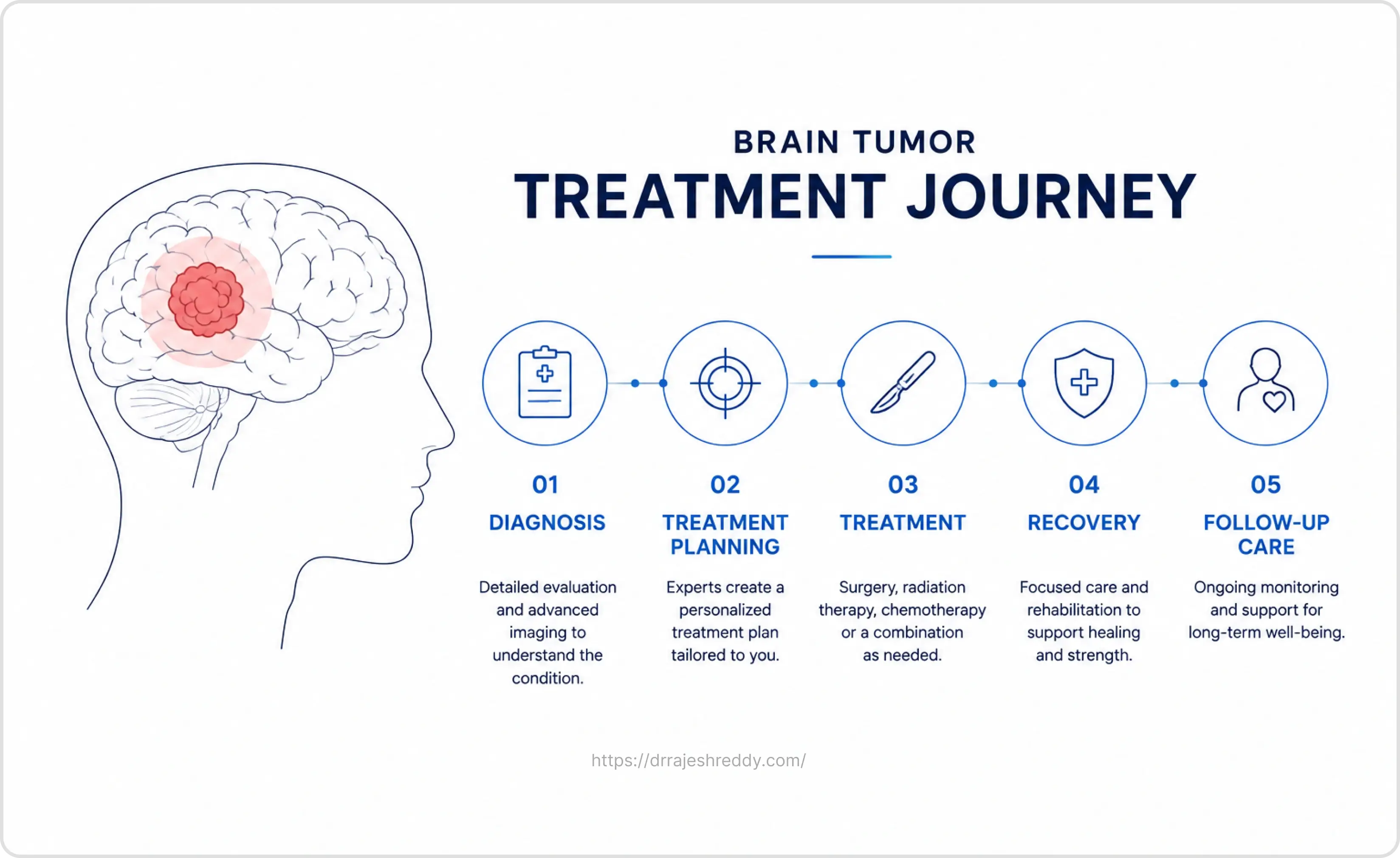
When Should You See a Neurosurgeon?
Not every headache or episode of memory difficulty is a brain tumor — but certain symptom patterns warrant prompt neurological evaluation and an MRI:
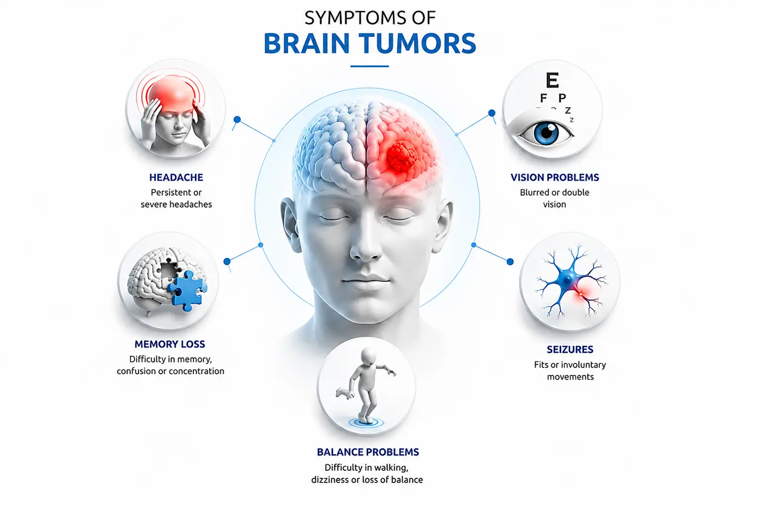
- New-onset seizures in an adult with no prior epilepsy history
- Headaches that are new, progressively worsening, or worse in the morning
- Unexplained progressive weakness, numbness, or coordination problems
- Sudden visual changes — double vision, loss of peripheral vision, or blurring
- Personality or cognitive changes noticed by family members
- Hormonal symptoms in the context of a suspected pituitary tumor (irregular periods, unexplained weight gain, abnormal hair growth)
- An incidental finding on an MRI done for another reason
Expert Advice:
If you have received an MRI report suggesting a brain lesion or mass and are unsure what it means, a consultation with Dr. Rajesh Reddy will provide clarity on the diagnosis, what further investigations are needed, and whether and when treatment is required. Early evaluation does not necessarily mean early surgery — but it does mean early peace of mind.
Frequently Asked Questions
Have Questions About Your Diagnosis? Consult Dr. Rajesh Reddy
Understanding which type of brain tumor you have is the essential first step in making informed treatment decisions. Dr. Rajesh Reddy Sannareddy provides comprehensive neurosurgical consultations — in person in Hyderabad or via the Dr. Reddy's Neuro Care app — to help patients and families navigate their diagnosis with clarity and confidence.
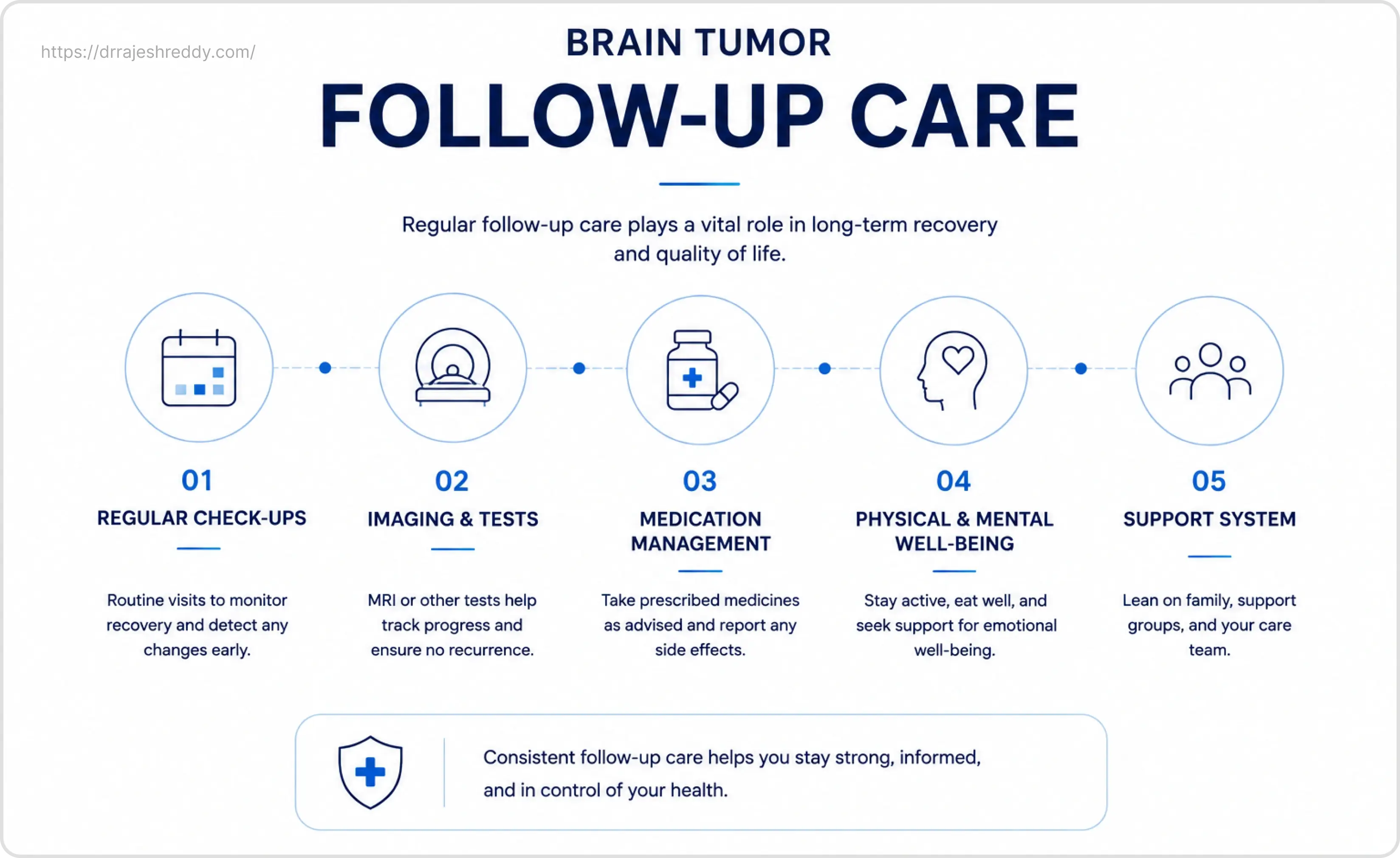
Related Articles
Continue reading to understand more about brain tumor symptoms, specific surgical approaches, and recovery:
Brain Tumor Symptoms: Warning Signs to Know
Learn key symptoms, when headaches or seizures warrant an urgent MRI, and red flags.
Read GuideGlioma Surgery in Hyderabad
Microsurgical & awake craniotomy techniques for high-grade and low-grade gliomas.
Read GuideMeningioma Treatment Without Open Surgery
Minimally invasive options, radiosurgery, and observation strategies for meningiomas.
Read GuidePituitary Tumor Surgery: Endoscopic Approach
Transsphenoidal endoscopic surgery through the nose with no visible facial scars.
Read GuideAwake Craniotomy: What to Expect
Understanding why awake brain surgery protects speech & motor functions during tumor removal.
Read GuideBrain Tumor Surgery Recovery: Week-by-Week
Step-by-step healing timeline, hospital discharge, neuro-rehab, and returning to activities.
Read GuideMinimally Invasive Brain Tumor Removal
Advanced neuronavigation, keyhole surgery, and endoscopic resection techniques.
Read GuideBrain Tumor Removal Surgery (Pillar Page)
Visit our comprehensive overview pillar page for brain tumor treatment in Hyderabad.
View Pillar Page