Introduction & What Is Awake Craniotomy?
Of all the procedures in neurosurgery, awake craniotomy is perhaps the one that patients find most difficult to comprehend. Brain surgery while you are awake? Talking to the surgeon while your skull is open? It sounds like something from science fiction — yet awake craniotomy is a well-established, routinely performed technique at centres of neurosurgical excellence around the world, and for many patients with brain tumors near critical functional areas, it is the procedure that makes the difference between preserving speech and motor function, or losing it.
This page is written specifically for patients who have been told they may need an awake craniotomy — and for their families. It answers the questions patients most urgently want answered: Why do I need to be awake? Will it hurt? What will I be asked to do? What if I panic? What are the risks? How is it different from regular brain surgery? The goal is to replace uncertainty and fear with understanding and confidence.
Dr. Rajesh Reddy Sannareddy, Senior Consultant Neurosurgeon in Hyderabad, performs awake craniotomy with a dedicated multidisciplinary team including a neuroanaesthesiologist, a neurophysiologist, and a speech-language therapist — the three pillars that make awake craniotomy both safe and effective. His training at the University Hospital, Zurich and visiting scholar experience at Barrow Neurological Institute, Phoenix give him deep exposure to awake craniotomy technique and intraoperative brain mapping in complex eloquent-area tumors.
What Is Awake Craniotomy and Why Is It Done?
Awake craniotomy is a type of brain surgery in which the patient is kept awake and interactive during the phase of the operation when the brain tumor is being removed. This allows the surgical team to monitor neurological function in real time — ensuring that critical brain areas responsible for speech, language, movement, vision, and cognition are preserved while the tumor is resected.
Why the Brain Can Be Operated on While the Patient Is Awake
The brain itself has no pain receptors. Once the scalp is numbed with local anaesthetic and the skull is opened — both of which are done while the patient is asleep or deeply sedated — the brain can be touched, stimulated, and operated on without causing pain. This fundamental neurological fact is what makes awake craniotomy possible.
The scalp, skull, and dura (the brain's outer covering) do contain pain-sensitive nerve fibres, which is why those phases of the operation are carried out under general anaesthesia or sedation. The awake phase occurs only during the period of brain mapping and tumor resection — when the brain itself is being accessed.
When Is Awake Craniotomy Recommended?
Awake craniotomy is indicated when a brain tumor is located in or adjacent to eloquent cortex — the areas of the brain that control functions that would severely impact the patient's quality of life if damaged. These include:
- Primary motor cortex — controls voluntary movement of the opposite side of the body
- Broca's area — controls speech production (in the dominant hemisphere, usually left)
- Wernicke's area — controls speech comprehension and language processing
- Primary sensory cortex — processes touch, pain, and positional sensation
- Supplementary motor area (SMA) — involved in planning and initiating movement
- Optic radiations — white matter tracts carrying visual information from the eyes to visual cortex
- Subcortical language pathways — including the arcuate fasciculus connecting Broca's and Wernicke's areas
These areas are most commonly at risk in gliomas — particularly low-grade gliomas, which often occur in young adults with tumors in the dominant frontal or temporal lobe. Awake craniotomy is also used for select metastases and cavernomas in eloquent locations.
The core principle of awake craniotomy is simple: you cannot fully protect what you cannot see. Pre-operative fMRI and DTI provide an estimate of where functional areas are — but intraoperative brain mapping during awake craniotomy provides the definitive, real-time map. This is why awake craniotomy consistently achieves greater tumor resection with lower rates of permanent neurological deficit compared to asleep surgery for eloquent-area tumors.
Awake Craniotomy
Communication. Cooperation. Better Outcomes.
Speak
Patient talks or responds to tasks
Map
Surgeon identifies critical brain areas
Protect
Essential functions are preserved
The Three Phases of Awake Craniotomy
Awake craniotomy is not an operation in which the patient is awake the entire time. It is structured into three distinct phases:
| Phase | Anaesthesia State | What Happens |
|---|---|---|
| Phase 1 — Opening | Asleep (general anaesthesia or deep sedation) | Scalp incision, craniotomy (bone flap removal), and dural opening performed while patient is fully anaesthetised and comfortable |
| Phase 2 — Mapping & Resection | Awake and interactive | Patient is gently woken. Cortical and subcortical stimulation mapping performed. Patient speaks, names objects, moves limbs, or counts while surgeon maps functional boundaries and resects the tumor |
| Phase 3 — Closure | Asleep again or sedated | Dura closed, bone flap replaced and secured with titanium plates, scalp closed. Patient re-sedated for comfort during this phase |
The awake phase — Phase 2 — typically lasts 1–2 hours, though this varies depending on the tumor's size, location, and the complexity of mapping required. The total operative time for an awake craniotomy is typically 5–8 hours.
Awake Craniotomy: The Three Phases
A safe, step-by-step approach to protect brain function
Asleep Phase
You are under general anesthesia. The surgeon opens a small window in the skull.
Awake Phase (Mapping)
You are gently awakened. The team maps important brain areas while you perform simple tasks.
Asleep Phase
You are put back to sleep. The surgeon removes the tumor while protecting vital areas.
Before the Operation: Preparation Is Everything
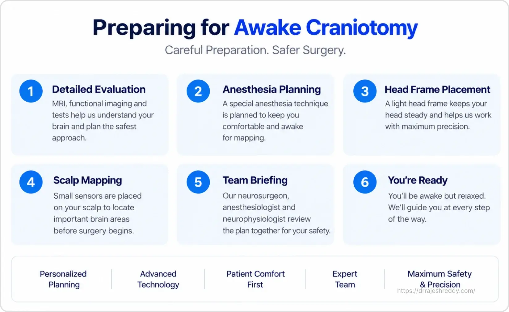
The success of an awake craniotomy depends as much on what happens before surgery as on the technique itself. A patient who understands what to expect, who has practiced the tasks they will be asked to perform, and who has built trust with the surgical team will cooperate more effectively during mapping — and achieve a better surgical result.
Neuropsychological and Speech Assessment
Before awake craniotomy, every patient undergoes a detailed neuropsychological and speech-language assessment. This establishes the patient's baseline — their pre-operative speech, language, naming, memory, and motor function. The same tasks that will be used intraoperatively are practiced during this pre-operative assessment, so nothing during surgery comes as a surprise.
The speech-language therapist designs a personalised set of mapping tasks based on which functions are at risk for each individual patient — picture naming cards, sentence repetition, object naming, counting, or finger-tapping sequences. Patients are always shown examples of what they will see during surgery and given the opportunity to practice until they are comfortable.
Pre-operative Imaging
- Functional MRI (fMRI) — maps the location of language and motor areas relative to the tumor
- Diffusion Tensor Imaging (DTI) with tractography — maps white matter fibre tracts including the arcuate fasciculus, corticospinal tract, and optic radiations
- High-resolution structural MRI — loaded into the neuronavigation system for intraoperative GPS guidance
- MR Spectroscopy — in selected cases to identify the most metabolically active region of the tumor
Anaesthetic Planning
A dedicated neuroanaesthesiologist meets with the patient before surgery to explain the anaesthetic plan. The technique most commonly used is asleep-awake-asleep (AAA) — general anaesthesia for the opening, gentle waking for the mapping and resection phase, and re-sedation for closure. Scalp nerve blocks with long-acting local anaesthetic ensure the scalp remains completely numb throughout.
Psychological Preparation
Many patients express anxiety not about the surgery itself, but about the awake phase — fear of claustrophobia, of losing control, of panic. Dr. Rajesh Reddy and the team address these concerns directly in the pre-operative consultation. Patients are told:
- You are in full control — if at any point you feel you cannot continue, surgery can be paused
- You will not see the surgical field — a sterile drape is positioned between your face and the operating site
- The environment is calm and supportive — the neuroanaesthesiologist and speech therapist remain by your side
- Mild anxiolytic medication can be given during the awake phase if anxiety increases
- The awake phase is temporary — most patients find it far more manageable than anticipated
Research consistently shows that patients who undergo awake craniotomy report lower anxiety and higher satisfaction with the procedure than they anticipated before surgery. Preparation and honest communication before surgery are the most powerful tools for making the awake phase a positive experience.
Intraoperative Brain Mapping: What Happens During the Awake Phase
Brain mapping during awake craniotomy uses a technique called direct electrical stimulation (DES) — passing a small, precisely controlled electrical current through a bipolar stimulator probe onto the brain surface or subcortical tissue. This current temporarily disrupts the function of the stimulated area for 1–4 seconds, allowing the surgeon to identify which points on the brain are responsible for which functions.
Cortical and Subcortical Mapping
Cortical Mapping (Surface): The surgeon systematically stimulates the exposed cortical surface in a grid pattern. If stimulation at a particular point causes a functional change — speech arrest, motor twitch, sensory response — that point is marked with a numbered tag placed on the brain surface.
Subcortical Mapping (Deep Pathways): As resection proceeds deeper into the brain, subcortical stimulation tracking maps white matter fibre tracts (arcuate fasciculus, corticospinal tract, optic radiations). This provides a real-time distance estimate from critical tracts, ensuring resection does not cross safe boundaries.
Intraoperative Brain Mapping
Real-time Testing. Real-time Protection.
Stimulate
Tiny pulses are delivered to small areas of the brain to test function.
Test
You'll be asked to speak, move, or name objects.
Protect
If a critical area is identified, the surgeon adjusts the plan to avoid it.
What We Map
- Motor (movement)
- Sensory (sensation)
- Speech & Language
- Vision & Auditory
Why It Matters
Real-time mapping helps remove the tumor while preserving the functions that matter most.
Mapping Tasks: What the Patient Is Asked to Do
| Brain Area at Risk | Mapping Task Given to Patient | What a Positive Response Means |
|---|---|---|
| Motor cortex (primary movement area) | Repeatedly lift fingers, tap foot, or squeeze a ball on the opposite side | Stimulation causes movement arrest or twitching — that point is marked as motor cortex |
| Broca's area (speech production) | Count aloud, name objects shown on picture cards, say days of the week | Stimulation causes speech arrest — patient stops mid-word or cannot initiate speech |
| Wernicke's area (speech comprehension) | Repeat simple sentences back; respond to verbal instructions | Stimulation causes errors in comprehension or repetition |
| Sensory cortex | Report any tingling or numbness felt during stimulation | Stimulation produces contralateral tingling — identifies sensory cortex boundary |
| Subcortical language tracts (arcuate fasciculus) | Continuous speech or naming task during deep resection | Slowed or disrupted speech during deep stimulation — identifies subcortical language pathway |
| Visual cortex / optic radiation | Report any visual disturbance or phosphenes | Stimulation causes visual phenomenon — identifies optic radiation boundary |
Awake Craniotomy vs. Standard Craniotomy: A Direct Comparison
Patients frequently ask whether awake craniotomy is truly necessary — whether a standard (asleep) craniotomy with intraoperative monitoring could achieve a similar result. The comparison below addresses this directly:
| Feature | Awake Craniotomy | Standard (Asleep) Craniotomy |
|---|---|---|
| Patient state during resection | Awake, interactive, performing tasks | Fully anaesthetised throughout |
| Functional mapping | Real-time cortical and subcortical stimulation mapping | Pre-operative fMRI and DTI only — no intraoperative confirmation |
| Tumour resection extent | Greater — resection taken closer to functional boundaries safely | More conservative margins around eloquent areas |
| Risk of permanent deficit | Lower — functional boundaries confirmed intraoperatively | Higher — relies on pre-operative imaging estimation |
| Duration | Longer — typically 5–8 hours including mapping phase | Shorter — typically 3–5 hours |
| Best suited for | Tumours in speech, motor, language, or visual areas | Tumours in non-eloquent areas; patients unable to cooperate |
| Patient experience | Unusual but well-tolerated with preparation; manageable | No experience during surgery; wakes after procedure |
For tumors in truly eloquent locations, the evidence consistently favours awake craniotomy: studies show that awake craniotomy achieves greater extent of resection with lower rates of permanent neurological deficit compared to asleep surgery for the same tumour locations. The initial discomfort of the awake experience is a small price for the preservation of a lifetime of speech and movement.
Risks of Awake Craniotomy: An Honest Assessment
Awake craniotomy is generally safe when performed by an experienced team with dedicated neuroanaesthesia support. However, patients deserve a clear understanding of its specific risks:
Procedure-Specific Risks
Intraoperative seizure (3–8%): Direct electrical stimulation can occasionally trigger a seizure during mapping. This is managed immediately by irrigating the brain surface with cold saline — which terminates the seizure within seconds — and pausing stimulation. The patient is given intravenous anti-epileptic medication if needed. This is a known and manageable complication, not an emergency.
Patient conversion (2–5%): In approximately 2–5% of cases, the patient becomes too anxious, fatigued, or distressed to continue cooperating with mapping tasks. In this situation, the surgeon converts to a standard asleep craniotomy and proceeds with available pre-operative imaging data.
Temporary neurological deficit: Most patients experience some temporary worsening of the function that was mapped — speech may be slightly more effortful, or limb movement slightly weaker — in the first few days after surgery. This almost always resolves within weeks as post-operative swelling subsides and the brain adapts.
Permanent neurological deficit (2–5%): The risk of permanent, significant neurological deficit after awake craniotomy in experienced hands is approximately 2–5% — significantly lower than the equivalent risk with asleep surgery for the same tumour location.
Anxiety and psychological distress: Some patients find the awake phase more distressing than anticipated. Thorough pre-operative preparation significantly reduces — though cannot entirely eliminate — this risk.
General Surgical Risks
- Bleeding — intracranial haemorrhage during or after surgery
- Infection — wound or intracranial infection
- CSF leak — uncommon
- Anaesthetic complications — rare with dedicated neuroanaesthesia
- Deep vein thrombosis — preventable with compression stockings and early mobilisation
The Awake Craniotomy Multidisciplinary Team
- Dr. Rajesh Reddy Sannareddy (Neurosurgeon): Leads the procedure, performs stimulation mapping, and resects the tumor.
- Neuroanaesthesiologist: Manages the asleep-awake-asleep transition, maintains patient comfort, and administers scalp blocks.
- Speech-Language Therapist / Neuropsychologist: Sits beside the patient during the awake phase, administers mapping tasks, and monitors cognition.
- Neurophysiologist: Runs continuous intraoperative neurophysiological monitoring (MEPs, SSEPs).
- Scrub Nurse & Operating Room Team: Ensures a sterile, calm, and quiet environment.
Recovery After Awake Craniotomy
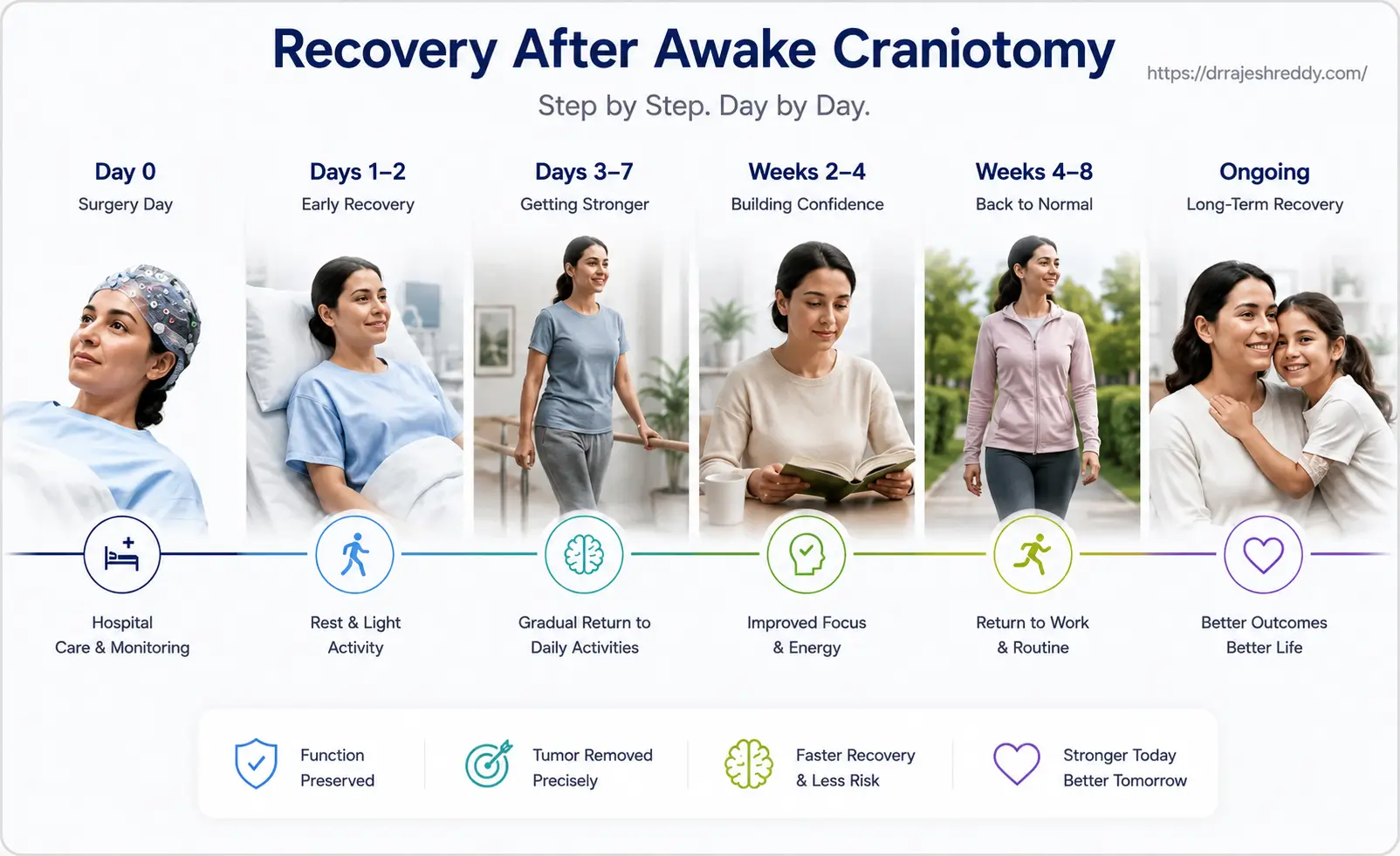
Immediate Post-Operative (Day 0–2)
- Most patients are remarkably alert after awake craniotomy — they have not received the same depth or duration of general anaesthesia as standard craniotomy patients.
- Neurological assessment performed immediately on return to recovery — speech, movement, and sensation tested.
- Some temporary worsening of mapped functions is normal and expected in the first 24–48 hours due to post-operative brain swelling.
- Steroids (dexamethasone) given to control swelling; anti-epileptic medication continued.
- Pain is usually well-controlled with simple analgesics — the brain itself does not generate pain, and the scalp wound is managed with regular analgesic medication.
Days 3–7 (Ward & Discharge)
- Most patients with uncomplicated awake craniotomy are fit for discharge by day 4–5.
- Speech and motor function typically begin to improve from day 2–3 as swelling reduces.
- Speech therapy assessment before discharge — and outpatient speech therapy arranged if needed.
- Post-operative MRI at 24–72 hours to confirm extent of resection.
Weeks 2–8
- Fatigue is the most prominent complaint — the brain requires significant energy for healing and functional reorganisation.
- Gradual return to activity — light walks from week 1; driving typically from week 4–6 (after seizure-risk assessment).
- Return to desk work typically possible within 4–6 weeks for most patients.
- For glioma patients: oncology referral and radiation/chemotherapy planning initiated at the 4–6 week mark.
3–6 Months
- Most functional deficits from awake craniotomy — particularly language — continue to improve for up to 6 months as the brain reorganises.
- Neuropsychological reassessment at 3 months to track recovery and guide rehabilitation.
- Return to full professional and social activities for most patients.
Why Choose Dr. Rajesh Reddy for Awake Craniotomy in Hyderabad?
- Double board certified MCh Neurosurgery (Exam Topper) & DNB Neurosurgery
- Visiting Scholar, Barrow Neurological Institute (Phoenix, Arizona) — global centre of excellence for awake craniotomy
- Visiting Scholar — Okayama University, Japan; Klinikum Stuttgart, Germany
- Integration of fMRI, DTI tractography, and connectomics in pre-surgical planning
- Established protocols for motor, language, sensory, and visual pathway mapping
- Dedicated neuroanaesthesia and speech therapy partnership