
Introduction
An arteriovenous malformation — AVM — is one of the most complex conditions in cerebrovascular medicine. Unlike a brain aneurysm, which is a localised bulge in a single artery, an AVM is a tangled mass of abnormal blood vessels connecting arteries directly to veins — bypassing the normal capillary network that regulates pressure, flow, and oxygen exchange. Present from birth, an AVM may be silent for decades, or it may announce itself catastrophically through bleeding into the brain, a first seizure, or a progressive neurological deficit.
The treatment of brain AVMs sits at the intersection of three neurosurgical disciplines — open microsurgery, endovascular embolisation, and stereotactic radiosurgery — and often requires all three working in a carefully planned sequence. Few neurological conditions demand more from a treating team, and few offer more complete cures when managed optimally.
Dr. Rajesh Reddy Sannareddy, Senior Consultant Endovascular Neurosurgeon in Hyderabad, brings a uniquely integrated skill set to AVM management: open microsurgical resection, endovascular embolisation using modern liquid embolic agents (Onyx, NBCA, Squid, PHIL), and coordination with radiosurgery teams for multimodal treatment of complex AVMs. His FINR fellowship in Zurich and visiting scholar experience at Barrow Neurological Institute, Phoenix — among the world's highest-volume cerebrovascular centres — have shaped his approach to these rare and challenging lesions.
What Is a Brain AVM?
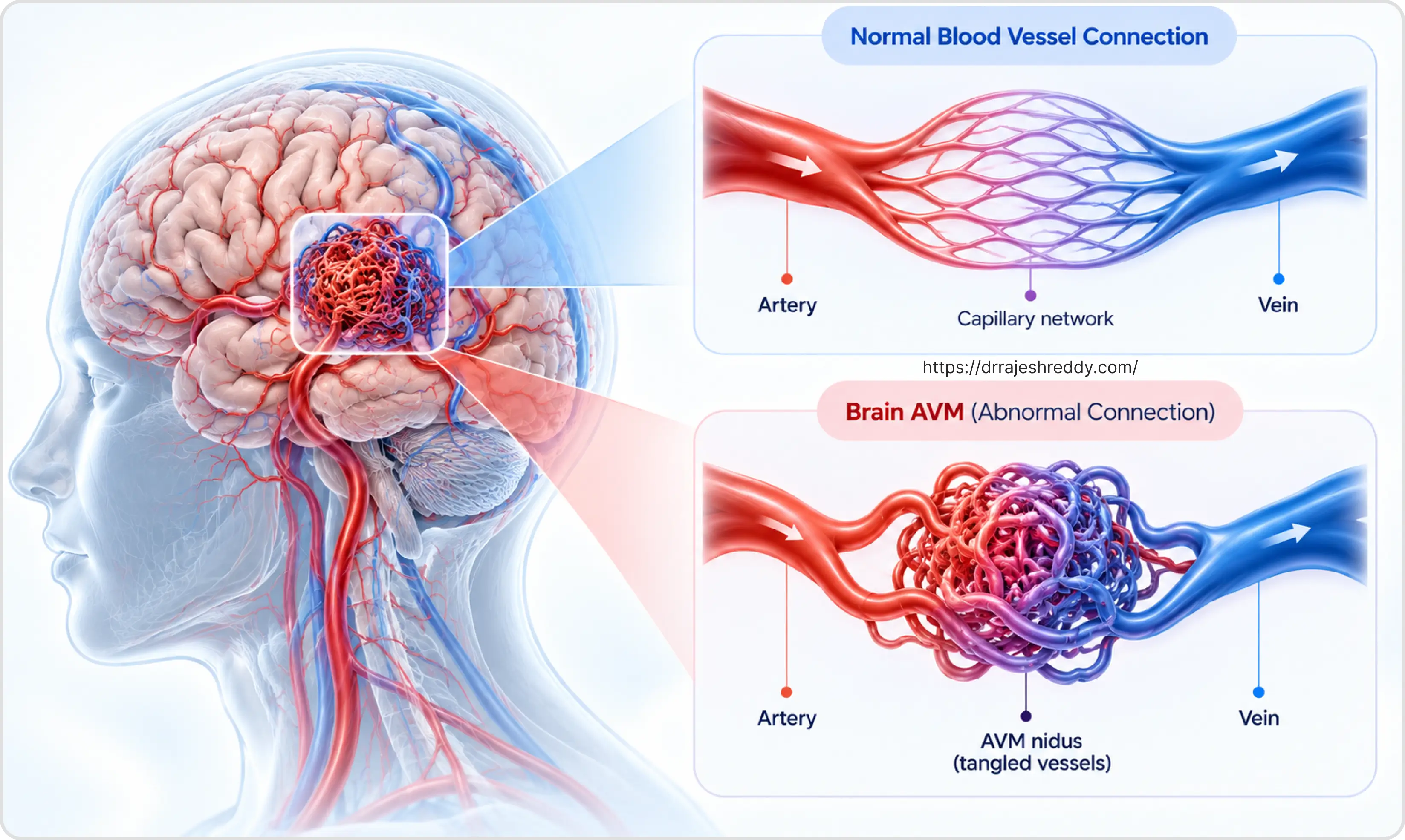
A cerebral arteriovenous malformation is a congenital (present from birth) abnormality of blood vessel development in which arteries connect directly to veins through a tangle of abnormal vessels — called the nidus — without an intervening capillary bed. Normally, capillaries slow blood down from high-pressure arterial flow to low-pressure venous flow, allowing oxygen exchange and protecting the veins from arterial pressure. In an AVM, this pressure regulation is absent: high-pressure arterial blood surges directly into thin-walled venous structures that are not designed to handle it.
Over time, this haemodynamic stress causes the AVM to enlarge, the feeding arteries to dilate, and the draining veins to become arterialized (thickened and prone to rupture). Flow-related aneurysms — true aneurysms that form on AVM feeding arteries due to increased flow — occur in approximately 10% of AVM patients and increase the overall bleeding risk significantly.
How Common Are Brain AVMs?
Brain AVMs are rare — affecting approximately 0.01–0.1% of the population, or roughly 1–2 per 100,000 people. They are typically diagnosed in the 2nd–4th decade of life, reflecting either the time it takes for the AVM to become symptomatic or the increasing use of brain MRI for other conditions. Unlike aneurysms, which are more common in women, AVMs have a roughly equal sex distribution.
AVM vs. Brain Aneurysm: Understanding the Difference
AVMs and brain aneurysms are both cerebrovascular conditions that can cause haemorrhagic stroke — but they are fundamentally different lesions with different origins, presentations, and treatment approaches:
| Feature | AVM | Cerebral Aneurysm |
|---|---|---|
| Origin | Congenital — present from birth; vessels develop abnormally | Acquired — develops over lifetime from arterial wall weakness |
| Age of presentation | Typically 20–40 years (younger) | Typically 40–60 years (older) |
| Primary presentation | Haemorrhagic stroke (50%), seizures (25%), headache, focal deficit | Thunderclap headache (SAH); incidental discovery; third nerve palsy |
| Annual bleed risk (untreated) | 2–4% per year | 0.5–2% (unruptured); much higher if ruptured |
| Type of bleed | Usually intracerebral (ICH); subarachnoid less common | Subarachnoid haemorrhage (SAH) — the primary event |
| Curative treatment | Yes — microsurgery or SRS can achieve complete cure | Yes — coiling, clipping, or flow diversion achieves permanent exclusion |
| Associated aneurysm | Flow-related aneurysms on feeding arteries present in ~10% | N/A (the primary lesion) |
| Radiosurgery role | Effective for AVMs < 3 cm — 80% obliteration at 3 years | Nil |
Symptoms of Brain AVM
AVMs can present in a wide variety of ways depending on their size, location, and whether they have bled:
Haemorrhage — The Most Common Presentation
Approximately 50% of AVMs present with intracranial haemorrhage — most commonly intracerebral haemorrhage (ICH) rather than subarachnoid haemorrhage (SAH). AVM bleeding typically presents as a sudden headache with rapid neurological deterioration — weakness, speech difficulty, loss of consciousness — corresponding to the location of the haematoma. The annual risk of haemorrhage from an unruptured AVM is approximately 2–4%, rising significantly after a first bleed (approximately 6–18% per year in the year following haemorrhage).
Seizures
Approximately 25–30% of AVMs present with new-onset seizures — either focal (affecting one limb or one side of the face) or generalised tonic-clonic seizures. Seizures occur because the AVM irritates surrounding cortical tissue through abnormal blood flow, arterial steal phenomena, or haemosiderin deposition from previous microbleeds. Any new-onset seizure in an adult under 40 should prompt an MRI of the brain to rule out an underlying AVM or other structural cause.
Progressive Neurological Deficit
Some AVMs cause progressive weakness, sensory loss, or visual disturbance without obvious haemorrhage — through arterial steal (blood being diverted from normal brain to the AVM), venous hypertension, or gradual cortical injury from repeated microbleeds too small to cause acute symptoms. These presentations can mimic brain tumour or demyelinating disease and require MRI and DSA for definitive diagnosis.
Headaches
Chronic headaches — not thunderclap, but persistent and sometimes pulsatile — may be an AVM presentation, particularly for large AVMs with significant haemodynamic impact or for AVMs located in the posterior fossa. Headaches alone are an uncommon isolated AVM presentation.
Diagnosing a Brain AVM
MRI Brain with Contrast
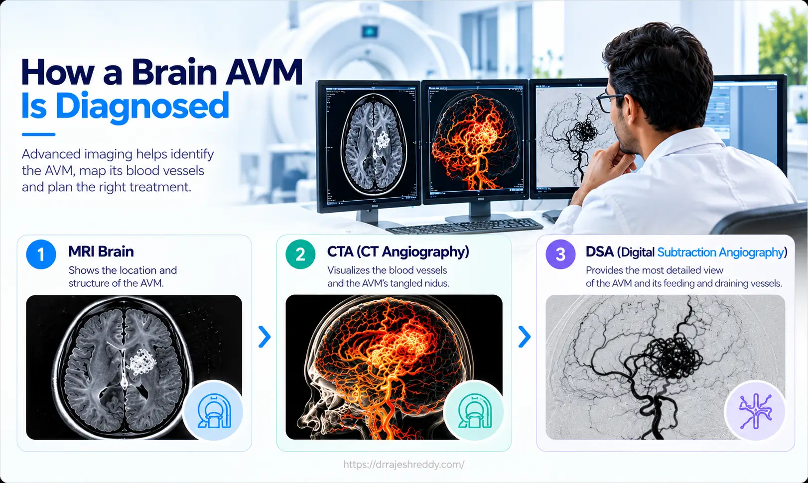
MRI is the primary diagnostic tool for AVM detection. The AVM nidus appears as a cluster of flow voids — dark tangles of high-flow vessels — on T2 sequences. Surrounding haemosiderin (from previous bleeds) appears as dark 'staining' on gradient-echo sequences. Gadolinium contrast enhancement helps delineate the nidus boundaries and the draining veins. Functional MRI (fMRI) and DTI tractography are used for AVMs near eloquent cortex to assess surgical risk.
CT Angiography (CTA)
CTA provides a rapid three-dimensional map of the AVM's feeding arteries, nidus, and draining veins — particularly useful in the emergency setting of AVM haemorrhage where DSA may not be immediately available. CTA has largely replaced diagnostic-only DSA for initial AVM characterisation.
Digital Subtraction Angiography (DSA) — Mandatory Before Treatment
DSA remains the gold standard for AVM characterisation before any treatment. 3D rotational DSA provides real-time dynamic information that static CT or MRI cannot: which arteries are feeding the AVM (arterial phase), the morphology of the nidus, which veins are draining it and in which direction, and — critically — whether any flow-related aneurysms are present on the feeding arteries. This information determines the Spetzler-Martin grade, the treatment approach, and the embolisation strategy.
Grading AVM Complexity: The Spetzler-Martin Scale
The Spetzler-Martin (SM) grading scale is the most widely used tool for assessing AVM surgical risk. It assigns a score of 1–5 based on three factors — AVM size, location in eloquent versus non-eloquent brain, and the pattern of venous drainage:
| SM Grade | Size | Eloquence | Deep Venous Drainage | Surgical Recommendation |
|---|---|---|---|---|
| Grade I | Small (<3cm) | Non-eloquent | No | Excellent surgical candidate — microsurgery preferred |
| Grade II | Small-medium | Non-eloquent or eloquent | No or Yes | Good surgical candidate — low operative risk |
| Grade III | Medium (3–6cm) OR eloquent OR deep venous | Variable | Variable | Intermediate risk — multimodal approach; case-by-case |
| Grade IV | Large (>6cm) + eloquent or deep venous | Eloquent | Yes | High surgical risk — embolisation + radiosurgery preferred |
| Grade V | Large + eloquent + deep venous | Eloquent | Yes | Very high risk — conservative or palliative management; surgery rarely curative |
The SM grade directly correlates with surgical morbidity — Grade I and II AVMs have excellent surgical outcomes with minimal risk; Grade IV and V AVMs carry very high surgical risk and are typically managed with non-surgical or staged multimodal approaches. Grade III AVMs occupy the most complex territory, where treatment decisions require careful individual case assessment and multidisciplinary discussion.
AVM Treatment Options: Surgery, Embolisation, Radiosurgery
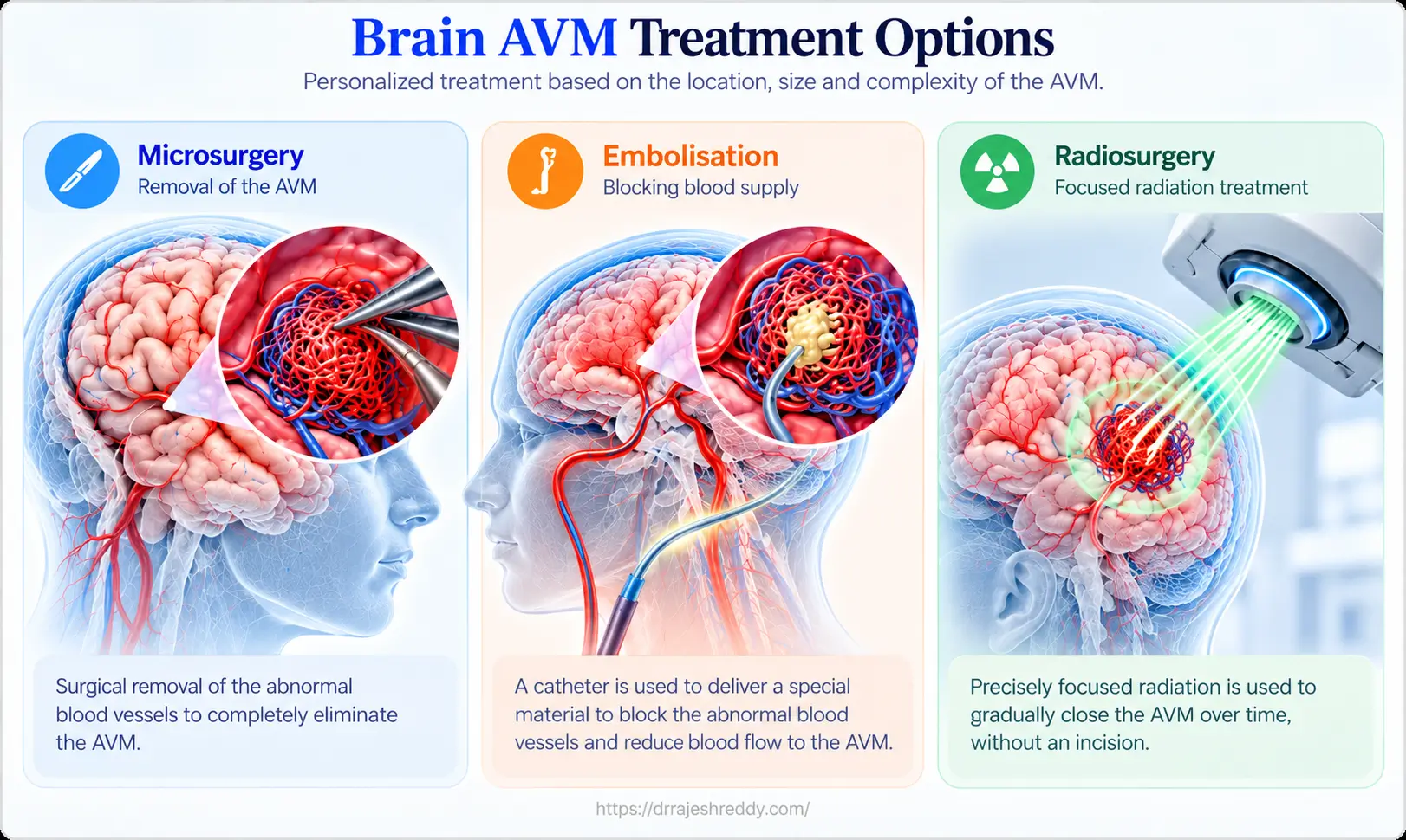
AVM treatment requires matching the right approach — or combination of approaches — to each AVM's specific anatomy, grade, and clinical presentation:
| Treatment | How It Works | Best For | Key Advantage |
|---|---|---|---|
| Microsurgical Resection | Open craniotomy; AVM nidus dissected and resected under microscope | SM Grade I–III; ruptured AVMs with haematoma; accessible location | Immediate cure — confirmed intraoperatively with angiography |
| Endovascular Embolisation | Catheter delivers embolic agents (NBCA glue, Onyx, Squid, PHIL) into AVM feeding arteries | Pre-surgical adjunct to reduce blood loss; palliative; rarely curative alone | Reduces AVM size and flow; makes surgery safer; can cure small AVMs |
| Stereotactic Radiosurgery (SRS) | Focused radiation causes progressive thrombosis of AVM nidus over 2–3 years | SM Grade I–III AVMs < 3 cm; eloquent location where surgery risky | No incision; outpatient; obliteration in ~80% of small AVMs at 3 years |
| Combined Approach | Embolisation + surgery, or embolisation + SRS, or all three | Large or complex AVMs (SM Grade III–IV) | Staged treatment reduces risk at each stage; achieves cure in high-grade AVMs |
| Conservative Management | Observation; seizure / BP management; no direct AVM treatment | Very high-grade AVMs (SM V); elderly unfit patients; unruptured low-risk AVMs | Avoids procedural risk in cases where treatment risk exceeds natural history risk |
Microsurgical AVM Resection
For low-grade AVMs (SM I–II) — and selected SM Grade III AVMs in accessible, non-eloquent locations — microsurgical resection offers the most durable cure: complete removal of the AVM nidus in a single procedure, with immediate cure confirmed by intraoperative DSA. The surgery involves a craniotomy, meticulous microsurgical dissection to identify all feeding arteries (which are individually coagulated and divided), circumferential dissection of the nidus, and finally occlusion of the draining veins — which are preserved until last, as premature venous occlusion causes the AVM to swell and bleed. Intraoperative ICG fluorescence angiography and intraoperative DSA confirm complete resection before wound closure.
Endovascular Embolisation
Embolisation is performed by navigating microcatheters into the AVM's feeding arteries under biplane fluoroscopic guidance and injecting liquid embolic agents — most commonly Onyx (ethylene vinyl alcohol copolymer), NBCA (N-butyl cyanoacrylate glue), Squid or PHIL (precipitating hydrophobic injectable liquid) — that solidify within the AVM vessels and permanently block flow. Modern liquid embolics — particularly Onyx — allow controlled, extended injections that can penetrate deeply into the AVM nidus, achieving more complete embolisation than was possible with older coil-based or particle techniques. Complete cure by embolisation alone is achievable in select cases in highly experienced hands.
Stereotactic Radiosurgery (SRS)
SRS delivers a high, precisely focused dose of radiation to the AVM nidus in a single session, causing progressive endothelial damage, vessel wall thickening, and eventual thrombosis of the AVM over 2–3 years. Complete obliteration is achieved in approximately 70–85% of AVMs under 3 cm at 3 years. SRS is the treatment of choice for small, deep, or eloquently located AVMs (particularly thalamic, basal ganglia, and brainstem AVMs) where open surgery carries unacceptably high risk. The main limitation is the 2–3 year latency period during which the AVM remains patent and carries its full bleeding risk — in approximately 5% of cases the AVM bleeds during this waiting period.
The Combined Multimodal Approach
For large or complex AVMs (SM Grade III–IV), the optimal approach combines all three modalities in a planned sequence: targeted embolization of the weak spot (intranidal aneurysm), followed by microsurgical resection or SRS for the residual nidus. This staged approach reduces the risk at each individual procedure and achieves cure in AVMs that would be untreatable by any single modality.
Dr. Rajesh Reddy's integrated endovascular and microsurgical expertise allows him to plan and execute both the embolisation and surgical phases — ensuring seamless coordination between the two most critical stages of multimodal AVM treatment.
AVM Embolisation: What the Procedure Involves
AVM embolisation is performed under general anaesthesia in a biplane neurointerventional suite. A guide catheter is placed in the internal carotid or vertebral artery, and a microcatheter is navigated into each AVM feeding artery in turn. Before injection, a test injection confirms the catheter is in the correct position and that the embolic material will reach the nidus without refluxing into normal arteries.
Onyx — the most commonly used liquid embolic — is injected slowly and continuously under real-time fluoroscopic guidance, watched as it fills the AVM vessels in a controlled front. The injection continues as long as the Onyx is progressing into the nidus; it is stopped when reflux threatens normal arteries or when the run is complete. The procedure may involve 1–4 embolisation sessions for a large AVM, each addressing different feeding arteries, typically spaced 4–6 weeks apart to allow healing between sessions.
Embolisation of a brain AVM is one of the most technically demanding endovascular procedures in neurosurgery — requiring precise control of liquid embolic injection over extended periods, detailed knowledge of AVM angioarchitecture, and the ability to recognise and manage complications in real time. The quality of the embolisation directly determines the safety of the subsequent surgery.
Why Dr. Rajesh Reddy for AVM Treatment in Hyderabad?
AVM management requires a specialist who bridges three disciplines: endovascular embolisation, microsurgical resection, and coordination with radiosurgery. Very few neurosurgeons in India offer all three under one roof.
- FINR — Fellowship in Interventional Neuroradiology, University Hospital, Zurich — endovascular AVM embolisation using Onyx, NBCA, Squid and PHIL in a high-volume European cerebrovascular centre
- Visiting Scholar — Barrow Neurological Institute, Phoenix, Arizona (one of the world's highest-volume AVM treatment programmes); Okayama University, Japan; Klinikum Stuttgart, Germany
- Double board certified neurosurgeon (MCh + DNB) — microsurgical AVM resection including intraoperative DSA confirmation of complete cure
- Integrated embolisation + surgery capability — plans and performs both phases of multimodal AVM treatment without handoff between specialists
- Experience with flow-related aneurysms associated with AVMs — aneurysm coiling and AVM embolisation combined in a single session when anatomy allows
- Coordination with stereotactic radiosurgery for multimodal Grade III–IV AVM management
- Over 5,000 neurosurgical and endovascular procedures across a 15+ year career
- Clinical acumen and judgement gained through years of training with leading global experts