
Introduction
Few diagnoses divide patients into two such different clinical realities as a brain aneurysm. One patient arrives in the emergency department unconscious, with a catastrophic headache and blood on their CT scan — a ruptured aneurysm, a neurosurgical emergency in which every hour matters. Another patient receives a phone call from their doctor asking them to come in to discuss a finding on an MRI done for something entirely unrelated — an unruptured aneurysm, requiring careful thought and expert guidance, but not panic.
These two scenarios — ruptured and unruptured aneurysm — are related by anatomy but separated by clinical urgency, management approach, treatment risk, and long-term prognosis. Understanding the differences between them is essential for patients and families navigating either situation. It is also the foundation of the treatment decision-making process that Dr. Rajesh Reddy Sannareddy uses with every aneurysm patient he sees.
Dr. Rajesh Reddy is a Senior Consultant Neurosurgeon and Interventional Neuroradiologist in Hyderabad with a Fellowship in Interventional Neuroradiology (FINR) from the University Hospital, Zurich and Visiting Scholar experience at Barrow Neurological Institute, Phoenix. His ability to offer both endovascular and microsurgical treatment means that the choice between them is made on clinical merit — not on which technique a specialist happens to perform.
Ruptured vs. Unruptured Brain Aneurysm: A Side-by-Side Comparison
The table below summarises the key differences across twelve dimensions — from how each is discovered to what treatment looks like and what patients can expect in the long term:
| Feature | Ruptured Aneurysm (SAH) | Unruptured Aneurysm |
|---|---|---|
| How discovered | Sudden catastrophic event — thunderclap headache, collapse, or emergency presentation | Incidentally on MRI/CT done for another reason, or via screening in high-risk patients |
| Presenting symptom | Thunderclap headache (worst ever, instantaneous), vomiting, LOC, neck stiffness | Usually none; occasionally mass effect symptoms (drooping eyelid, vision loss) |
| Urgency | Neurosurgical emergency — minutes to hours matter | Elective evaluation — days to weeks acceptable for most |
| Immediate risk | Rebleeding risk 4% in first 24 hrs; vasospasm (days 4–14); hydrocephalus; death | Annual rupture risk typically 0.5–2% depending on size, location, and risk factors |
| Investigations | CT brain → LP (if CT negative) → CTA → DSA — all urgent | MRA or CTA followed by DSA before any planned intervention — scheduled |
| Treatment urgency | Secure the aneurysm within 24–72 hours of rupture | Elective — timing planned based on risk-benefit assessment |
| Primary treatment goal | Prevent catastrophic rebleeding; manage SAH complications | Prevent first rupture; preserve neurological function |
| Preferred treatment | Endovascular coiling preferred in most SAH cases (ISAT evidence) | Individualised — coiling, clipping, flow diversion, or observation depending on anatomy and risk |
| Hospital stay | ICU 7–14 days; total 3–6 weeks | 1–5 days (endovascular) or 5–7 days (surgical) |
| Mortality | ~40–50% overall (including pre-hospital deaths) | Procedural mortality < 1% (endovascular) to 1–3% (surgical) |
| Long-term outcome | ~50% of survivors have permanent disability; prolonged recovery | Most patients return to normal life within weeks |
| Follow-up | Intensive — vasospasm monitoring, rehabilitation, MRA at 6 and 18 months | MRA surveillance at 6 and 18 months post-procedure; annual if observed |
A Brief Recap: What Is a Brain Aneurysm?
A brain (intracranial) aneurysm is a weakened, bulging section of a blood vessel wall within the brain — most commonly at the branching points of the large arteries forming the Circle of Willis at the base of the brain. The aneurysm is essentially a bubble formed where the muscular wall of the artery has thinned or failed, leaving only the inner and outer layers to hold back arterial blood pressure.
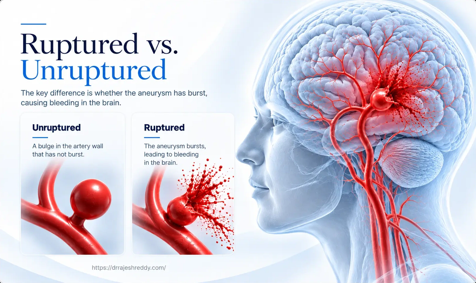
The critical question for every aneurysm is whether it has ruptured. A ruptured aneurysm has burst — releasing blood under high pressure into the subarachnoid space surrounding the brain, causing subarachnoid hemorrhage (SAH). An unruptured aneurysm is intact — it has not yet burst, though it carries some risk of doing so in the future.
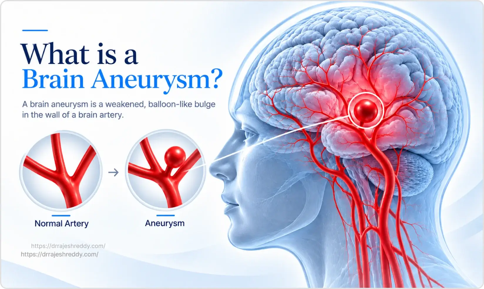
The same anatomical lesion — a brain aneurysm — produces two completely different clinical situations depending on whether it has ruptured. This is why the management of ruptured and unruptured aneurysms follows entirely different pathways, with different urgency, different treatment goals, and different outcome expectations.
The Ruptured Aneurysm: A Life-Threatening Emergency
What Happens When an Aneurysm Ruptures
When an aneurysm ruptures, blood escapes from the arterial circulation under high systolic pressure into the subarachnoid space — the fluid-filled space between the brain and the arachnoid membrane. This causes an instantaneous, massive spike in intracranial pressure, which is responsible for the hallmark symptom: the thunderclap headache, reaching maximal severity within 1–2 seconds.
The escaped blood spreads through the basal cisterns and across the brain surface, irritating the meninges and triggering an intense inflammatory response. The body's attempts to control the bleeding cause arterial spasm in adjacent vessels — the beginning of the vasospasm process that will threaten the brain in the days to follow. Meanwhile, if the initial haemorrhage is large enough, blood may also enter the brain itself (intracerebral haematoma) or the ventricular system (intraventricular haemorrhage), compounding the brain injury.
CRITICAL WARNING: REBLEEDING DANGER
After initial rupture, the soft clot sealing the aneurysm can give way at any moment. Rebleeding carries a mortality of 70–80%. Securing the aneurysm within 24–72 hours is vital.
Why Rebleeding Is the Most Immediate Danger
After the initial rupture, the aneurysm is sealed by a soft clot — a temporary, fragile plug that can give way under normal arterial blood pressure. Rebleeding — a second haemorrhage before the aneurysm is surgically or endovascularly secured — is the most lethal early complication of SAH. The mortality of rebleeding is approximately 70–80%.
The risk is highest in the first 24 hours (approximately 4%) and remains at 1–2% per day until the aneurysm is treated. This is why early aneurysm treatment — within 24–72 hours of rupture — is the global standard of care. Every hour of delay is a window of rebleeding risk.
The most important determinants of outcome after aneurysm rupture (in order of priority) are:
- The initial response of an individual to the bleed, determined by level of alertness and neurological deficits (if any)
- Location of the aneurysmal bleed and its relationship to the surrounding functional areas of the brain
- How quickly the aneurysm is secured (patients treated within 24 hours have significantly lower rebleeding rates and better neurological outcomes)
- Medical facility at which the patient receives treatment
Complications of Ruptured Aneurysm / SAH
A ruptured aneurysm is not just a single event — it sets off a cascade of complications over the following days and weeks that require intensive, expert management:
| Complication | Timing After SAH | Frequency | Management |
|---|---|---|---|
| Rebleeding | Highest risk first 24 hrs (4%); then 1–2% per day until aneurysm secured | 20–30% if untreated within 2 weeks | Secure aneurysm within 24–72 hours; bed rest; avoid straining; control BP |
| Cerebral Vasospasm | Days 4–14 (peak day 7–10) | 30–70% angiographic; 20–30% symptomatic | Nimodipine; euvolaemia; TCD monitoring; endovascular rescue (angioplasty/vasodilators) |
| Hydrocephalus (acute) | Hours to days | 15–20% | External ventricular drain (EVD); later VP shunt if persistent |
| Hyponatraemia | Days 3–14 | 30–50% | Careful fluid management; fludrocortisone; hypertonic saline in severe cases |
| Neurogenic cardiac dysfunction | First 24–48 hours | 25–30% ECG changes; 10–15% significant | Cardiology consultation; echo; avoid aggressive fluid restriction |
| Seizures | At time of rupture or delayed | 10–20% | Anti-epileptic prophylaxis; EEG monitoring in unconscious patients |
| Delayed cerebral ischaemia (DCI) | Days 4–14 | 20–30% | Augment BP; correct hypovolaemia; endovascular rescue if refractory |
| VP Shunt (chronic hydrocephalus) | Weeks to months | 15–20% | Ventriculoperitoneal shunt placement |
Managing these complications requires a dedicated neurosurgical ICU with 24/7 monitoring, transcranial Doppler surveillance for vasospasm, and the ability to perform endovascular rescue procedures (angioplasty, intra-arterial vasodilators) when vasospasm becomes symptomatic and does not respond to medical management.
Outcome After Ruptured Aneurysm
The overall prognosis after aneurysm rupture and SAH depends heavily on the clinical grade at presentation (Hunt & Hess / WFNS scale):
- Good-grade SAH (Hunt & Hess I–II): approximately 70–80% of patients achieve good neurological outcome with early treatment
- Moderate-grade SAH (Hunt & Hess III): approximately 50–60% achieve good outcome; cognitive and fatigue-related symptoms often persist for months
- Poor-grade SAH (Hunt & Hess IV–V): outcomes are guarded; approximately 20–40% achieve meaningful functional recovery; a proportion are left with significant disability or die
Even patients who make a good physical recovery after SAH frequently experience prolonged cognitive symptoms — memory difficulties, concentration problems, fatigue, depression, and anxiety — that are not visible on imaging but significantly impact quality of life. These are now well-recognised features of post-SAH syndrome and should be proactively addressed in follow-up care.
The Unruptured Aneurysm: Careful Evaluation, Not Panic
An unruptured brain aneurysm is a fundamentally different clinical situation. There is no emergency, no blood in the brain, and no immediate threat to life. What there is, is a decision to be made — and time to make it thoughtfully.
The core question for every unruptured aneurysm is: does the risk of treating it outweigh the risk of leaving it? This is not a simple calculation. It requires integrating the aneurysm's size, location, shape, the patient's age and health, their individual risk factors, and the technical risks of the proposed treatment.

How Unruptured Aneurysms Are Discovered
- Incidental discovery: The most common scenario. An MRI or CT done for a headache, head injury, sinus symptoms, or another neurological condition reveals an aneurysm that has been present — silently — for an unknown period. This is understandably alarming for patients, but the key message is that an incidental aneurysm is not an emergency: there is time to evaluate it carefully.
- Screening: Patients with a family history of brain aneurysm or SAH (particularly those with two or more affected first-degree relatives), or with conditions known to be associated with aneurysms (polycystic kidney disease, neurofibromatosis Type 1, connective tissue disorders), may undergo proactive screening with MRA — discovering aneurysms before they cause any symptom.
- Symptomatic unruptured aneurysm: A minority of unruptured aneurysms cause symptoms through mass effect — compressing adjacent structures. A third nerve palsy (drooping eyelid + dilated pupil), progressive visual loss, or facial pain may indicate an enlarging aneurysm that requires more urgent evaluation, though still not the same emergency as rupture.
The Natural History of Unruptured Aneurysms: What Does the Evidence Say?
Several large international studies — including the International Study of Unruptured Intracranial Aneurysms (ISUIA) and the UCAS Japan study — have mapped the natural history of unruptured aneurysms. Key findings:
- Small aneurysms (< 7 mm) in the anterior circulation have an annual rupture risk of approximately 0.1–0.5% — meaning most will never rupture in the patient's lifetime
- Aneurysms ≥ 7 mm carry significantly higher annual rupture risk — increasing sharply with size
- Posterior circulation aneurysms (basilar tip, PICA) rupture more frequently than anterior circulation aneurysms of the same size
- Irregular shape, daughter sac, or high aspect ratio are independent predictors of rupture risk, regardless of size
- Growth on serial imaging is the single most important indicator that an aneurysm should be treated — any documented growth warrants intervention
A small aneurysm with low-risk features in an elderly patient may be entirely appropriate for observation. The same size aneurysm in a 35-year-old smoker with a family history of SAH and an irregular daughter sac warrants a very different conversation. Dr. Rajesh Reddy individualises every risk assessment — there is no single size or location cutoff that applies to all patients.
Assessing Rupture Risk: Low vs. High Risk Features
The table below summarises the features that define lower and higher rupture risk for unruptured aneurysms — the basis for the treatment decision conversation:
| Factor | Low Rupture Risk | Higher Rupture Risk |
|---|---|---|
| Aneurysm size | < 5 mm | > 7 mm; especially > 10 mm (large) and > 25 mm (giant) |
| Location | Anterior circulation (MCA, ACA, ICA) | Posterior circulation (basilar tip, PICA, PICA); also PComm |
| Shape / morphology | Smooth, round, regular | Irregular, lobulated, daughter sac present |
| Aspect ratio | Low (dome height / neck width < 1.6) | High aspect ratio > 1.6 — tall narrow-necked aneurysms |
| Previous SAH from another aneurysm | No prior SAH | Prior SAH from a different aneurysm — increased risk in remaining aneurysms |
| Symptoms | Asymptomatic | Symptomatic — CN III palsy, headache, visual symptoms |
| Growth on serial imaging | Stable size over 12–24 months | Any demonstrable growth — even small increment warrants intervention |
| Patient age | Elderly (> 70) — shorter life expectancy reduces cumulative risk | Younger patient — longer life expectancy increases cumulative rupture risk |
| Smoking | Non-smoker | Active smoker — significantly elevated risk |
| Hypertension | Controlled BP | Uncontrolled hypertension |
| Family history of SAH | No family history | Two or more first-degree relatives with SAH — genetic predisposition |
How Treatment Differs: Ruptured vs. Unruptured
Treatment of Ruptured Aneurysm
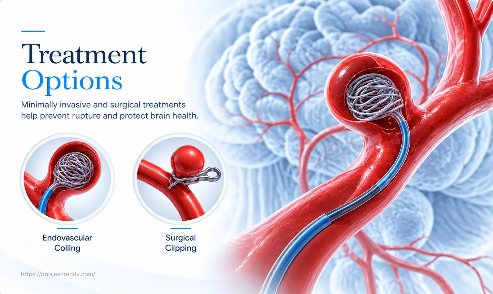
For a ruptured aneurysm, the first priority is always to secure the aneurysm as rapidly as possible to prevent rebleeding. The two main treatment options — endovascular coiling and microsurgical clipping — are both aimed at permanently excluding the aneurysm from the circulation.
- Endovascular coiling: Preferred in most ruptured aneurysm cases. The ISAT trial demonstrated significantly better neurological outcomes for coiling versus clipping in patients with ruptured aneurysms suitable for both treatments. Coiling avoids craniotomy and general anaesthesia for a prolonged open surgery in an already critically ill patient — reducing physiological stress at a vulnerable time.
- Microsurgical clipping: Preferred when the aneurysm anatomy is not suitable for coiling (wide neck, complex morphology, incorporated branch vessels), when there is an associated intracerebral haematoma requiring surgical evacuation, or when the aneurysm is at the MCA bifurcation — where clip geometry and surgical access are often superior to catheter access.
The choice between coiling and clipping for ruptured aneurysms is determined at the emergency multidisciplinary meeting, weighing the aneurysm anatomy on CTA, the patient's clinical grade, and the available expertise. Dr. Rajesh Reddy's ability to perform both procedures allows him to make this decision based on what is genuinely best for the patient.
Treatment of Unruptured Aneurysm
For unruptured aneurysms, the decision is more nuanced — and includes the option of observation. Treatment is not always the right answer. When treatment is indicated, the full range of options is available:
- Observation: For small, low-risk aneurysms — particularly in elderly or medically unfit patients. Regular MRA surveillance (typically annually for 2 years, then every 2 years if stable) monitors for growth, which is the trigger for reassessment.
- Endovascular coiling: First choice for most saccular aneurysms suitable for catheter access. Shorter recovery (1–3 days), no open surgery, and excellent long-term occlusion rates for appropriately selected aneurysms.
- Flow diversion (Pipeline device / SILK / DERIVO / p64 / Surpass / FRED): Preferred for large, giant, or fusiform aneurysms where coiling is impractical. Particularly effective for paraclinoid ICA aneurysms.
- Intrasaccular flow diversion devices (WEB, Contour, Artisse, Seal): Particularly suited to wide-neck bifurcation aneurysms (MCA, basilar tip, AComm) — a single intrasaccular device requiring no stent and no dual antiplatelet therapy.
- Microsurgical clipping: Preferred for younger patients (< 50) where the lifelong durability of clipping is advantageous; for MCA aneurysms where surgical anatomy is favourable; and for wide-neck aneurysms not suitable for catheter-based treatment.
Modifiable Risk Factors: What You Can Do Right Now
Whether your aneurysm is ruptured or unruptured, there are modifiable risk factors that significantly influence both rupture risk and overall cerebrovascular health. Addressing these is part of the management plan regardless of the chosen treatment strategy:
Blood Pressure Control
Hypertension is the single most important modifiable risk factor for brain aneurysm formation, growth, and rupture. High blood pressure exerts continuous mechanical stress on already-weakened arterial walls. Target blood pressure for aneurysm patients is typically < 130/80 mmHg. If you are not already on antihypertensive medication and your blood pressure is elevated, discuss treatment with your doctor immediately — this is one of the most impactful steps you can take.
Smoking Cessation
Cigarette smoking doubles the risk of aneurysm rupture. Nicotine and the toxic compounds in cigarette smoke promote arterial wall inflammation, enzymatic degradation of the vessel wall, and reduced endothelial repair — all of which weaken the aneurysm wall. Smoking cessation is strongly recommended for all aneurysm patients, whether managed with observation or post-treatment surveillance. The risk reduction from quitting is measurable and significant.
Avoiding Exertional Triggers
Heavy lifting, vigorous straining (including during defaecation), and explosive physical exertion cause transient spikes in blood pressure and intracranial pressure that can trigger rupture. Patients with known unruptured aneurysms awaiting treatment are advised to avoid heavy exertion, heavy resistance training, and activities involving sustained breath-holding and straining. Light to moderate aerobic exercise is generally safe and beneficial for cardiovascular health.
Alcohol and Cocaine Avoidance
Chronic heavy alcohol use is associated with increased aneurysm risk. Cocaine use causes acute hypertensive surges that are a recognised trigger for aneurysm rupture. Both should be avoided entirely by patients with known aneurysms.
Should Family Members Be Screened?
Brain aneurysms can run in families. The risk of an intracranial aneurysm in a first-degree relative of someone who has had an aneurysm or SAH is 3–7 times higher than the general population risk of approximately 2–3%.
When Screening Is Recommended
- Two or more first-degree relatives (parents, siblings, children) with confirmed brain aneurysm or SAH — screening with MRA from age 30 is recommended
- One first-degree relative with SAH — screening may be considered from age 30–40
- Diagnosed genetic conditions associated with aneurysms: Autosomal Dominant Polycystic Kidney Disease (ADPKD), neurofibromatosis Type 1, Ehlers-Danlos syndrome (vascular type), Marfan syndrome, Coarctation of aorta.
- Patients who have themselves had one aneurysm — 20–30% have additional aneurysms that should be identified and assessed
The Screening Investigation
MRA (magnetic resonance angiography), preferably on a 3T MRI, is the standard screening tool — non-invasive, no radiation, no contrast injection, and highly sensitive for aneurysms down to 2–3 mm. If a screening MRA is positive, further characterisation with CTA or DSA follows. A negative MRA at age 30 does not provide lifelong reassurance — repeat screening at 5–10 year intervals may be appropriate for high-risk individuals.