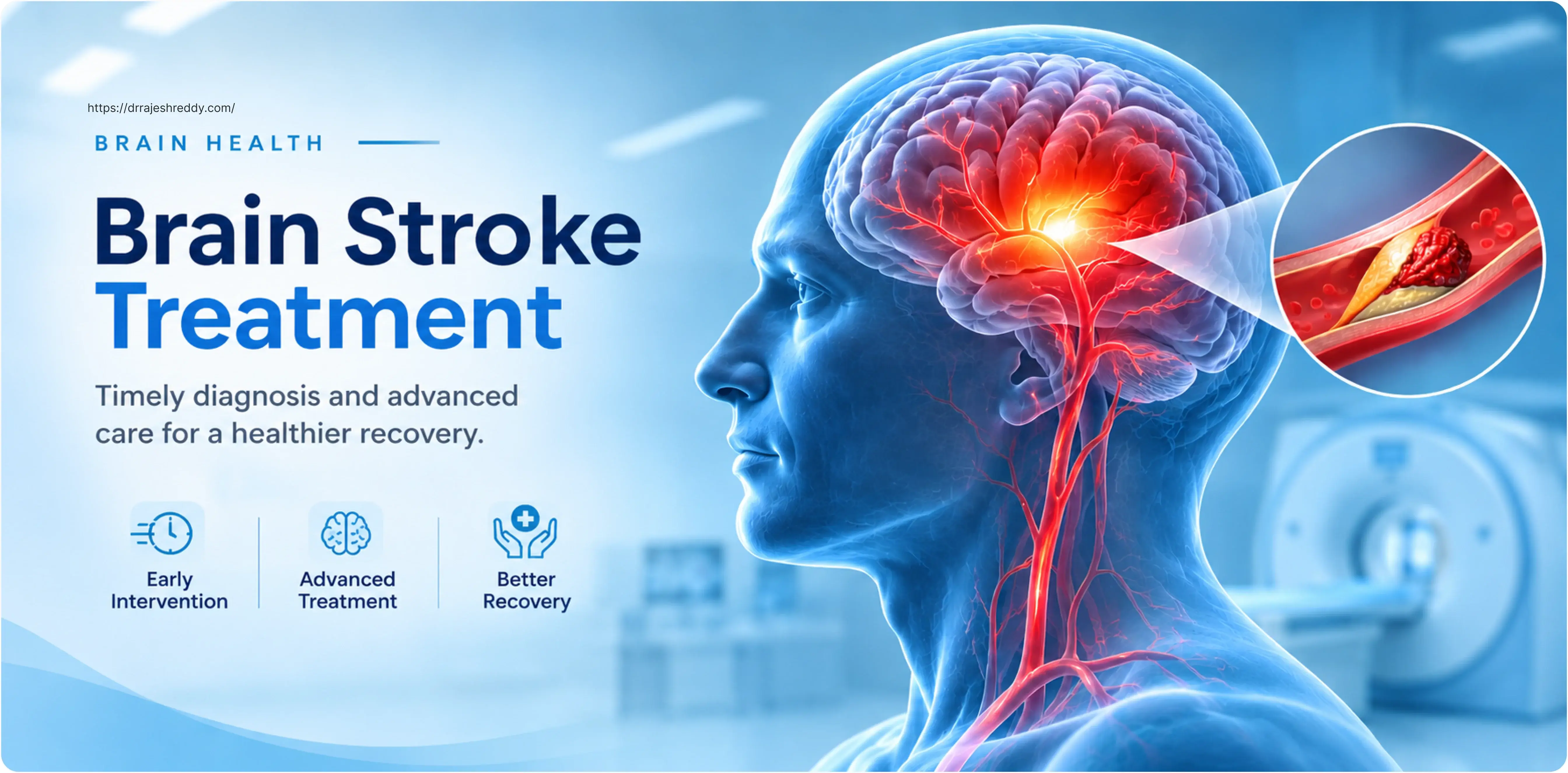
Every two seconds, someone in the world has a stroke. In India, stroke is now the second leading cause of death and the third leading cause of disability — and Hyderabad, as one of India's largest metropolitan cities, sees a substantial burden of acute stroke every day. The tragedy of stroke is that it is both largely preventable and, when it does occur, often treatable — but only if the patient reaches a capable centre quickly enough.
Dr. Rajesh Reddy Sannareddy is a Senior Consultant Neurosurgeon in Hyderabad specialising in cerebrovascular disease — the full spectrum of conditions in which the brain's blood supply is disrupted. His practice encompasses emergency intervention for acute stroke (mechanical thrombectomy, haemorrhage management), surgical procedures to prevent future stroke (carotid endarterectomy, aneurysm treatment), and coordination of the multidisciplinary rehabilitation that determines how much function a stroke survivor recovers.
This pillar page is the entry point to Dr. Rajesh Reddy's stroke content hub. It provides a comprehensive overview of stroke — what it is, the different types, how each is treated, and when to seek emergency care. The cluster articles linked below go into clinical depth on each topic. Whether you are a patient who has just been told they have had a TIA, a family member of someone in the acute stroke unit, or a clinician looking to refer a patient for thrombectomy assessment, this resource is designed to answer your questions.
STROKE IS A MEDICAL EMERGENCY
If you or someone near you suddenly develops face drooping, arm weakness, speech difficulty, or any other sudden neurological symptom — call 108 immediately. Do not drive yourself to hospital. Time from symptom onset to treatment directly determines how much brain is saved and how much function is recovered.
What Is a Brain Stroke?
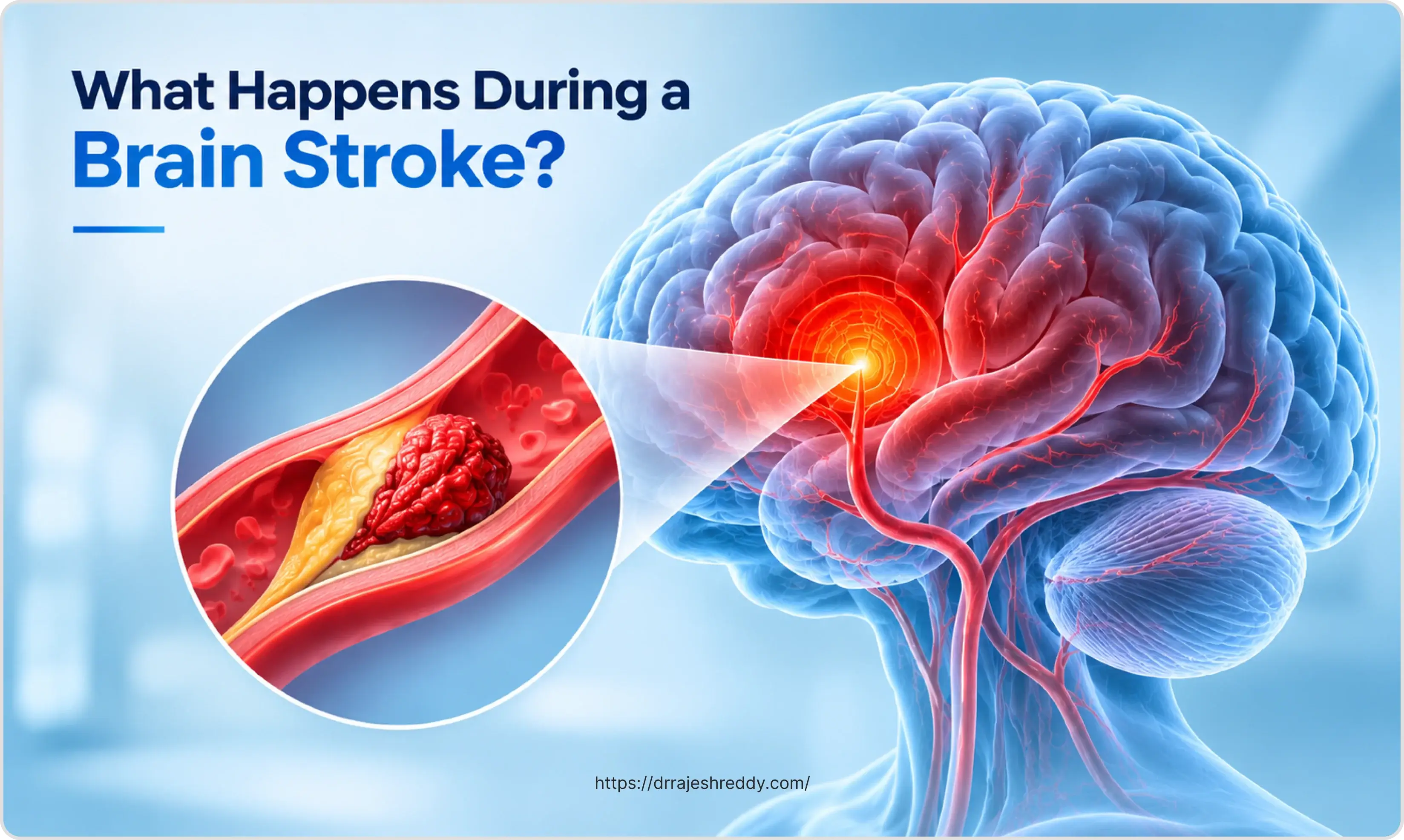
A stroke occurs when the blood supply to part of the brain is suddenly cut off. Brain cells begin to die within minutes of losing their blood supply — the brain consumes approximately 20% of the body's oxygen despite being only 2% of its weight, and it has no reserve energy supply. The territory of brain affected by the stroke determines the symptoms: a stroke affecting the motor cortex causes limb weakness or paralysis; a stroke affecting the speech areas causes aphasia; a stroke affecting the cerebellum causes imbalance and vertigo.
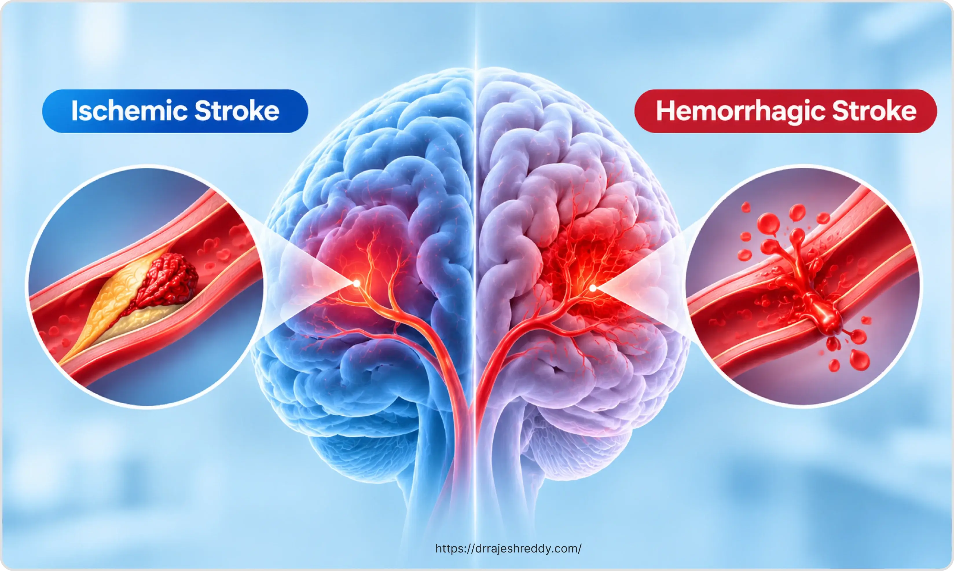
Strokes are broadly divided into two categories: ischaemic strokes, in which a blood vessel is blocked by a clot, and haemorrhagic strokes, in which a blood vessel ruptures and bleeds into or around the brain. These two categories require fundamentally different treatments — clot-dissolving drugs that are life-saving in ischaemic stroke are dangerous in haemorrhagic stroke — which is why rapid brain imaging (CT scan) is performed as the very first step in emergency stroke management.
The Scale of the Problem in India
India has approximately 1.8 million new strokes every year — roughly one stroke every 17 seconds. The age-adjusted stroke incidence in India has been rising, and strokes are occurring in younger patients than in Western populations: approximately 20% of strokes in India occur in patients under 40 years of age. Hypertension — which affects an estimated 30% of Indian adults — is the single most important modifiable risk factor for both ischaemic and haemorrhagic stroke. Other major risk factors prevalent in the Indian population include diabetes (affecting 11% of adults), dyslipidaemia, atrial fibrillation, tobacco use (smoking and smokeless tobacco), and physical inactivity.
Despite this burden, stroke care in India has improved markedly. The establishment of dedicated stroke units in major centres, the availability of mechanical thrombectomy in cities like Hyderabad, and increased public awareness of FAST symptoms have all contributed to better outcomes. The challenge remains getting patients to treatment within the therapeutic window — the critical period within which treatment can save brain tissue and reduce disability.
Recognising a Stroke: The BEFAST Signs
The classic FAST mnemonic — Face, Arms, Speech, Time — has been expanded to BEFAST to include two additional symptoms more common in posterior circulation strokes, which affect the back of the brain:
- B — Balance: Sudden loss of balance or coordination; stumbling or falling without explanation
- E — Eyes: Sudden vision loss in one or both eyes; double vision; vision that cuts off on one side
- F — Face: One side of the face drooping; uneven smile; face feels numb
- A — Arms: One arm drifts down when both are raised; arm weakness or numbness
- S — Speech: Slurred speech; inability to find words; not understanding what others are saying
- T — Time: Time to call 108 immediately — note the time symptoms started; this information is critical for treatment decisions
Other stroke symptoms that people miss: sudden severe headache with no known cause ('the worst headache of my life' — a subarachnoid haemorrhage warning sign); sudden confusion; sudden difficulty swallowing; sudden severe dizziness. Any sudden neurological symptom that is new and unexplained should be treated as a stroke until proven otherwise.
Types of Stroke: Understanding the Differences
Not all strokes are the same, and the distinction between stroke types is essential because the treatments are different — sometimes opposite. The table below summarises the major stroke types, their mechanisms, and their emergency treatments.
| Type | Mechanism | Proportion of Strokes | Emergency Treatment |
|---|---|---|---|
| Ischaemic stroke — thrombotic | Blood clot forms on an atherosclerotic plaque inside a brain artery, blocking blood flow | ~40–50% of all strokes | IV thrombolysis (tPA) if within 4.5 hours; thrombectomy if large vessel occlusion |
| Ischaemic stroke — embolic | Clot from the heart (atrial fibrillation, valve disease) or proximal artery travels and lodges in a brain vessel | ~25–30% of all strokes | IV thrombolysis and/or mechanical thrombectomy; anticoagulation for AF |
| Ischaemic stroke — lacunar | Small vessel occlusion deep in the brain (basal ganglia, internal capsule, brainstem) from hypertension | ~15–20% of all strokes | Antiplatelet therapy; aggressive BP control; thrombolysis may be used |
| Intracerebral haemorrhage (ICH) | Rupture of a small penetrating artery, causing bleeding directly into brain tissue; hypertension is the main cause | ~10–15% of all strokes | BP control; reversal of anticoagulation; surgical evacuation in selected cases; monitoring in neurocritical care |
| Subarachnoid haemorrhage (SAH) | Rupture of an intracranial aneurysm into the subarachnoid space; often presents as 'thunderclap headache' | ~3–5% of all strokes | Aneurysm securing (coiling or clipping); nimodipine; hydrocephalus management |
| Transient ischaemic attack (TIA) | Temporary blockage of blood flow with symptoms resolving within 24 hours (usually minutes); high early stroke risk | Not counted as stroke but precedes ~10–15% of strokes | ABCD² score–guided urgent assessment; dual antiplatelet; imaging; risk factor control |
The Stroke Treatment Window: Why Every Minute Counts
The phrase 'time is brain' captures a biological reality: in a large vessel ischaemic stroke, approximately 1.9 million neurons die every minute without treatment. The effective treatment window for different interventions differs:
- IV thrombolysis (clot-dissolving injection): 0–4.5 hours from symptom onset (standard); extended to up to 6 hours in selected patients with favourable CT perfusion mismatch profile per 2026 updated AHA/ASA and ESO guidelines. The earlier within this window, the better the outcome. 'Door-to-needle' time — from hospital arrival to treatment — is a key quality metric, with the target being under 60 minutes
- Mechanical thrombectomy (endovascular clot removal): 0–24 hours in selected patients, based on brain perfusion imaging. For patients with a large area of brain at risk but limited irreversible damage, the window extends to 24 hours from last known well time
- Haemorrhagic stroke — blood pressure control: Within the first few hours; rapid reduction to systolic <140 mmHg within 1 hour of presentation reduces haematoma expansion in ICH
2026 Guideline Update — BP in Acute Ischaemic Stroke
AHA/ASA and ESO 2026 guidelines do NOT recommend aggressive blood pressure lowering in the first 24–48 hours of acute ischaemic stroke. Permissive hypertension — allowing BP up to 220/120 mmHg in non-thrombolysed patients — is preferred to maintain perfusion of the ischaemic penumbra. Post-tPA target remains <180/105 mmHg. Aggressive BP lowering risks extending the infarct by reducing pressure to the penumbral zone.
- Carotid endarterectomy after TIA or minor stroke: Within 48 hours for highest-risk patients; within 2 weeks at the latest — delay significantly reduces the benefit
What to do if you suspect a stroke: Call 108 immediately. Do not wait to see if symptoms improve — TIAs and minor strokes often 'recover' temporarily before a devastating stroke occurs within hours. Do not give the patient anything to eat or drink — swallowing may be impaired. Note the exact time symptoms began — this is the most important piece of information for the emergency team.
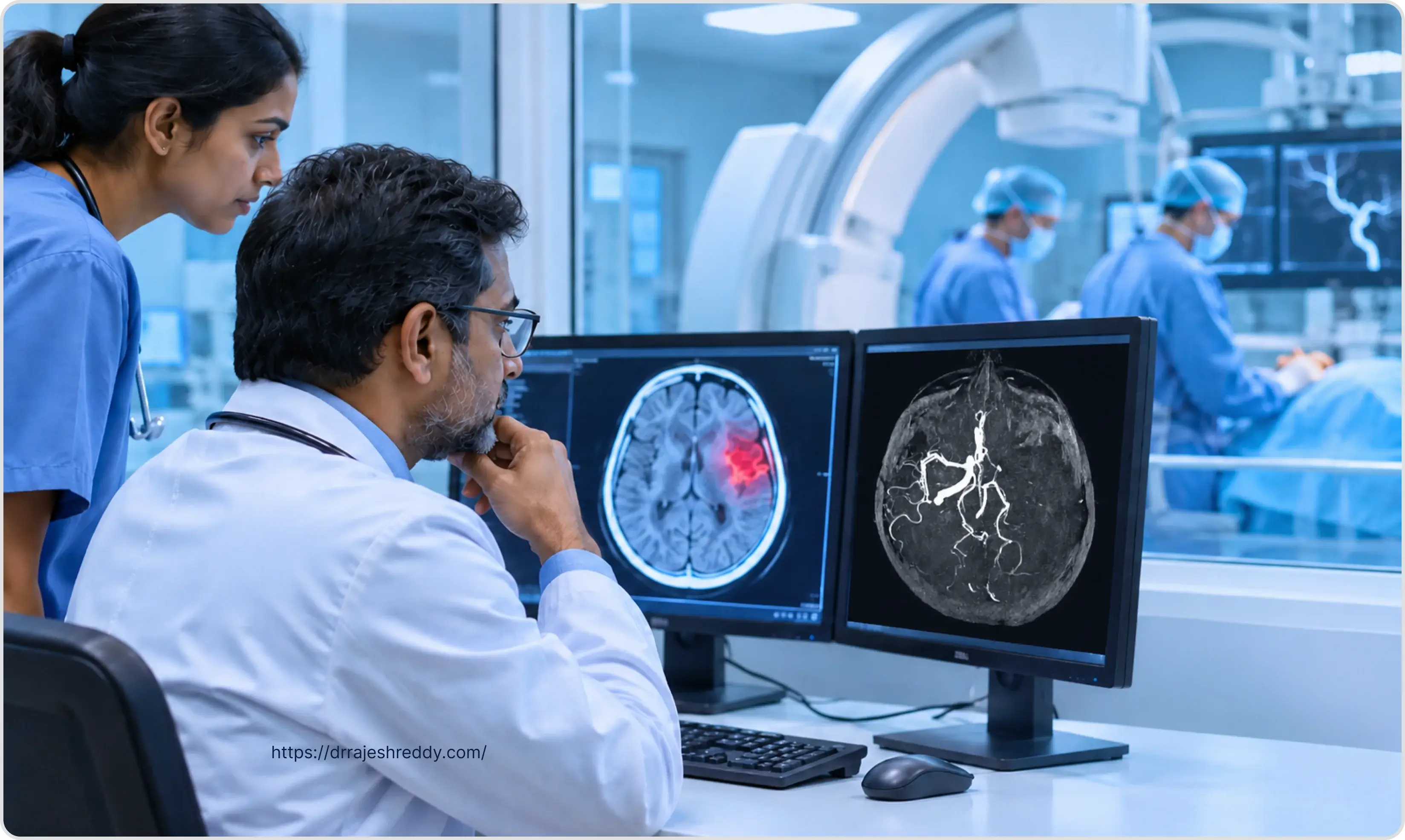
Brain Stroke Treatment Options
Dr. Rajesh Reddy's stroke treatment practice covers the full range of interventions — from emergency endovascular procedures in the acute phase through surgical procedures to prevent recurrent stroke and the long-term multidisciplinary rehabilitation that maximises recovery.
| Treatment | Indication | What It Does |
|---|---|---|
| IV thrombolysis (tPA / tenecteplase) | Ischaemic stroke within 4.5 hours of onset; no haemorrhage on CT | Dissolves the clot pharmacologically; restores blood flow; best outcomes when given within 60–90 minutes of arrival |
| Mechanical thrombectomy | Large vessel occlusion (ICA, MCA, basilar artery); up to 24 hours in selected patients | Catheter-based removal of clot from large brain artery; best available treatment for major stroke; independent outcome in ~50% of treated patients |
| Intracerebral haemorrhage management | Spontaneous ICH; hypertensive ICH; anticoagulant-associated ICH | BP reduction to systolic <140 mmHg; anticoagulation reversal; surgical clot evacuation in selected cases; neurocritical care monitoring |
| Aneurysm treatment (coiling/clipping) | Subarachnoid haemorrhage; unruptured aneurysm found incidentally | Prevents re-bleeding; endovascular coiling (preferred) or surgical clipping depending on aneurysm anatomy |
| Carotid endarterectomy / stenting | Symptomatic carotid stenosis >50%; asymptomatic stenosis >70% | Removes atherosclerotic plaque from carotid artery; reduces future stroke risk by up to 65% in symptomatic stenosis |
| Decompressive craniectomy | Malignant middle cerebral artery infarction with severe brain swelling | Life-saving; removes part of skull to allow swollen brain to expand without compressing vital structures |
| Stroke unit care | All stroke patients | Coordinated multidisciplinary care (neurologist, neurosurgeon, physio, speech therapist, occupational therapist, nurses) shown to independently reduce death and disability |
Stroke in Younger Adults: A Growing Concern in India
The stereotype of stroke as a disease of the elderly does not reflect the Indian reality. Dr. Rajesh Reddy regularly treats patients in their 30s and 40s — and occasionally younger — with stroke. In young adults, the causes of stroke differ from those in older patients:
- Cardioembolic stroke from rheumatic heart disease and atrial fibrillation — rheumatic heart disease remains far more prevalent in India than in high-income countries
- Hypercoagulable states — antiphospholipid syndrome, protein C/S deficiency, factor V Leiden — often undiagnosed
- Cervical artery dissection — spontaneous or following chiropractic manipulation or minor trauma; a leading cause of stroke under 45
- Paradoxical embolism through patent foramen ovale (PFO) — a hole in the heart present in approximately 25% of the population; found in higher proportions of young stroke patients
- Illicit drug use — cocaine, amphetamines
- Oral contraceptive use combined with migraine with aura and smoking — a significantly elevated risk combination in young women
- Vasculitis — central nervous system vasculitis, Takayasu's arteritis
- Hyperhomocysteinaemia — elevated plasma homocysteine is an underappreciated but treatable risk factor for stroke in young adults; it damages the vascular endothelium and promotes thrombosis; a simple, inexpensive blood test; treated with B6, folate (B9), and B12 supplementation; should be part of the routine young-stroke workup in every patient without an obvious alternative cause
Young stroke requires a thorough investigation protocol — not just the standard CT and ECG, but also cardiac MRI, prolonged cardiac monitoring for paroxysmal AF, coagulation screen, connective tissue disease workup, and in some cases digital subtraction angiography (DSA). Dr. Rajesh Reddy coordinates this workup and ensures the cause of the stroke — not just the event — is identified and addressed.
Cerebral Venous Sinus Thrombosis (CVST): The Often-Missed Stroke
Most stroke resources focus exclusively on arterial stroke. Cerebral venous sinus thrombosis (CVST) is a distinct and frequently overlooked form of stroke caused by thrombosis of the cerebral venous sinuses or cortical veins — not arterial occlusion. Because venous drainage is blocked, the brain develops venous congestion, oedema, and haemorrhagic infarction that does not follow arterial vascular territories and can appear anywhere in the brain. CVST is most common in young women — particularly during pregnancy and the postpartum period — and in patients with thrombophilia, oral contraceptive use, dehydration, infections (mastoiditis, sinusitis), or haematological disorders.
The clinical presentation is protean and easily confused with migraine, encephalitis, or idiopathic intracranial hypertension: progressive headache (the most common symptom), focal neurological deficits, seizures, visual disturbance, and encephalopathy. Diagnosis is confirmed on MRI with MR venography (MRV) — conventional CT may appear normal or show only subtle signs, and CT angiography targeting arteries will miss CVST entirely.
CVST Treatment Is Counterintuitive
CVST is treated with ANTICOAGULATION (low molecular weight heparin, then warfarin or a DOAC) — NOT thrombolysis or antiplatelet therapy. This applies even when haemorrhagic infarction is present on imaging. Missing the diagnosis or treating it as arterial ischaemic stroke can be fatal. Any young patient — particularly a young woman — presenting with new severe headache and neurological symptoms should have CVST excluded with an MRV before arterial causes are assumed.
CVST is a topic most neurology and stroke resources under-represent. Dr. Rajesh Reddy's practice includes the full workup and management of CVST — from initial MRV diagnosis through anticoagulation and monitoring for complications. A dedicated cluster article on CVST is available in this content hub.
Stroke Prevention: Before the First Stroke and After
Primary Prevention
Primary prevention targets people who have never had a stroke but carry risk factors. The most impactful interventions are:
- Blood pressure control: Target systolic BP <130 mmHg in high-risk patients; hypertension is responsible for approximately 50% of all strokes in India
- Atrial fibrillation detection and treatment: AF is a major cause of embolic stroke; anticoagulation (DOACs or warfarin) reduces stroke risk by approximately 70% in AF patients with risk factors
- Diabetes management: HbA1c <7% target; diabetes doubles stroke risk
- Dyslipidaemia treatment: High-intensity statin therapy reduces ischaemic stroke risk independently of cholesterol level in high-cardiovascular-risk patients
- Tobacco cessation: Smoking approximately doubles stroke risk; risk returns to baseline within 5 years of cessation
- Healthy weight, regular aerobic exercise, and dietary modification — particularly salt reduction for blood pressure control
Secondary Prevention
Secondary prevention is the treatment given after a stroke or TIA to prevent recurrence — the period of highest risk. The first 48–72 hours after a TIA or minor stroke carry a 10–15% risk of a major stroke. Secondary prevention measures include:
- Antiplatelet therapy (aspirin + clopidogrel for the first 21 days after a minor stroke or TIA, then single agent long-term) — reduces recurrent ischaemic stroke risk by approximately 25%
- Anticoagulation for AF-related stroke — DOAC (apixaban, rivaroxaban) preferred over warfarin in most patients; initiated 1–2 weeks after the stroke depending on its severity
- Statin therapy — high-intensity (atorvastatin 40–80 mg); LDL target <70 mg/dL
- Aggressive blood pressure reduction — target systolic <130 mmHg in most ischaemic stroke patients
- Carotid endarterectomy or stenting — for significant symptomatic carotid stenosis; must be performed urgently (within 2 weeks) for maximum benefit
- Lifestyle modification — same as primary prevention but even more critical

Dr. Rajesh Reddy's Stroke Care Approach
Stroke care at its best is multidisciplinary and time-critical. Dr. Rajesh Reddy's approach integrates:
- 24/7 availability for emergency thrombectomy assessment — patients referred from any Hyderabad hospital for large vessel occlusion can be assessed and treated without delay
- Neurosurgical expertise for haemorrhagic stroke — both hypertensive intracerebral haemorrhage (where surgical evacuation is appropriate in selected cases) and subarachnoid haemorrhage from aneurysm rupture
- Surgical stroke prevention — carotid endarterectomy for symptomatic carotid stenosis, performed within the two-week window that provides maximum benefit
- Post-stroke rehabilitation coordination — physiotherapy, speech therapy, occupational therapy, and neuropsychological support, with the goal of maximising neurological recovery through neuroplasticity
- Long-term follow-up — recurrent stroke prevention requires sustained medical management; Dr. Rajesh Reddy's team monitors risk factor control and medication adherence at regular intervals
Consult Dr. Rajesh Reddy — Stroke Specialist in Hyderabad
If you have been diagnosed with a TIA, a minor stroke, or significant carotid stenosis — or if you have a family history of stroke and are concerned about your risk — book a consultation with Dr. Rajesh Reddy for a structured cerebrovascular assessment. Early intervention saves lives and prevents disability.
For appointments call 9000224265 or visit Medicover Hospital, Financial District, Hyderabad.
Frequently Asked Questions
References
The clinical statements on this page are supported by peer-reviewed literature. Links open the publisher record for verification.
- GBD 2019 Stroke Collaborators. Global, regional, and national burden of stroke and its risk factors, 1990–2019: a systematic analysis for the Global Burden of Disease Study 2019. Lancet Neurol. 2021;20(10):795–820. → PubMed
- Hacke W, et al. (ECASS III). Thrombolysis with alteplase 3 to 4.5 hours after acute ischemic stroke. N Engl J Med. 2008;359(13):1317–1329. → PubMed
- Goyal M, et al. (HERMES collaborators). Endovascular thrombectomy after large-vessel ischaemic stroke: a meta-analysis of individual patient data from five randomised trials. Lancet. 2016;387(10029):1723–1731. → PubMed
- Hemphill JC, et al. (AHA/ASA). Guidelines for the management of spontaneous intracerebral hemorrhage: a guideline for healthcare professionals from the American Heart Association/American Stroke Association. Stroke. 2015;46(7):2032–2060. → PubMed
- Rothwell PM, et al. (Carotid Endarterectomy Trialists Collaboration). Endarterectomy for symptomatic carotid stenosis in relation to clinical subgroups and timing of surgery. Lancet. 2004;363(9413):915–924. → PubMed
- Stroke Unit Trialists\' Collaboration. Organised inpatient (stroke unit) care for stroke. Cochrane Database Syst Rev. 2013;(9):CD000197. → PubMed
- Powers WJ, et al. (AHA/ASA). Guidelines for the early management of patients with acute ischemic stroke: 2019 update to the 2018 guidelines. Stroke. 2019;50(12):e344–e418. → PubMed