
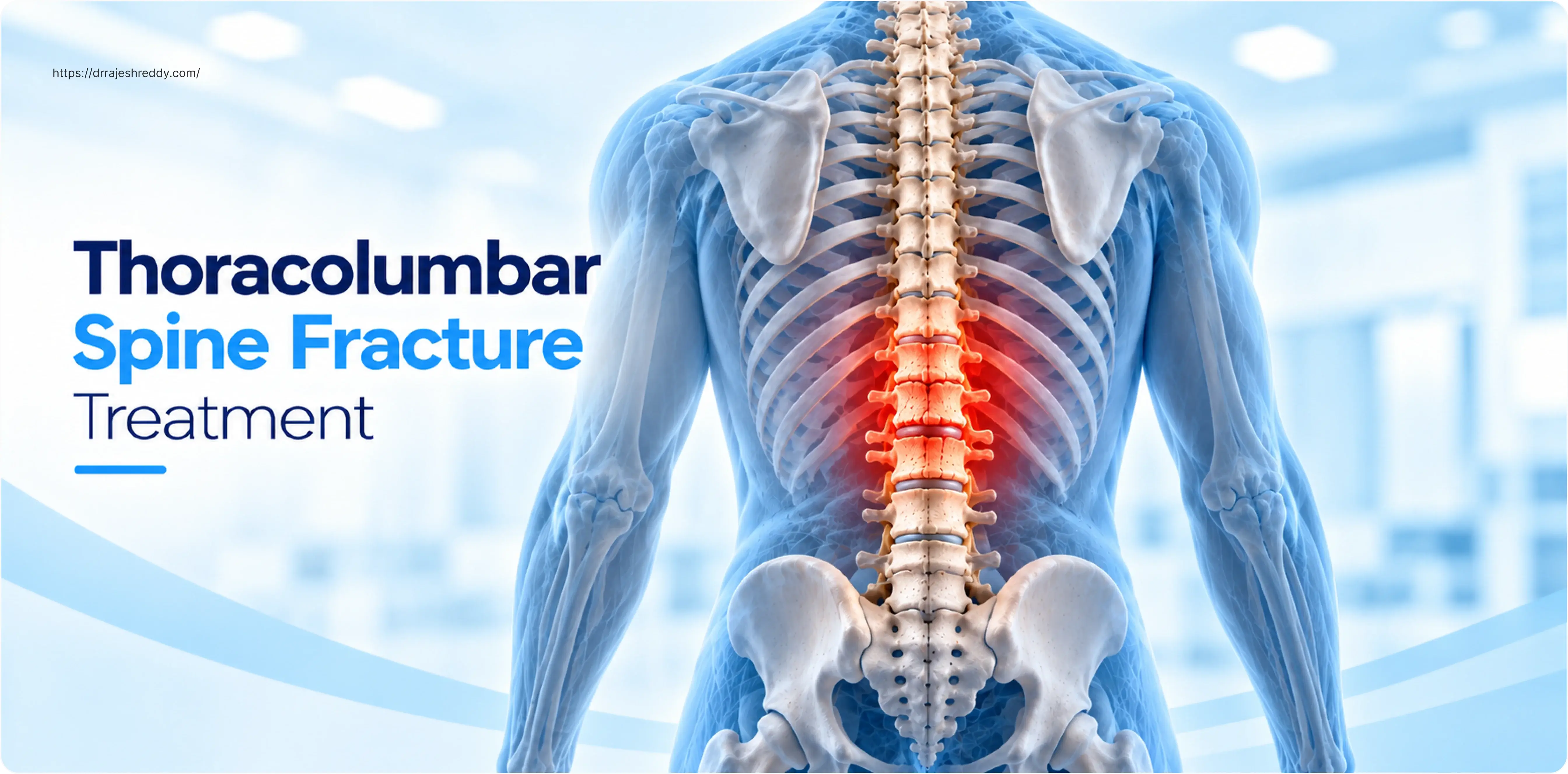
The thoracolumbar junction — the transition zone between T11 and L2 — is the most commonly fractured region of the spine in adults. It is where the rigid, rib-braced thoracic spine meets the more mobile lumbar spine, concentrating mechanical stress during forward flexion, axial loading, and trauma. A fall from height, a road traffic accident, a sports injury, or even a simple fall in an osteoporotic elderly patient can produce fractures at this junction ranging from stable wedge compressions that heal with bracing alone, to unstable burst fractures and fracture-dislocations that demand urgent surgical intervention.
The central question in thoracolumbar fracture management is one that patients and families ask repeatedly: does this fracture need surgery, or can it be treated with a brace? The answer is not always obvious — and it is one that requires systematic assessment of the fracture pattern, the degree of canal compromise, the integrity of the posterior ligamentous complex, and the patient's neurological status. Getting this decision right matters enormously: unnecessary surgery carries its own risks, while under-treating an unstable fracture risks neurological deterioration and progressive deformity.
Dr. Rajesh Reddy Sannareddy, Senior Consultant Neurosurgeon in Hyderabad, uses validated scoring systems — principally the TLICS (Thoracolumbar Injury Classification and Severity) score — alongside his clinical and imaging assessment to guide every thoracolumbar fracture treatment decision. He offers the full range of treatment options: conservative brace management, minimally invasive percutaneous fixation (MISS), and open decompression with instrumented fusion — ensuring the decision is based on what is best for the patient, not on what is most technically convenient.
The Thoracolumbar Junction: Why T11–L2 Is So Vulnerable
The thoracolumbar junction represents a biomechanical transition between two very different spinal segments:
- The thoracic spine (T1–T10) is inherently rigid — the rib cage provides circumferential bracing that limits flexion, extension, and rotation, making this segment resistant to fracture under most everyday loads.
- The lumbar spine (L1–L5) is mobile but supported by large paraspinal muscles and wide vertebral bodies built for load bearing.
- The junction (T11–L2) has neither the rib cage protection of the upper thoracic spine nor the muscular support of the mid-lumbar spine — it is the hinge point where the two systems meet.
During the axial loading and forward flexion of a high-energy impact — a fall from a roof, a motorcycle accident, a collapsing structure — this junction absorbs the concentrated mechanical energy of the entire spinal column above it. The result is a fracture pattern ranging from simple anterior compression to catastrophic multi-column burst fractures with spinal canal compromise.
Types of Thoracolumbar Fractures
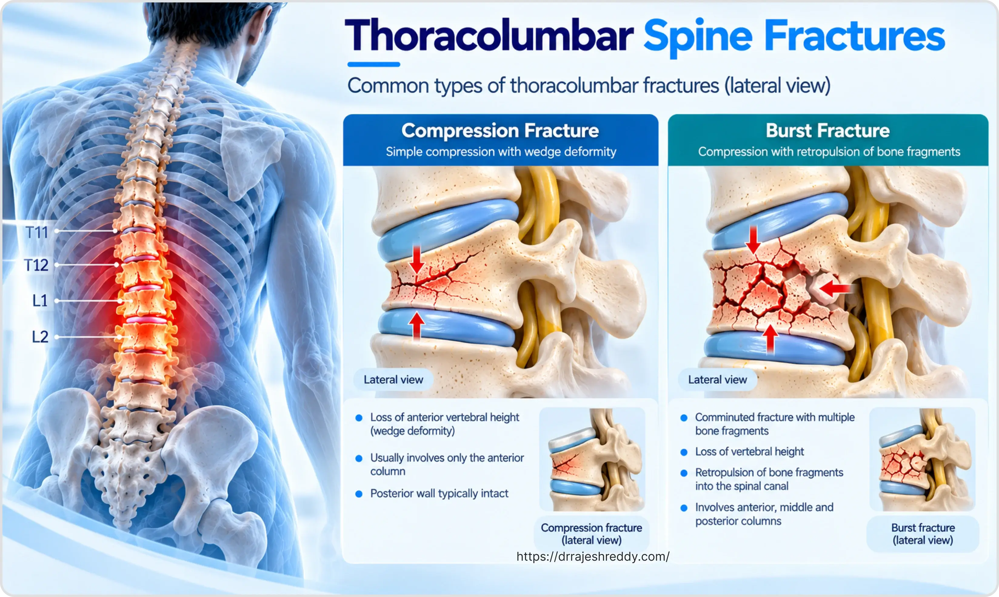
Compression Fracture
The most common thoracolumbar fracture — particularly in osteoporotic patients. The anterior column is compressed, producing a wedge-shaped loss of vertebral height. The middle column (posterior vertebral wall) is intact, making the fracture stable. Neurological deficit is rare unless multiple levels are affected and cumulative kyphosis becomes significant. Most compression fractures are managed conservatively with a TLSO brace and early mobilisation.
Burst Fracture
A burst fracture involves failure of both the anterior and middle columns — the vertebral body comminutes under axial load, and fragments of the posterior vertebral wall retropulse into the spinal canal. Canal compromise ranges from minimal to complete occlusion. The critical clinical questions are: is the posterior ligamentous complex (PLC) intact, is the patient neurologically intact, and what is the degree of canal compromise? These three factors — along with the fracture morphology score — determine the TLICS score and the treatment recommendation.
Burst fractures of L1 are the single most common surgically treated thoracolumbar injury in young adults. They typically result from falls from height (construction workers, roofers) or road traffic accidents, and often occur in isolation or as part of polytrauma.
Chance Fracture (Flexion-Distraction)
The Chance fracture is a horizontal splitting of the vertebra through bone, disc, or a combination — caused by the spine flexing sharply over a fulcrum (classically a lap seat belt in a forward collision). The injury distracts the posterior elements (spinous process, lamina, facets) while compressing the anterior column. In purely bony Chance fractures, healing with a hyperextension brace is often possible. In ligamentous Chance fractures — where the posterior ligamentous complex is torn — surgical posterior fusion is required. Associated abdominal visceral injuries are common with seat-belt Chance fractures and should be actively sought.
Fracture-Dislocation
The most severe thoracolumbar injury — a fracture with translation or rotation of one vertebra over another, disrupting all three columns. Neurological deficit is common and may be complete. Emergency surgical stabilisation is required to restore alignment, decompress the neural elements, and prevent further injury. These injuries are typically associated with high-energy polytrauma.
The TLICS Score: The Evidence-Based Decision Tool
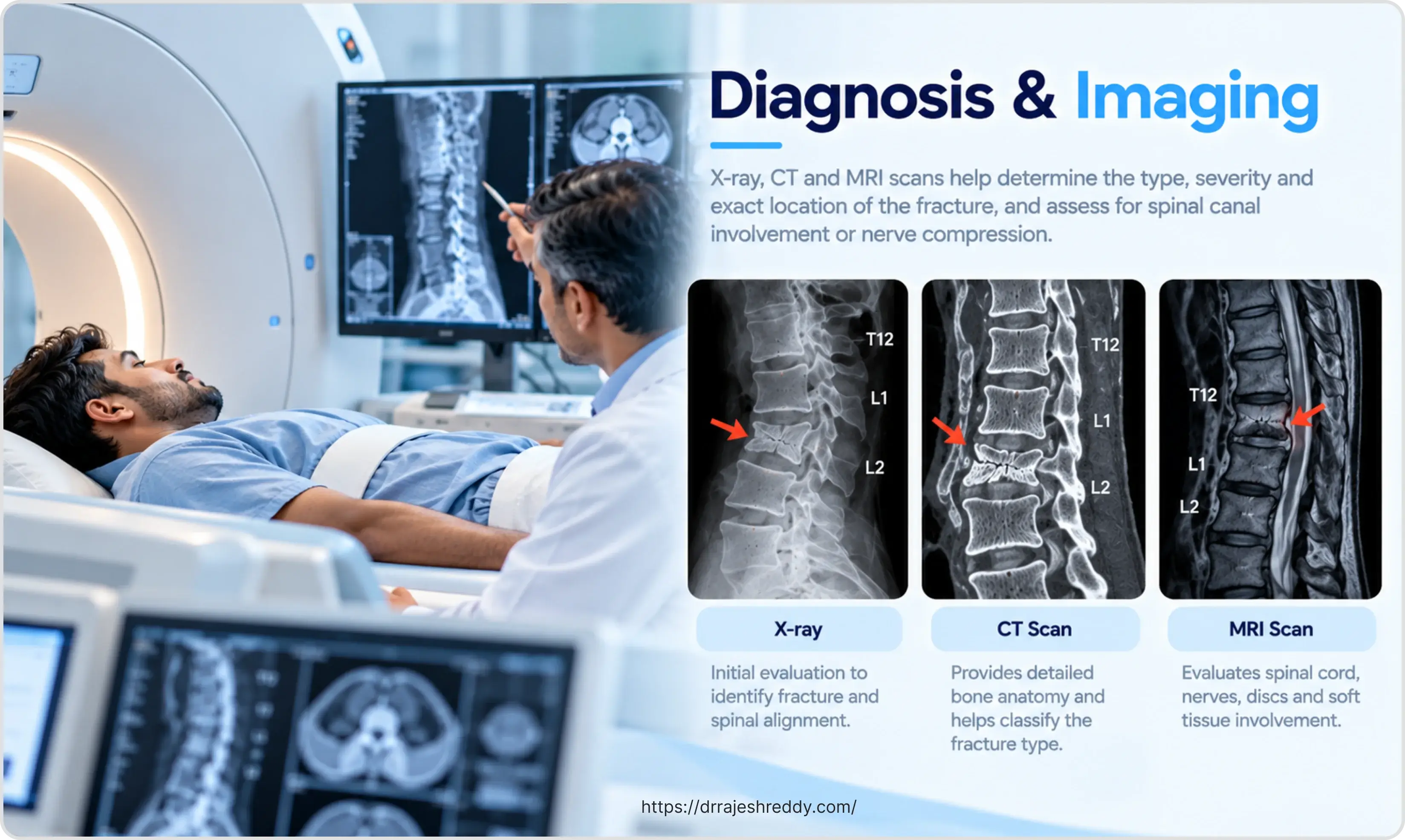
The Thoracolumbar Injury Classification and Severity (TLICS) score provides an objective, reproducible framework for the surgery vs. conservative management decision. It scores three parameters — fracture morphology, posterior ligamentous complex integrity, and neurological status — and adds the scores:
| Parameter | Score | Details |
|---|---|---|
| Fracture Morphology | ||
| — Compression | 1 | Anterior wedging; no middle column involvement |
| — Burst | 2 | Middle column failure; retropulsion possible |
| — Translation / rotation | 3 | Fracture-dislocation; multi-column failure |
| — Distraction | 4 | Chance fracture; seat-belt injury |
| Posterior Ligamentous Complex (PLC) | ||
| — Intact | 0 | No PLC injury on MRI |
| — Suspected / indeterminate | 2 | MRI equivocal; clinical suspicion |
| — Disrupted | 3 | Confirmed PLC tear on MRI (STIR sequence) |
| Neurological Status | ||
| — Intact | 0 | No deficit |
| — Nerve root injury | 2 | Radiculopathy only |
| — Complete cord injury | 2 | ASIA A — complete |
| — Incomplete cord injury | 3 | ASIA B–D — incomplete |
| — Cauda equina syndrome | 3 | Bladder/bowel dysfunction |
• TLICS ≤ 3: Non-operative management is recommended and safe.
• TLICS 4: Clinical judgement zone (conservative or surgical depending on patient factors).
• TLICS ≥ 5: Surgical stabilisation and/or decompression is recommended.
The posterior ligamentous complex (PLC) is the critical determinant of instability in thoracolumbar fractures. It consists of the supraspinous ligament, interspinous ligament, ligamentum flavum, and facet joint capsules. MRI STIR sequences are the most sensitive way to assess PLC integrity — a disrupted PLC on MRI adds 3 points to the TLICS score and usually tips the decision toward surgery even if the neurological status and fracture morphology would otherwise suggest conservative management.
Surgery vs. Conservative Management: The Decision Framework
The table below maps the key clinical and imaging factors against their influence on the treatment decision:
| Factor | Favours Conservative Treatment | Favours Surgical Treatment |
|---|---|---|
| TLICS score | ≤ 3 — non-operative management safe | ≥ 5 — surgery recommended; 4 = grey zone |
| Neurological status | Neurologically intact | Any neurological deficit (cord, root, or cauda equina) |
| Fracture stability | Anterior column only; < 50% height loss | Two or more columns involved; > 50% height loss |
| Canal compromise | < 30–40% canal narrowing | > 50% canal narrowing with neural compression |
| Kyphosis (deformity) | < 20–25° regional kyphosis | > 30° kyphosis; progressive deformity on follow-up imaging |
| Posterior ligamentous complex | Intact on MRI | Disrupted on MRI STIR — key indicator of instability |
| Patient age and health | Elderly, frail, significant comorbidities | Young, fit patient with unstable injury |
| Fracture level | Mid-thoracic (T4–T10) — protected by rib cage | Thoracolumbar junction (T11–L2) — highest stress concentration |
| Patient compliance | Reliable for brace and follow-up | Non-compliant — surgery provides more dependable stability |
No single factor determines the treatment in isolation. A burst fracture with 40% canal compromise in a neurologically intact patient with an intact PLC (TLICS = 2) is a very different clinical situation from the same fracture in a patient with an incomplete cord injury and a disrupted PLC (TLICS = 7). Dr. Rajesh Reddy integrates all of these factors — along with the patient's age, functional demands, occupation, and values — to arrive at an individualised recommendation.
Conservative Treatment: Brace Management in Detail
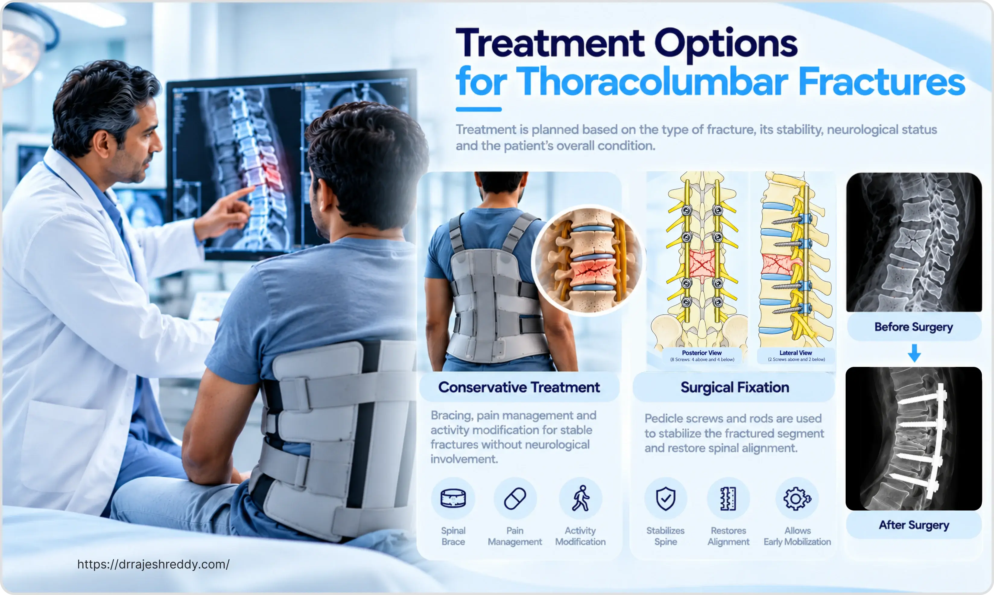
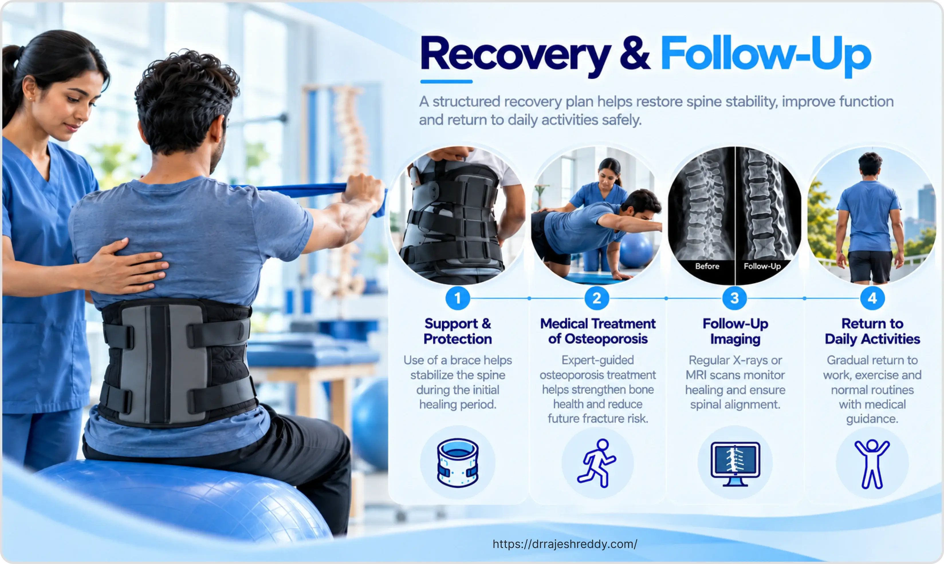
For thoracolumbar fractures that meet conservative management criteria — stable injury, neurologically intact, TLICS ≤ 3 — a thoracolumbar sacral orthosis (TLSO) brace is the primary treatment. Conservative management does not mean no treatment — it requires a structured programme of bracing, monitoring, and rehabilitation.
The TLSO Brace
A custom-fitted TLSO brace extends from the mid-thorax to the pelvis, limiting flexion and rotation at the thoracolumbar junction while allowing the patient to mobilise. It is worn for 8–12 weeks, during which the fracture heals and the anterior column reconstitutes. Key points for patients:
- The brace must be worn whenever upright — walking, sitting, standing — for the full prescribed duration
- It may be removed for lying flat, bathing (with care), and skin inspection
- Skin care under the brace is important — check for pressure areas daily
- Do not attempt to lift anything heavier than 2–3 kg while in the brace
- Sleep on a firm mattress; use the log-roll technique for getting in and out of bed
Monitoring During Conservative Treatment
Conservative management requires close follow-up — it is not a passive process. Serial X-rays at 2 weeks, 6 weeks, and 12 weeks assess:
- Progressive kyphosis — if regional kyphosis increases by > 5° between appointments, the fracture is failing to maintain alignment and surgical intervention should be reconsidered.
- Vertebral height restoration or loss — some remodelling occurs during healing; progressive height loss indicates instability.
- Any new neurological symptoms — any new weakness, numbness, or bladder/bowel change during conservative management is an indication for emergency imaging and surgical reassessment.
Surgical Treatment: Minimally Invasive vs. Open
Percutaneous Minimally Invasive Spine Surgery (MISS)
For neurologically intact patients with unstable thoracolumbar fractures, percutaneous pedicle screw fixation has become the surgical approach of choice at experienced centres. Through four small incisions (each approximately 1.5 cm), pedicle screws are placed above and below the fracture under fluoroscopic or CT navigation guidance, connected by percutaneous rods that span and stabilise the fracture. The paraspinal muscles are not stripped from the spine — they are dilated rather than cut, preserving muscle function and dramatically reducing post-operative pain.
The advantages over open surgery are substantial:
- Blood loss: 100–300 mL versus 500–1500 mL for open fixation
- Operating time: 60–90 minutes versus 2–4 hours
- Hospital stay: 2–4 days versus 5–7 days
- Post-operative pain: significantly less; simple analgesics usually sufficient
- Return to activity: 4–6 weeks versus 8–12 weeks
- Infection risk: lower with smaller incisions
MISS does not decompress the spinal canal directly — it relies on ligamentotaxis (indirect reduction through distraction across the fracture via the posterior tension band) to reduce retropulsed fragments. For neurologically intact patients with < 50% canal compromise, this indirect reduction is typically sufficient. For patients with neurological deficit and direct canal compromise, open decompression is required.
Open Decompression and Instrumented Fusion
Open posterior surgery is indicated when: direct decompression of the spinal canal is required (neurological deficit from retropulsed bone); the fracture involves multi-level instability; or the deformity is severe enough to require direct reduction and reconstruction. Through a midline incision, the paraspinal muscles are retracted to expose the posterior spine — laminectomy removes the posterior arch, decompressing the canal, and pedicle screws above and below provide rigid fixation.
For severe burst fractures where the anterior column has lost > 50% of height and the vertebral body cannot be reconstructed posteriorly alone, an anterior corpectomy and cage reconstruction is added — either as a same-day combined procedure or as a staged anterior approach 2–5 days later.
Implant Removal Considerations
Percutaneous pedicle screws are typically left permanently in neurologically intact patients — there is no clinical benefit to removal and it would require a second procedure. In selected younger patients where screws are causing adjacent segment stress or instrument-related symptoms, removal can be considered after confirmed fusion (typically 12–18 months). Dr. Rajesh Reddy will advise on the implant strategy appropriate to each patient's age, injury, and functional demands.
Recovery: Side-by-Side Timeline
Recovery timelines differ significantly between conservative and surgical management:
| Phase | Conservative (Brace) | Surgical (MISS or Open) |
|---|---|---|
| Hospital | 2–5 days (pain management, brace fitting, physiotherapy) | MISS: 2–4 days; Open: 5–7 days |
| Week 1–2 | Brace worn full-time; walking with physiotherapy; no lifting | Walking day 1–2; brace fitted; wound care; discharge when mobile |
| Week 2–6 | Increasing walking; light household activity; brace at all times | Increasing activity within brace; light daily tasks; desk work from week 4–6 |
| Week 6–12 | X-ray review; brace weaned if healing confirmed; physiotherapy starts | X-ray confirms screw position; physiotherapy intensifies; brace weaned |
| Month 3–6 | Physiotherapy; return to light work; avoid heavy lifting | Return to most activities; physical work 3–6 months |
| Month 6–12 | Full activity for most patients; heavy work 6–12 months | Full activity; heavy work 6–12 months |
Frequently Asked Questions
Not necessarily, but it depends on several factors that go beyond the simple presence of a burst fracture. The TLICS scoring system considers three things: the fracture morphology (burst = 2 points), the integrity of the posterior ligamentous complex on MRI (if disrupted = 3 points), and your neurological status (intact = 0 points). An isolated burst fracture with an intact PLC and no neurological deficit typically scores 2 — well within the conservative management range. If the PLC is disrupted or there is significant canal compromise, the score rises and surgery becomes the recommended option. Dr. Rajesh Reddy will review your CT and MRI and calculate a personalised assessment.
The posterior ligamentous complex (PLC) is a set of ligaments running along the back of the spine — the supraspinous ligament, interspinous ligament, ligamentum flavum, and facet joint capsules. Together they act as a tension band that prevents the spine from collapsing forward. If the PLC is intact after a burst fracture, the spine retains its ability to resist flexion forces and conservative management is feasible. If the PLC is torn — visible on MRI as bright signal on STIR sequences — the spine has lost its posterior tension band and will progressively collapse forward under daily loads, regardless of how good the brace is. This is why PLC assessment on MRI is the most important single imaging finding in the surgery vs. brace decision.
For most thoracolumbar compression or burst fractures managed conservatively, the TLSO brace is worn continuously (whenever upright) for 8–12 weeks. At the 6-week X-ray review, if the fracture is healing and alignment is maintained, the brace duration may be confirmed or the weaning process started. If X-rays show progressive kyphosis or height loss, the brace duration is extended and surgical options are reconsidered. After brace removal, a structured physiotherapy programme begins to restore core strength and spinal stability.
A Chance fracture is caused by forward flexion over a fulcrum — classically a lap seat belt in a frontal collision. Rather than compressing the vertebra under axial load (like a burst fracture), a Chance fracture splits the vertebra horizontally through distraction of the posterior elements. In purely bony Chance fractures (bone splits through the pedicles and body), healing with a hyperextension brace is often excellent. In ligamentous Chance fractures (the posterior ligamentous complex tears rather than bone breaking), surgical posterior fusion is required as soft tissue does not heal reliably with immobilisation. Associated bowel and abdominal injuries are common and must be sought in any patient with a Chance fracture.
Yes — in the majority of cases. Patients with neurologically intact thoracolumbar fractures — whether managed conservatively or surgically — are mobilised (encouraged to walk with supervision) within 1–2 days of injury or surgery. Walking with the brace is an important part of recovery and helps prevent the deconditioning and complications of bed rest. The key restriction is avoiding bending, twisting, and lifting rather than walking itself. Patients with neurological deficits have a more complex mobilisation programme managed in coordination with physiotherapy and rehabilitation specialists.
This depends on the treatment approach and the nature of your work. For conservative management: desk-based work is typically possible from week 6–8 within the brace; manual or physically demanding work requires 3–6 months of recovery after brace removal. After minimally invasive surgical fixation: desk work from week 4–6; physical work 3–6 months. After open surgery: desk work 6–8 weeks; physical work 6–12 months. All return-to-work plans are individualised based on fracture healing on X-ray, neurological recovery, and the specific physical demands of the job.
Consult Dr. Rajesh Reddy for Thoracolumbar Fracture Assessment in Hyderabad
Whether you have recently sustained a thoracolumbar fracture and need an expert assessment of whether surgery is required — or you have been recommended surgery and want a second opinion — Dr. Rajesh Reddy Sannareddy provides comprehensive spine fracture consultations in Hyderabad with access to the full range of treatment options. Book via the Dr. Reddy's Neuro Care app on Google Play or call 079951 11695.