
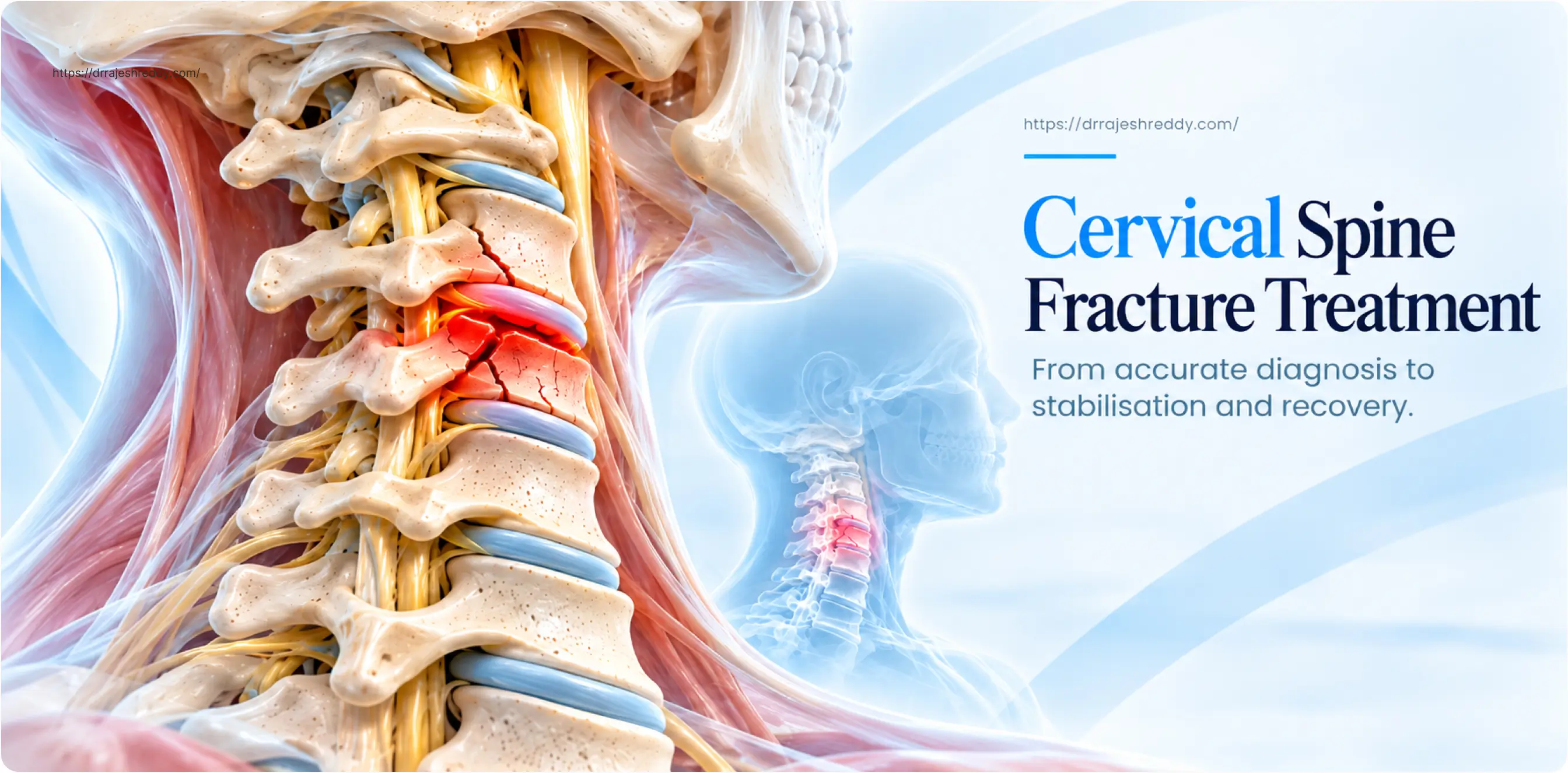
The cervical spine — the seven vertebrae between the base of the skull and the upper chest — is the most mobile, and most vulnerable, segment of the spinal column. It houses the cervical spinal cord within a relatively narrow canal, and its injuries carry the highest neurological stakes of any spinal fracture. A displaced cervical fracture at or above C4 can sever the connection between the brain and the phrenic nerve — causing immediate respiratory arrest. A fracture-dislocation at C5–C6 can leave a previously healthy person unable to move their arms or legs.
Yet the cervical spine's range of injuries is wide. At one end, a clay-shoveller's fracture of a spinous process requires nothing more than a soft collar and pain relief. At the other, a teardrop fracture-dislocation in a road traffic accident requires emergency surgical decompression and stabilisation within hours. Between these extremes lie odontoid fractures, Jefferson fractures, hangman's fractures, burst fractures, and subaxial fracture-dislocations — each with its own biomechanical characteristics, neurological risk, and treatment requirements.
Dr. Rajesh Reddy Sannareddy, Senior Consultant Neurosurgeon in Hyderabad, manages the full spectrum of cervical spine injuries — from emergency decompression and stabilisation in the acute setting to elective treatment of craniocervical instability and atlantoaxial subluxation. His training at Barrow Neurological Institute, Phoenix, Okayama University, Japan, and Klinikum Stuttgart, Germany has given him direct exposure to the highest-volume cervical spine trauma programmes internationally.
The Cervical Spine: A Brief Anatomy
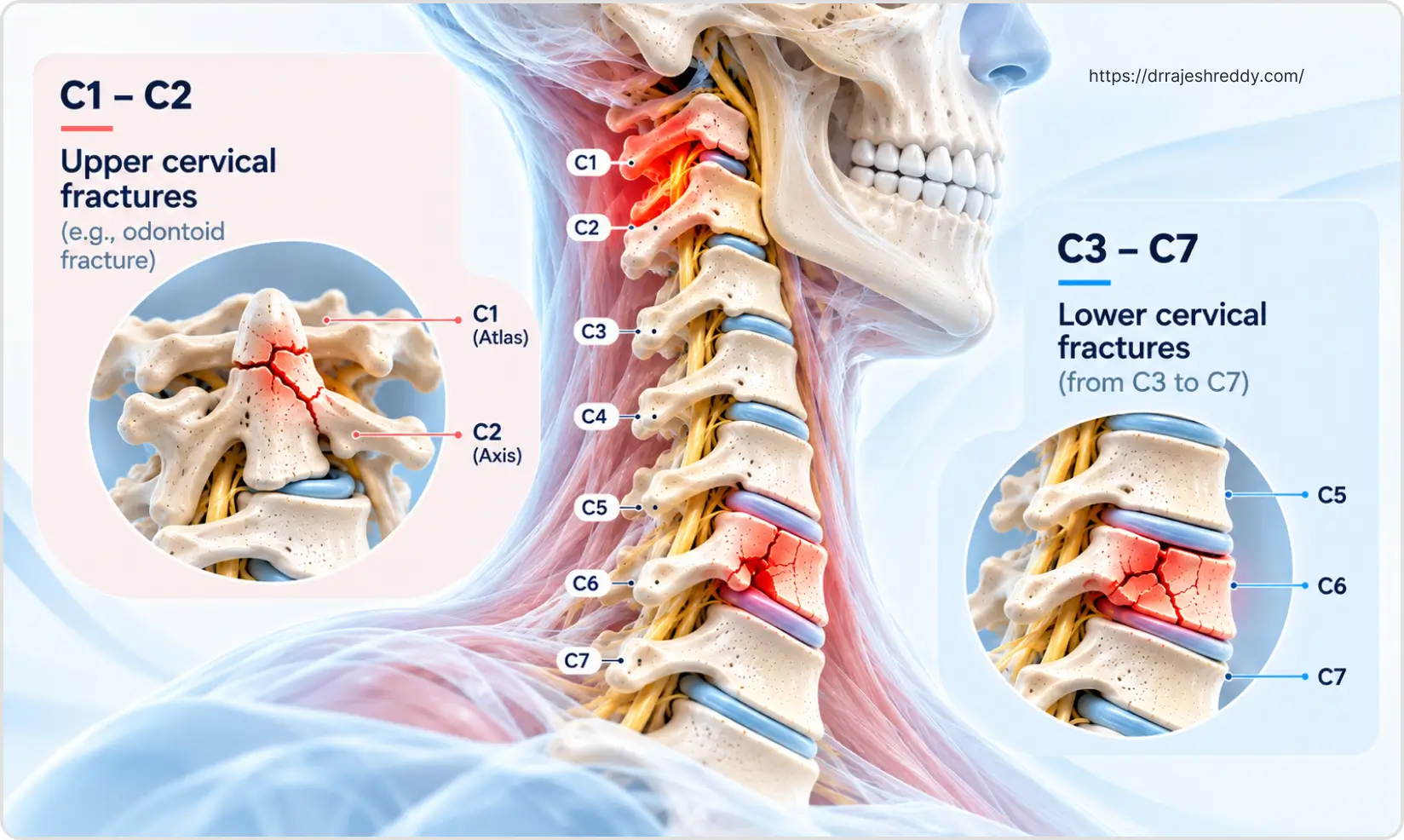
Understanding cervical fracture management requires a basic grasp of cervical anatomy. The cervical spine has two anatomically and functionally distinct regions:
Upper Cervical Spine (Occiput–C1–C2)
The craniocervical junction and the first two cervical vertebrae — the atlas (C1) and axis (C2) — form a unique functional unit that provides the majority of cervical rotation (C1–C2, approximately 50°) and flexion-extension (occiput–C1, approximately 25°). The atlas is a ring without a vertebral body; the axis has the odontoid process (dens) projecting upward into the ring of the atlas, held in place by the transverse ligament. Instability at this junction — from fracture, ligamentous injury, or congenital anomaly — risks compression of the upper cervical cord and brainstem.
Subaxial Cervical Spine (C3–C7)
C3 through C7 have conventional vertebral bodies, discs, facet joints, and posterior elements. The subaxial cervical canal is narrower than the upper cervical region, and the cord is more vulnerable to compression from displaced bone or disc material. The brachial plexus nerve roots — which supply the arms — exit at C5–T1, so subaxial injuries commonly cause arm weakness and radiculopathy in addition to or instead of cord injury.
Types of Cervical Spine Fractures
The table below summarises the most important cervical fracture and dislocation types, their levels, neurological risk, and typical management:
| Fracture / Injury | Level | Neurological Risk | Typical Management |
|---|---|---|---|
| Jefferson fracture (C1 burst) | C1 (atlas) | Variable; unstable if transverse ligament torn | Halo vest 8–12 weeks; surgery if ligament disrupted (posterior C1–C2 fusion) |
| Odontoid fracture Type I | Tip of dens (C2) | Low; stable | Cervical collar 6–8 weeks |
| Odontoid fracture Type II | Base of dens — most common | Moderate; atlantoaxial instability risk | Surgery preferred (odontoid screw or posterior C1–C2 fusion); collar for elderly poor surgical candidates |
| Odontoid fracture Type III | Base through C2 body | Low-moderate; often heals with immobilisation | Halo vest or collar; surgery if displaced |
| Hangman's fracture (C2 pedicles) | C2 bilateral pedicle fractures | Low; often neurologically intact (canal widens at C2) | Collar / halo; surgery if severe angulation or disc disruption |
| Subaxial compression fracture (C3–C7) | Any level C3–C7 | Low if stable; higher if middle column involved | Rigid collar; surgery if unstable or canal compromise |
| Subaxial burst fracture | C3–C7 | Moderate-high; cord compression from retropulsed fragments | ACDF (anterior decompression); posterior fusion if posterior instability |
| Flexion-teardrop fracture | C5–C6 most common | High — anterior cord syndrome; often profound deficit | Emergency surgical decompression and stabilisation |
| Fracture-dislocation | Any subaxial level | Very high — often complete cord injury | Emergency reduction and surgical stabilisation |
| Clay-shoveller's fracture | C6–T1 spinous process | None — stable | Conservative; collar; pain management |
Upper Cervical Injuries: C1 and C2
Jefferson Fracture (C1 Burst Fracture)
A Jefferson fracture results from an axial load on the top of the head — classically from diving into shallow water or a fall onto the crown. The ring of C1 bursts outward at its weakest points, typically in 2–4 places. On an open-mouth odontoid X-ray, the lateral masses of C1 are seen overhanging the superior articular surface of C2 (the 'rule of Spence'). The critical determinant of stability and neurological risk is the integrity of the transverse atlantal ligament — which holds the dens against the anterior arch of C1.
- Transverse ligament intact: The fracture is relatively stable. A halo vest for 8–12 weeks allows healing in the majority of cases with good outcome. Neurological deficit is uncommon.
- Transverse ligament torn (atlantoaxial instability): The fracture is highly unstable. The dens can migrate posteriorly and compress the upper cervical cord or medulla. Posterior C1–C2 fusion is required to stabilise the craniocervical junction.
Odontoid Fractures (C2 Dens Fractures)
Odontoid fractures are among the most common cervical fractures in the elderly — often from a low-energy fall in a patient with osteoporosis. They are classified by Anderson and D'Alonzo into three types:
- Type I: Fracture of the tip of the dens — stable; treated with a rigid collar.
- Type II: Fracture at the base of the dens — the most common and most challenging type. High non-union rate with conservative treatment (up to 60–70% in displaced fractures). Surgical treatment is generally preferred: odontoid screw fixation (anterior approach, preserves rotation) or posterior C1–C2 fusion (more reliable, but sacrifices rotation).
- Type III: Fracture extends through the body of C2 — generally good healing potential with a halo vest or collar. Surgery for significant displacement.
Type II odontoid fractures in elderly patients present a particular dilemma: the non-union rate is high with conservative treatment, but surgical risk is elevated due to age and comorbidities. Dr. Rajesh Reddy individualises this decision — weighing bone quality, fracture displacement, patient functional status, and the relative risks of halo immobilisation (aspiration pneumonia, falls) versus posterior fusion under general anaesthesia.
Hangman's Fracture (Traumatic Spondylolisthesis of C2)
The hangman's fracture — bilateral fractures through the pedicles (pars interarticularis) of C2 — is caused by hyperextension and axial load, classically in motor vehicle accidents and diving injuries. Despite its dramatic name, patients with hangman's fractures are often neurologically intact: the bilateral pedicle fractures actually widen the spinal canal at C2, paradoxically decompressing the cord at the moment of injury.
Management depends on the degree of angulation and translation at C2–C3:
- Minimal displacement: rigid cervical collar for 12 weeks — excellent healing in most cases
- Moderate displacement with disc disruption at C2–C3: halo vest or anterior C2–C3 ACDF
- Severe displacement or instability: posterior C2–C3 or C1–C3 fusion
Subaxial Cervical Fractures (C3–C7)
Compression Fractures
Simple anterior compression fractures of the subaxial cervical spine — where only the anterior column is involved — are stable injuries that can typically be managed with a rigid collar for 6–8 weeks. The fracture involves wedging of the anterior vertebral body with the posterior wall intact. Neurological deficit is uncommon. Serial X-rays at 6 and 12 weeks confirm healing.
Burst Fractures
A cervical burst fracture involves both the anterior and middle columns — the posterior vertebral wall comminutes and fragments may retropulse into the spinal canal, compressing the cord. The injury typically results from an axial load combined with flexion. Neurological deficit ranges from a central cord syndrome (disproportionate arm weakness) to complete cord injury, depending on the degree of canal compromise.
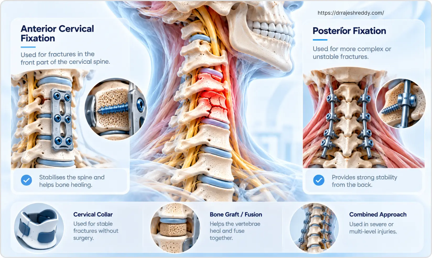
Surgical treatment is mandatory for burst fractures with neurological deficit or significant canal compromise (> 50%). Anterior cervical corpectomy and fusion (ACCF) is the standard approach — the fractured vertebral body is removed, the canal directly decompressed, and a titanium mesh cage with anterior plate and screws restores height and alignment.
Flexion-Teardrop Fracture
The flexion-teardrop fracture is one of the most serious cervical injuries — a small triangular fragment avulsed from the anteroinferior corner of a vertebral body associated with severe posterior ligamentous disruption and cord injury. It typically produces an anterior cord syndrome: complete motor paralysis and loss of pain/temperature sensation below the level, with preserved proprioception and vibration. Emergency surgical decompression and stabilisation is required.
Facet Fractures and Dislocations
Unilateral facet dislocation (jumped facet) typically causes a nerve root injury at the level of dislocation — arm pain, weakness, and numbness — but incomplete cord injury. Bilateral facet dislocation causes complete cervical cord injury in the majority of cases. Both require urgent closed reduction (where safe) followed by surgical stabilisation.
MRI before any reduction attempt is essential to rule out an associated disc herniation — a herniated disc anterior to the cord can be pushed further into the cord during reduction, worsening the injury. If a herniated disc is present, anterior decompression before reduction is performed.
The sequence matters: MRI before reduction, then anterior discectomy if disc herniation is present, then reduction and posterior stabilisation. Attempting closed reduction without first excluding a herniated disc in the presence of cord injury is a preventable cause of neurological deterioration.
Surgical Approaches for Cervical Fractures
The choice of surgical approach depends on where the compressive pathology lies, the injury pattern, and the level of instability:
| Approach | Best Suited For | Key Points |
|---|---|---|
| Anterior Cervical Discectomy and Fusion (ACDF) | Single or two-level disc-bone compression; teardrop fractures; burst fractures with anterior cord compression | Removes compressive pathology directly; cage + plate restores height; 95%+ fusion rate; 4–6 week collar post-op |
| Anterior Cervical Corpectomy and Fusion (ACCF) | Multi-level burst fractures; retrovertebral bone compression spanning > 1 disc level | Removes entire fractured vertebral body; titanium mesh cage + plate; longer construct; may need posterior supplementation |
| Posterior Cervical Fusion with lateral mass / pedicle screws | Fracture-dislocations; posterior ligamentous instability; multi-level posterior element fractures | Reduces dislocation; rigid posterior fixation; laminectomy for cord decompression if needed; faster and simpler than anterior for dislocations |
| Combined anterior-posterior | Complex fractures with anterior compression + posterior instability; failed single-approach fixation | Staged or same-sitting combined; most stable construct; used for severe multi-level injuries |
| Odontoid screw fixation | Type II odontoid fracture with intact transverse ligament; recent fracture with good bone quality | Single anterior screw through C2 dens; preserves C1–C2 rotation; technically demanding |
| Posterior C1–C2 fusion | Type II odontoid in elderly (poor bone for screw); chronic non-union; transverse ligament tear | Eliminates atlantoaxial instability permanently; sacrifices ~50% of cervical rotation; gold standard for atlantoaxial instability |
| Occipitocervical fusion | Craniocervical instability (C0–C2); atlanto-occipital dislocation; occipital condyle fractures with instability; failed C1–C2 fusion; destructive lesions involving C1–C2 | Extends fixation from occiput to C2 or below; uses occipital plate + cervical screws; sacrifices all upper cervical motion; indicated when instability involves the occiput |
In complex injuries requiring both anterior decompression and posterior stabilisation, Dr. Rajesh Reddy performs either staged or same-sitting combined anterior-posterior procedures, depending on the patient's haemodynamic stability and the urgency of decompression.
Vertebral Artery Injury in Cervical Fractures
Vertebral artery injury (VAI) occurs in approximately 20–30% of cervical fractures involving the transverse foramen — the bony canal through which the vertebral artery passes at each cervical level. VAI is most commonly associated with fractures involving C3–C6 transverse foramen, unilateral facet dislocations, and severe fracture-dislocations.
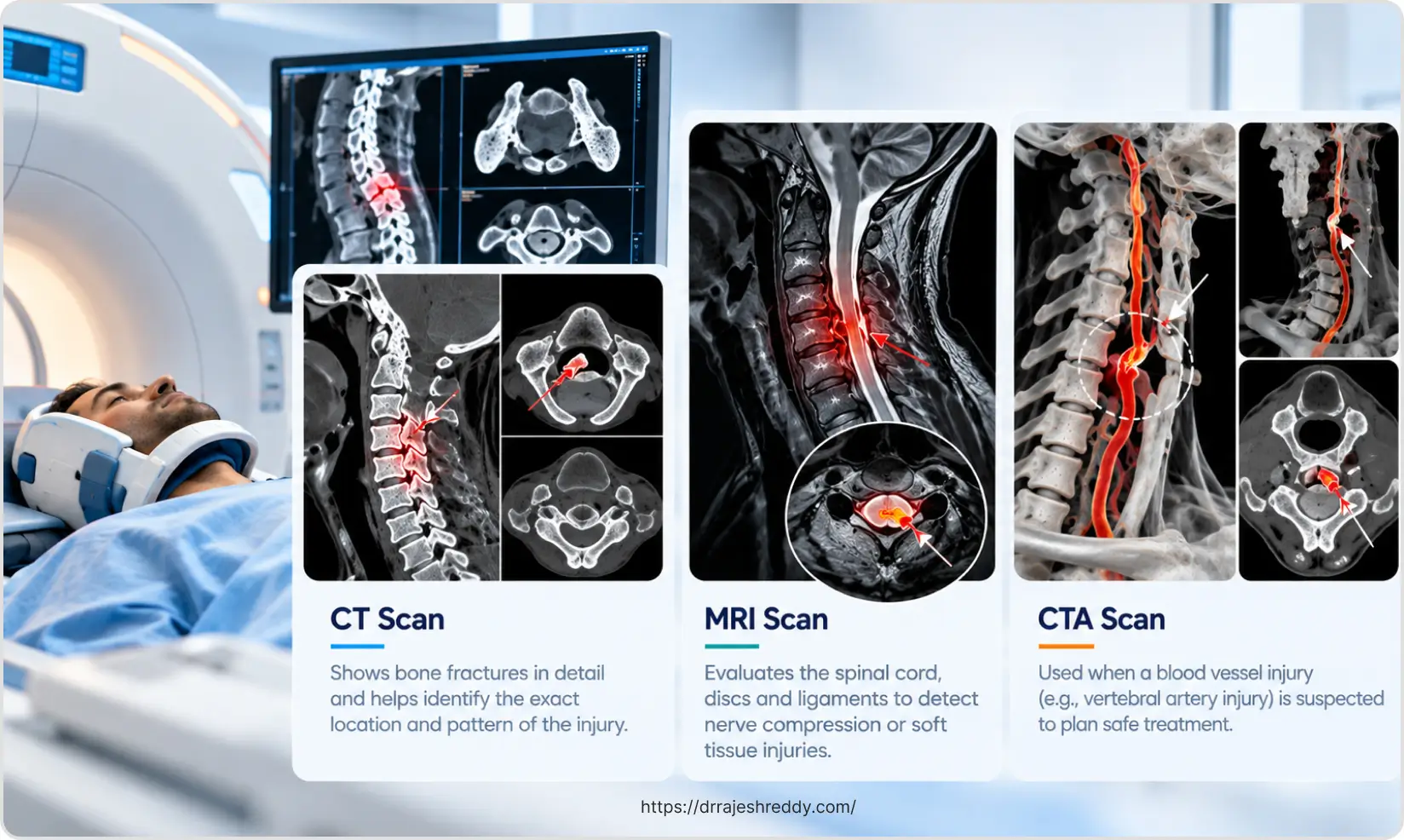
Most VAIs are clinically silent — the contralateral vertebral artery provides sufficient collateral flow. However, a minority cause posterior circulation stroke (vertebrobasilar ischaemia) — presenting as dizziness, diplopia, nystagmus, ataxia, or Wallenberg syndrome in the days after injury. All cervical fractures involving the transverse foramen should have CT angiography of the neck vessels to identify VAI. Anticoagulation or antiplatelet therapy may be recommended for high-risk VAIs.
Recovery After Cervical Fracture Treatment
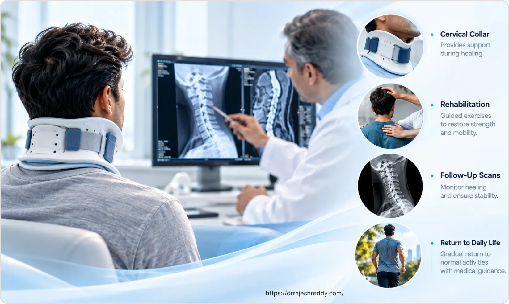
After Conservative Management (Collar / Halo)
- Cervical collar: worn continuously for 6–12 weeks; collar hygiene and skin care are important
- Halo vest: 8–12 weeks; significantly limits daily function but provides superior immobilisation
- Physiotherapy: gentle isometric neck strengthening commences after collar/halo removal
- Serial imaging: X-rays at 6 and 12 weeks confirm healing; CT or MRI if any new symptoms
- Return to work: desk work 4–8 weeks after collar removal; driving after neurosurgical clearance
After ACDF / ACCF
- Hospital stay: 2–4 days
- Soft cervical collar for 4–6 weeks post-operatively
- Swallowing: mild dysphagia is common for 1–2 weeks after anterior cervical surgery; soft diet initially
- Hoarseness: mild voice change from retraction of the recurrent laryngeal nerve; typically resolves within 2–4 weeks
- Return to work: desk work 4–6 weeks; driving after 6-week review; physical work 3–6 months
- Fusion confirmed on X-ray at 3 and 6 months
After Posterior Cervical Fusion
- Hospital stay: 3–5 days
- Rigid cervical collar for 6–12 weeks depending on construct length
- Physiotherapy: gentle mobilisation from week 2 within the brace; formal physiotherapy from 6–8 weeks
- Return to work: desk work 6–8 weeks; driving after neurosurgical clearance; physical work 4–6 months
Frequently Asked Questions
Neck pain after a significant trauma — road traffic accident, fall from height, diving, or sports injury — must be treated as a potential cervical fracture until imaging proves otherwise. Attend the nearest emergency department immediately. Do not attempt to assess neck movement or remove any collar that has been placed. A CT scan of the cervical spine (and MRI if there is any neurological symptom) will definitively identify or exclude a fracture. Most people with post-traumatic neck pain do not have a fracture — but the consequences of a missed unstable cervical fracture are severe enough that imaging is mandatory after any significant mechanism.
The odontoid process (dens) is a peg-like projection of the second cervical vertebra (C2) that fits into the ring of C1 — held in place by the transverse ligament. An odontoid fracture disrupts this joint. Type I (tip fracture) and Type III (through C2 body) fractures generally heal with collar or halo immobilisation. Type II fractures (base of dens) are the most problematic — they have a high non-union rate with conservative treatment (up to 60–70% in displaced cases in younger patients) and typically require surgical fixation. In elderly patients with poor bone quality, the decision between surgery and prolonged immobilisation is carefully individualised.
Many cervical fractures — particularly stable compression fractures, clay-shoveller's fractures, Type I and III odontoid fractures, and minimally displaced hangman's fractures — heal reliably with collar or halo vest immobilisation over 8–12 weeks. Surgery is required when: the fracture is unstable; there is neurological deficit from cord or root compression; conservative treatment has failed or the fracture is not uniting; or the degree of deformity is unacceptable. Dr. Rajesh Reddy will advise on the least invasive approach that achieves stability and neurological protection for your specific injury.
ACDF (Anterior Cervical Discectomy and Fusion) is a surgical procedure performed through a small incision on the side of the neck — the damaged disc and any bony fragments are removed, a cage or bone graft is placed in the disc space, and a plate and screws are used to hold the construct while fusion occurs. After a one or two-level fusion, most patients retain very good neck range of motion — the remaining mobile levels compensate well. After three or more levels fused, some reduction in range of motion is expected. Physiotherapy after collar removal helps restore the motion available.
After ACDF or anterior cervical corpectomy, a soft cervical collar is typically worn for 4–6 weeks. After posterior cervical fusion, a rigid collar is usually required for 6–12 weeks depending on the number of levels fused and the underlying injury. The collar is worn to protect the fusion while bone healing occurs — removing it early risks non-union. Dr. Rajesh Reddy will confirm the collar-wearing schedule at each post-operative review and adjust based on X-ray evidence of fusion progress.
Atlantoaxial instability refers to abnormal movement between C1 (atlas) and C2 (axis) — either from a fracture of the odontoid, rupture of the transverse ligament, or a combination. The dens can move posteriorly toward the spinal cord with neck movement, risking cord compression and potentially causing sudden tetraplegia or even death with a fall or minor trauma. The standard treatment for symptomatic atlantoaxial instability is posterior C1–C2 fusion — a procedure that permanently stabilises the C1–C2 joint. The trade-off is loss of approximately 50% of cervical rotation (which occurs mainly at C1–C2), but most patients adapt well and function normally for daily activities.
Emergency or Elective: Consult Dr. Rajesh Reddy for Cervical Spine Fracture
Whether you have sustained a recent cervical fracture and need emergency assessment, or have been told you have an unstable cervical injury and are seeking expert neurosurgical opinion — Dr. Rajesh Reddy Sannareddy provides comprehensive cervical spine consultations.
For emergencies, call 079951 11695. For elective consultations, book via the Dr. Reddy's Neuro Care app on Google Play.
Related Articles
Continue reading to explore spinal fracture types, surgical techniques, and recovery: