
A spine fracture diagnosis — whether from a road accident, a fall, a sports injury, or an underlying condition like osteoporosis — is frightening. Patients and their families often have more questions about recovery than about the fracture itself: How long will I be in hospital? When can I walk? Will I need physiotherapy? Will I ever get back to my normal life? When should I worry about something being wrong?
The answers depend on which vertebra was fractured, how severely, whether there was neurological involvement, and what treatment was chosen — conservative management in a brace, minimally invasive surgery, or open spinal fusion. But there are consistent principles that govern recovery from all spine fractures, and understanding what to expect at each stage makes the process less frightening and helps patients participate actively in their own rehabilitation.
Dr. Rajesh Reddy Sannareddy, Senior Consultant Neurosurgeon in Hyderabad, manages the full recovery journey for his spine fracture patients — from the immediate post-operative period through physiotherapy and long-term follow-up. This guide reflects his clinical approach and the evidence base for recovery after each major fracture type.
Factors That Determine Your Recovery Timeline
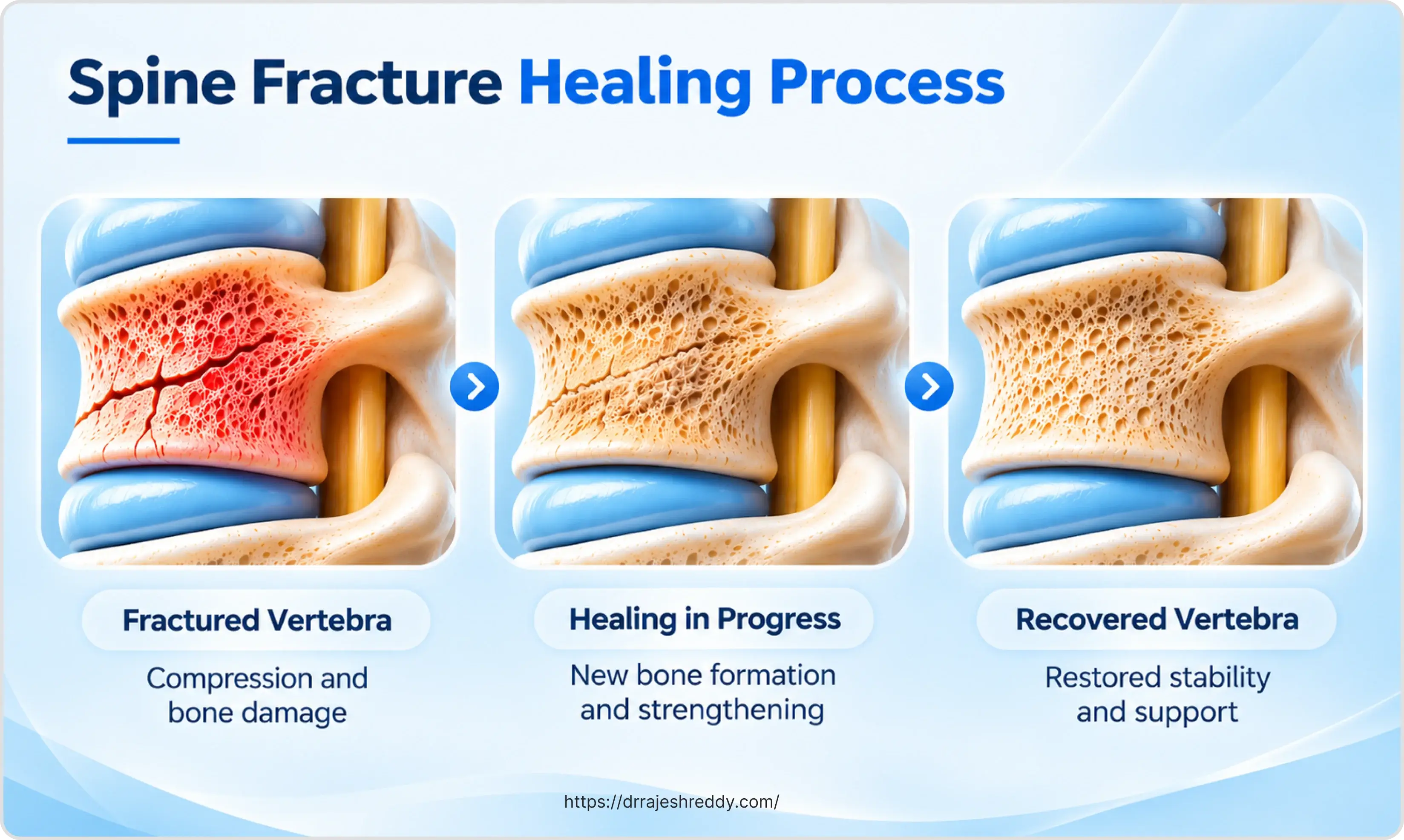
No two spine fracture recoveries are identical. The single most important factor is whether there was any neurological injury — damage to the spinal cord or nerve roots — because neurological recovery follows different principles from bone healing. Other key factors include:
- Type and level of fracture: Cervical fractures have different recovery trajectories from thoracolumbar fractures; stable fractures heal faster than unstable ones.
- Treatment chosen: Conservative management (brace alone) vs. surgery; and if surgery, minimally invasive vs. open; degree of instrumentation (single level vs. multi-level fusion).
- Patient age and bone quality: Younger patients with good bone density heal faster; osteoporotic bone heals more slowly and is at higher risk of adjacent fractures.
- Pre-existing health: Patients with diabetes, cardiovascular disease, or on immunosuppressants have slower healing and higher infection risk.
- Underlying cause: Traumatic fractures heal differently from pathological fractures (osteoporosis, cancer); pathological fractures require concurrent treatment of the underlying disease.
- Compliance with rehabilitation: Patients who engage consistently with physiotherapy and follow activity restrictions recover faster and with fewer complications.
The goal of Dr. Rajesh Reddy's post-operative care is not just technical surgical success — it is restoring the patient's function and quality of life. Recovery is a collaborative process between the surgeon, physiotherapist, patient, and family.
Recovery Timeline: Phase by Phase
A structured timeline helps patients track their healing milestones and sets realistic expectations for daily activities:
| Phase | Timeframe | Goals | What Dr. Rajesh Reddy Monitors |
|---|---|---|---|
| Immediate post-operative | 0–48 hours | Pain control; neurological stability; wound integrity; early mobilisation with physiotherapy | Neurological examination hourly then 4-hourly; drain output; wound; respiratory function in cervical cases |
| Early recovery | Days 2–6 (in hospital) | Sit to stand with assistance; short walking distances; bowel and bladder function established; DVT prevention | Daily wound review; progressive mobilisation milestones; VTE prophylaxis; C-reactive protein trend |
| Subacute recovery | Weeks 1–6 (home/rehab) | Independent ambulation; stair climbing; return to light activities of daily living; pain reducing to manageable levels | Outpatient review at 2 and 6 weeks; wound healing; implant position on X-ray; pain trajectory; physiotherapy progress |
| Consolidation | Weeks 6–12 | Return to desk work and light occupation; driving (if cervical or lumbar surgery); core strengthening programme | X-ray or CT at 6–8 weeks to confirm fusion or fracture healing; neurological function; pain medication reduction |
| Long-term rehabilitation | 3–12 months | Return to full occupational and recreational activities; optimisation of bone health and fitness | 6-month CT or MRI as indicated; bone density review; optimisation of anti-osteoporosis treatment if applicable |
| Ongoing monitoring | Beyond 12 months | Maintenance of function; prevention of adjacent-segment problems; disease management (cancer or osteoporosis) | Annual review; imaging only if new symptoms; continued medical management of underlying condition |
Recovery by Fracture Type and Treatment
The table below gives realistic expectations for each major fracture and treatment category. These are averages — individual recoveries vary, and Dr. Rajesh Reddy will give specific guidance based on your case at each follow-up.
| Fracture Type | Typical Hospital Stay | Expected Return to Mobility | Return to Work / Driving |
|---|---|---|---|
| Stable cervical fracture (halo or collar) | 2–5 days (or outpatient if managed conservatively) | Immediate with collar; full mobility when collar removed (6–12 weeks) | Office work 4–8 weeks; driving when cleared by surgeon (typically 6–12 weeks) |
| Cervical fusion (anterior/posterior) | 3–5 days | Walk day 1–2; no heavy lifting for 6 weeks | Office work 4–6 weeks; manual work 3–6 months; driving 6–12 weeks |
| Thoracolumbar fracture (conservative brace) | 1–3 days or outpatient | Walk with brace immediately; brace for 6–12 weeks | Office work 4–8 weeks; manual work 3–4 months |
| Percutaneous pedicle screw fixation (MIS) | 2–4 days | Walk day 1–2; stairs day 3–4; early physiotherapy | Office work 2–4 weeks; manual work 6–12 weeks |
| Open posterior fusion (multi-level) | 4–7 days | Walk with assistance day 1–2; stairs by day 4–5 | Office work 6–8 weeks; manual work 3–6 months |
| Kyphoplasty / vertebroplasty (osteoporosis) | Day case or 1 night | Walk day of procedure; stairs day 1–2 | Light activities within days; office work within 1 week; heavy lifting restricted 6 weeks |
| Separation surgery (spinal metastasis) | 5–10 days | Walk with assistance early; discharge goal is independent ambulation | Depends on oncological treatment schedule; desk work when medically stable |
| Spinal cord injury (incomplete) | 2–6 weeks acute; rehabilitation months | Depends on injury level and completeness; goal is maximum functional independence | Highly variable; supported by multidisciplinary rehabilitation team |
What Happens in Hospital After Spine Surgery?
Day of Surgery
Most patients wake from spine surgery in the recovery room and are transferred to the ward or, for more complex procedures, the intensive care unit for overnight monitoring. Pain is managed with a combination of intravenous medication, nerve blocks, and oral analgesics from the outset — adequate pain control in the first 24 hours is essential for early mobilisation. A urinary catheter is usually in place and removed within 24–48 hours.
Day 1–2: Early Mobilisation
Early mobilisation — getting out of bed — is one of the most important steps in spine surgery recovery and is typically initiated on the first post-operative day by the physiotherapy team. This is not rushing recovery; it is best practice. Bed rest beyond 24–48 hours increases the risk of deep vein thrombosis, chest infection, pressure sores, and muscle deconditioning without any benefit to healing. Patients are surprised to find they can stand and walk short distances the day after surgery, even after a multi-level fusion.
Blood clot prevention (DVT prophylaxis) is a priority after spine surgery. Dr. Rajesh Reddy's protocol includes compression stockings from the day of surgery, a blood-thinning injection (low molecular weight heparin) starting 24–48 hours post-operatively, and early walking. Patients with higher clot risk may continue injections at home for 2–4 weeks.
Before Discharge: What You Need to Be Able to Do
Before going home, patients are expected to reach a set of milestones that the physiotherapy team assesses daily:
- Walk independently or with a walking aid for a short distance
- Climb at least 2–3 stairs safely (if there are stairs at home)
- Manage basic self-care — washing, dressing, toilet — without excessive pain
- Understand their activity restrictions and brace-wearing instructions if applicable
- Understand their medication schedule and when to take pain relief
- Know what warning signs to watch for
Physiotherapy and Rehabilitation After Spine Fracture
Physiotherapy is not optional after spine fracture — it is a core part of treatment. The physiotherapist's role evolves across the recovery phases:
Acute Phase (In Hospital and First 2 Weeks at Home)
- Safe mobilisation: Teaching correct techniques for moving, turning in bed (log-rolling), sitting up, and standing, which protect the operated or healing spine.
- Breathing exercises: Essential after chest or upper back surgery to prevent atelectasis and chest infections.
- Graduated walking programme: Distance and duration increased daily based on endurance.
- Pain management: Ice, heat, positioning, TENS where appropriate.
Subacute Phase (Weeks 2–6)
- Gentle core activation exercises: Restoring control of the deep stabilising muscles (transversus abdominis, multifidus) without loading the fracture site.
- Postural correction: Reversing the protective postures patients adopt after injury (rounded shoulders, guarded gait) which become problematic if maintained.
- Hydrotherapy (water therapy): May be introduced for suitable patients; buoyancy reduces spinal load and allows more movement.
- Scar management: Gentle massage of the surgical scar once healed to prevent deep tissue adhesions.
Consolidation Phase (Weeks 6–12)
- Progressive strengthening: Gluteal, hip, and core strengthening; gradually increasing resistance loads.
- Balance and proprioception training: Particularly important for patients with any residual neurological involvement.
- Functional activities: Returning to specific tasks (desk work, driving, household tasks) under expert guidance.
Long-Term Rehabilitation (3–12 Months)
- Return to sport or physically demanding work: Structured graded return with functional capacity evaluations.
- Management of persistent symptoms: Long-term core endurance and ergonomic optimization.
- Spinal cord injury rehabilitation (if applicable): Intensive, multidisciplinary programme coordinated at a specialised spinal rehabilitation centre.
Note on Kyphoplasty: Patients often ask whether physiotherapy is needed for kyphoplasty (cement injection for osteoporotic fractures). The answer is yes — physiotherapy after kyphoplasty focuses not on the fracture itself, which is mechanically stable immediately, but on addressing the muscle deconditioning, poor posture, and falls risk that contributed to the fracture in the first place. Without it, the risk of a subsequent fracture is significantly higher.

Pain Management During Recovery
Pain after a spine fracture — whether treated conservatively or surgically — is expected and manageable. The goal is not zero pain, but pain that is controlled well enough to allow mobility and rehabilitation.
Pain Is Worst in the First 2 Weeks
Surgical pain peaks in the first 48–72 hours and then steadily decreases over 2 weeks. By 4–6 weeks, most patients need only over-the-counter analgesics for activity-related discomfort. Pain that is not improving on this trajectory should be reviewed.
The Right Balance: Taking Enough to Move, Not So Much to Mask Problems
Adequate pain control enables physiotherapy and early mobilisation — both of which are essential for recovery. Undertreated pain causes patients to guard their spine, adopt harmful postures, and avoid the movement that heals. At the same time, pain that is completely masked may hide a warning sign. Dr. Rajesh Reddy's team calibrates the analgesic regimen at each review.
Common Pain Medications After Spine Surgery
- Paracetamol (acetaminophen): Regular dosing (1 g four times daily) is the foundation — not just a mild option; it significantly reduces opioid requirements when taken regularly.
- NSAIDs (ibuprofen, diclofenac): Effective for bone and soft tissue pain; avoid in patients with kidney disease, peptic ulcer history, or those on blood thinners; avoid or use cautiously in the first 6 weeks after fusion surgery as there is evidence they may impair bone fusion.
- Tramadol or codeine: Moderate opioids for breakthrough pain; constipation is a significant side effect — stool softeners should be prescribed alongside.
- Gabapentin / pregabalin: Used when there is a neuropathic (nerve) component to pain — shooting, burning, or electric-shock sensations down the arms or legs.
- Muscle relaxants: For muscle spasm around the fracture site, particularly in the early weeks.
Opioid medications (tramadol, codeine, morphine) are appropriate for short-term use after spine surgery but should be weaned as quickly as pain allows. Prolonged opioid use carries risks of dependence, cognitive effects, and constipation, and does not improve long-term recovery outcomes. Dr. Rajesh Reddy will guide the tapering schedule at each review.
Activity Restrictions During Recovery: The BLT Principles
Specific restrictions vary by fracture type and operation, but the following general principles apply to almost all spine fracture recoveries:
The BLT Principles (No Bending, No Lifting, No Twisting)
- Bending at the waist: Avoid deep forward bending for the first 6–12 weeks; bend at the hips and knees (squat technique) when picking something up from the floor.
- Lifting: Nothing heavier than 2–3 kg (a light bag) for the first 6 weeks; nothing heavy (more than 5–10 kg) for 3 months; specific limits depend on the level and extent of surgery.
- Twisting the spine: Avoid rotational movements that torque the operated segment; turn the whole body rather than rotating just the trunk.
Driving Guidelines
Driving requires the ability to perform an emergency stop — which demands a rapid twisting action and application of force. It is typically restricted until:
- Cervical surgery: 6–12 weeks, or until the surgeon confirms adequate neck mobility and control.
- Thoracolumbar surgery: 4–8 weeks for minimally invasive; 6–12 weeks for open fusion.
- Brace wearers: No driving while wearing a rigid brace — the brace restricts the movement required for safe control and emergency response.
Return to Work
Return to work timing depends heavily on the physical demands of the job. Sedentary (desk) work can usually resume within 2–6 weeks for most fracture types; manual or physically demanding work may require 3–6 months. A phased return — starting part-time or on light duties — reduces strain and improves compliance with recovery restrictions.

Warning Signs: When to Contact Dr. Rajesh Reddy Urgently
Most spine fracture recoveries proceed without serious complications. However, knowing the warning signs that require urgent attention can prevent a complication from becoming a catastrophe:
| Warning Sign | What It May Indicate | Action Required |
|---|---|---|
| New or worsening leg/arm weakness, or increasing numbness | Epidural haematoma, implant displacement, or cord swelling | Emergency: Call clinic helpline immediately or visit nearest emergency room |
| Loss of bladder or bowel control (new onset after surgery) | Spinal cord or cauda equina compression | Emergency: Seek immediate neurosurgical emergency care; do not wait |
| Wound redness, warmth, swelling, discharge, or opening | Surgical site infection or wound dehiscence | Urgent: Contact clinic within 24 hours; keep wound dry |
| High fever (>38.5°C) within 4 weeks of surgery | Deep infection, discitis, or systemic infection | Urgent: Contact clinic same day for blood work and evaluation |
| Sudden severe pain at or near the operated level | Implant failure, adjacent fracture, or pseudarthrosis | Urgent: Arrange X-ray and clinical review within 24–48 hours |
| Calf pain, swelling, or redness | Deep vein thrombosis (DVT) | Urgent: Seek Doppler ultrasound assessment immediately |
| Shortness of breath or acute chest pain | Pulmonary embolism (clot in lungs) | Emergency: Call ambulance (112 / 108) immediately |
| Progressive worsening of pain despite medication | Inadequate pain control, complication, or psychological stress | Non-urgent but important: Contact clinic to adjust medication regimen |
Emergency Helpline: Dr. Rajesh Reddy Sannareddy's clinic line is 9000224265. For acute neurological deterioration — sudden weakness, sensory loss, or bowel/bladder incontinence — do not wait for clinic hours. Present immediately to the nearest tertiary emergency department.
Bone Healing and Fusion: What the Follow-Up Imaging Shows
After a spinal fusion operation, the goal is osseous fusion — the implanted bone graft or bone substitute integrating with the patient's own vertebrae to form a continuous, solid bony bridge across the operated segment. This process takes time:
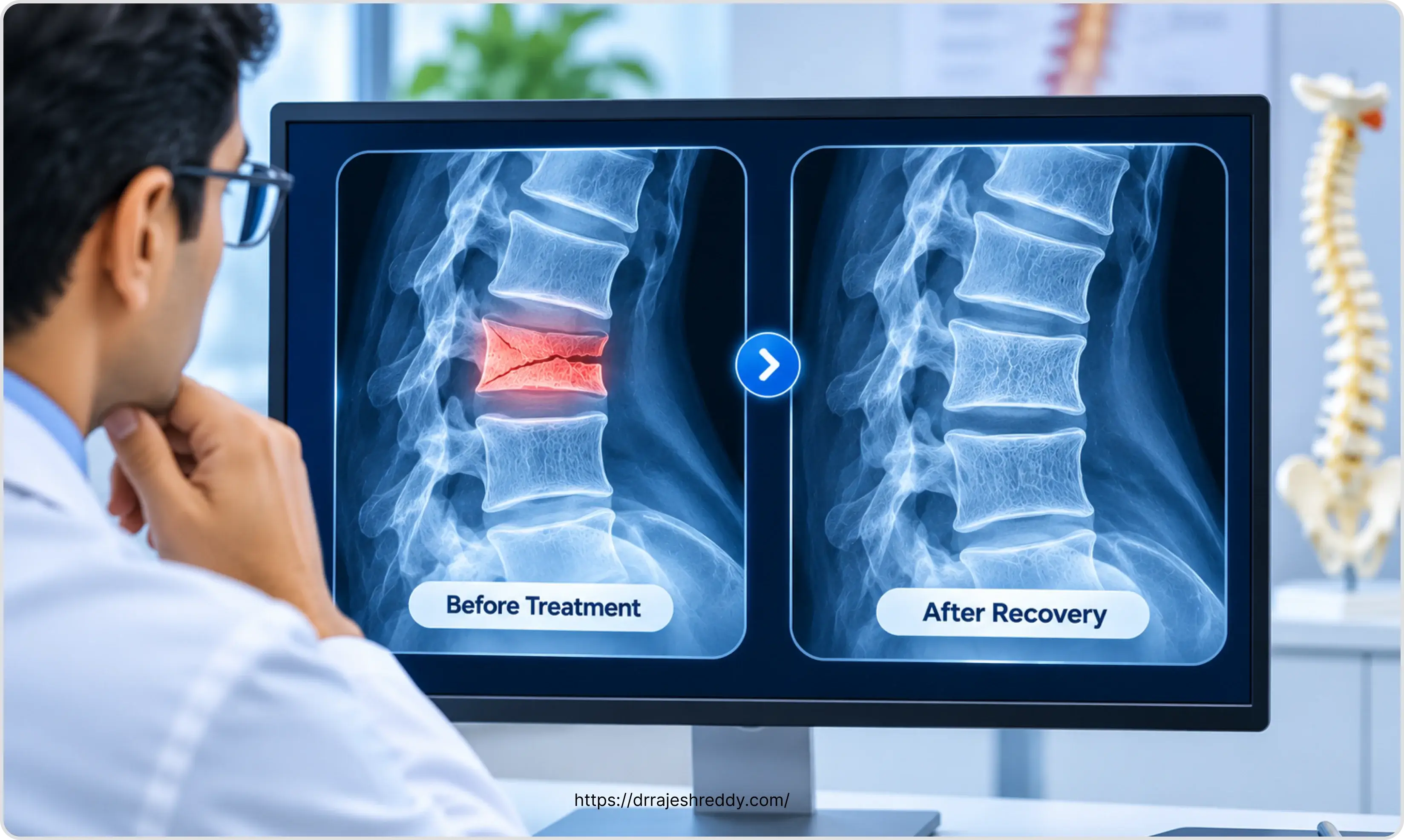
- Early consolidation is visible on CT from 6–8 weeks: Bony callus formation indicates the healing response is active.
- Solid fusion is typically confirmed on CT at 3–6 months post-operatively: Though it may take up to 12 months in some patients with compromised bone density.
- Plain X-rays: Show implant position (pedicle screws, rods, cages) and overall spinal alignment but cannot reliably assess micro-trabecular fusion — CT remains the gold standard.
- Follow-up MRI: Used at 6–12 months to assess residual cord or nerve compression, particularly if neurological symptoms persist or evolve.
For osteoporotic compression fractures treated with kyphoplasty or vertebroplasty, the cement cures within minutes. X-ray or CT at 6–8 weeks confirms cement position and adjacent vertebral integrity. There is no 'fusion' to wait for — the treated level is mechanically stable from day one.
For pathological fractures from cancer, imaging during recovery also monitors tumour response to systemic treatment and radiotherapy, and assesses the need for further intervention.
Long-Term Outcomes: What Recovery Looks Like at 12 Months
The large majority of patients who undergo spine fracture treatment — whether surgical or conservative — achieve a good functional outcome at 12 months. 'Good outcome' means:
- Pain reduced to a level that does not significantly limit daily activity (though mild residual stiffness at the operated level is common and not a sign of failure).
- Return to most or all pre-injury activities, modified where necessary.
- No progressive neurological deficit — and, for patients who had neurological injury, the maximum neurological recovery that their injury pattern allows.
- Radiological evidence of healed fracture or solid bony fusion.
The factors most associated with a worse long-term outcome are: late presentation with established neurological injury, multi-level or highly unstable injuries, poor bone quality, significant comorbidities, and inadequate rehabilitation. None of these are within the patient's control at the time of the fracture — but the commitment to rehabilitation afterwards is.
Studies consistently show that patients who complete their physiotherapy programme and attend all post-operative follow-up appointments have significantly better pain and function outcomes at 12 months than those who do not. Recovery from spine fracture is an active process, not a passive one — the surgeon provides the structural repair; the patient rebuilds the function around it.
Frequently Asked Questions About Spine Fracture Recovery
A stable vertebral compression fracture treated conservatively — with a brace, pain management, and physiotherapy — typically heals sufficiently in 6–12 weeks for most daily activities to resume. Complete bone healing (assessed on CT) may take 3–6 months. However, 'healed' does not mean the spine looks entirely normal on imaging — some degree of vertebral height loss and altered contour may persist. The clinical goal is pain-free function, which for most stable fractures is achieved by 3 months.
Leg pain (sciatica or radiculopathy) that was present before surgery often persists for weeks to months after the surgery that relieved the nerve compression. The nerve that was compressed takes time to recover — sometimes weeks, sometimes months — even after the structural cause has been removed. Persisting neurological symptoms in the early weeks after surgery are not a sign of surgical failure. However, new leg pain that was not present before surgery, or leg pain that is worsening rather than improving, should be reviewed promptly.
Most patients find sleeping flat uncomfortable in the early post-operative period. A semi-reclined position (pillow under the knees for lumbar surgery; slight head elevation for cervical surgery) is generally more comfortable. From a medical perspective, there is no absolute restriction on sleeping position — comfort is the guide, provided you are not sleeping in a position that creates sustained pressure on the wound. By 4–6 weeks most patients can sleep in their preferred position. Specific advice may differ for cervical surgery patients.
Long-haul air travel is generally deferred for 6 weeks after major spine surgery because the combination of immobility, dehydration, and reduced cabin pressure significantly increases the risk of deep vein thrombosis. Short domestic flights may be acceptable from 2–3 weeks after minimally invasive procedures, with adequate hydration and in-flight movement. Confirm with Dr. Rajesh Reddy before booking any travel in the first 3 months after surgery.
Nutrition plays a meaningful role in bone healing. The priorities are: adequate protein (1.2–1.5 g per kg body weight per day) as the building block for bone matrix; adequate calcium (1,000–1,200 mg per day from dairy, fortified foods, or supplements); adequate vitamin D (deficiency is extremely common in India and significantly impairs calcium absorption and bone healing — supplementation is almost always recommended); and zinc, vitamin C, and magnesium as cofactors in bone remodelling. Smoking significantly impairs bone healing and fusion — cessation is strongly recommended. Alcohol in excess impairs bone healing and increases fall risk.
The purpose of the brace is to restrict the specific movements that would stress the healing fracture or new surgical fusion during the most vulnerable period of bone healing. Whether a brace is prescribed and how strictly it needs to be worn varies by the type of surgery and the surgeon's judgement — not all spine surgery requires post-operative bracing. If Dr. Rajesh Reddy has prescribed one, it should be worn as instructed: typically during all activity outside of bed (washing and sleeping may be exceptions). Removing it prematurely increases the risk of implant displacement or non-union.
Recovery Guidance from Dr. Rajesh Reddy — Hyderabad's Spine Fracture Specialist
If you have questions about your recovery from a spine fracture — whether you are a patient of Dr. Rajesh Reddy or seeking a second opinion — contact the clinic for a comprehensive post-operative review at Medicover Hospital - Financial District, Hyderabad. Book via the Dr. Reddy's Neuro Care app on Google Play or call 9000224265.
Scientific References
The clinical statements on this page are supported by peer-reviewed literature:
- Lee JY, et al. Thoracolumbar injury classification and severity score: a new paradigm for the treatment of thoracolumbar spine trauma. J Orthop Sci. 2005;10(6):671–675. [PubMed]
- Fehlings MG, et al. Efficacy and safety of surgical decompression in patients with cervical spondylotic myelopathy: results of the AOSpine North America prospective multi-center study. J Bone Joint Surg Am. 2013;95(18):1651–1658. [PubMed]
- Weinstein JN, et al. Surgical versus nonoperative treatment for lumbar disk herniation: the Spine Patient Outcomes Research Trial (SPORT): a randomized trial. JAMA. 2006;296(20):2441–2450. [PubMed]
- van Middendorp JJ, et al. Diagnosis and prognosis of traumatic spinal cord injury. Global Spine J. 2011;1(1):1–8. [PubMed]
- National Institute for Health and Care Excellence (NICE). Spinal injury: assessment and initial management. NICE Guideline NG41. Published 2016; updated 2022. [NICE Guideline]
- World Health Organization. Rehabilitation 2030: A Call for Action. WHO. 2017. [WHO Link]