
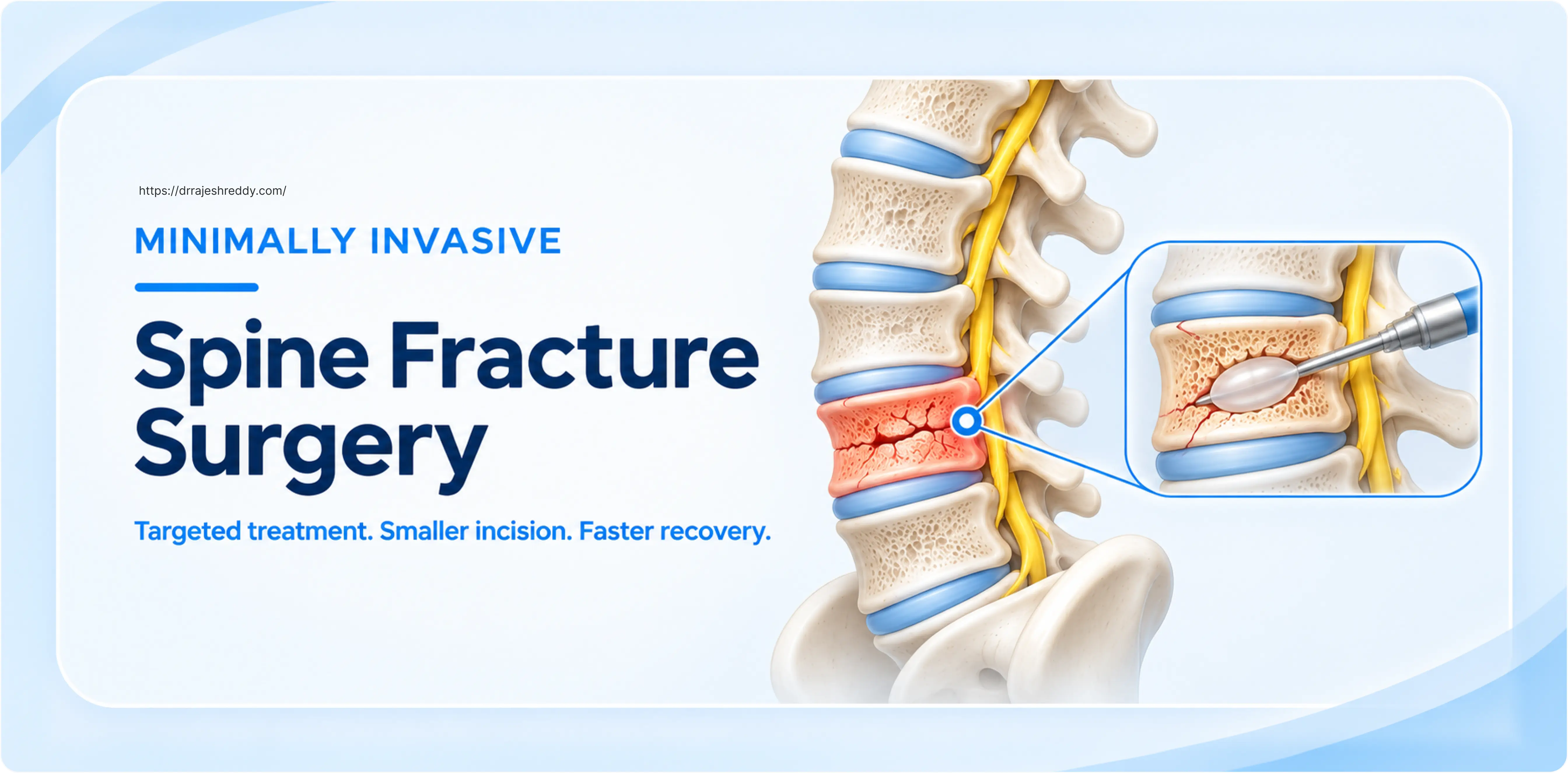
Spine fracture surgery has been transformed over the past two decades. Where a broken vertebra once almost always required a long incision, extensive muscle stripping, and a week or more in hospital, many of the same fractures can today be treated through incisions smaller than a centimetre — with patients sitting up the same evening and going home within 48 to 72 hours.
Minimally invasive spine (MIS) surgery uses specialised instruments, real-time imaging guidance, and tubular retractors to reach the spine through a narrow corridor rather than by cutting and peeling back the large paraspinal muscles. The mechanical result — screw fixation, cement augmentation, or decompression — is equivalent to open surgery, while the biological cost to the patient is dramatically lower: less blood loss, less post-operative pain, lower infection risk, and a faster return to independent function.
Dr. Rajesh Reddy Sannareddy, Senior Consultant Neurosurgeon in Hyderabad, performs the full range of minimally invasive spinal procedures for fracture management — from percutaneous pedicle screw fixation in trauma patients to balloon kyphoplasty for osteoporotic compression fractures to endoscopic decompression for nerve root compression after injury. His approach to each case is governed by one principle: the least invasive approach that achieves the necessary surgical goals. When MIS is appropriate, it is the first choice. When the complexity of the injury demands an open approach, he does not hesitate to recommend it.
What Makes a Spine Surgery 'Minimally Invasive'?
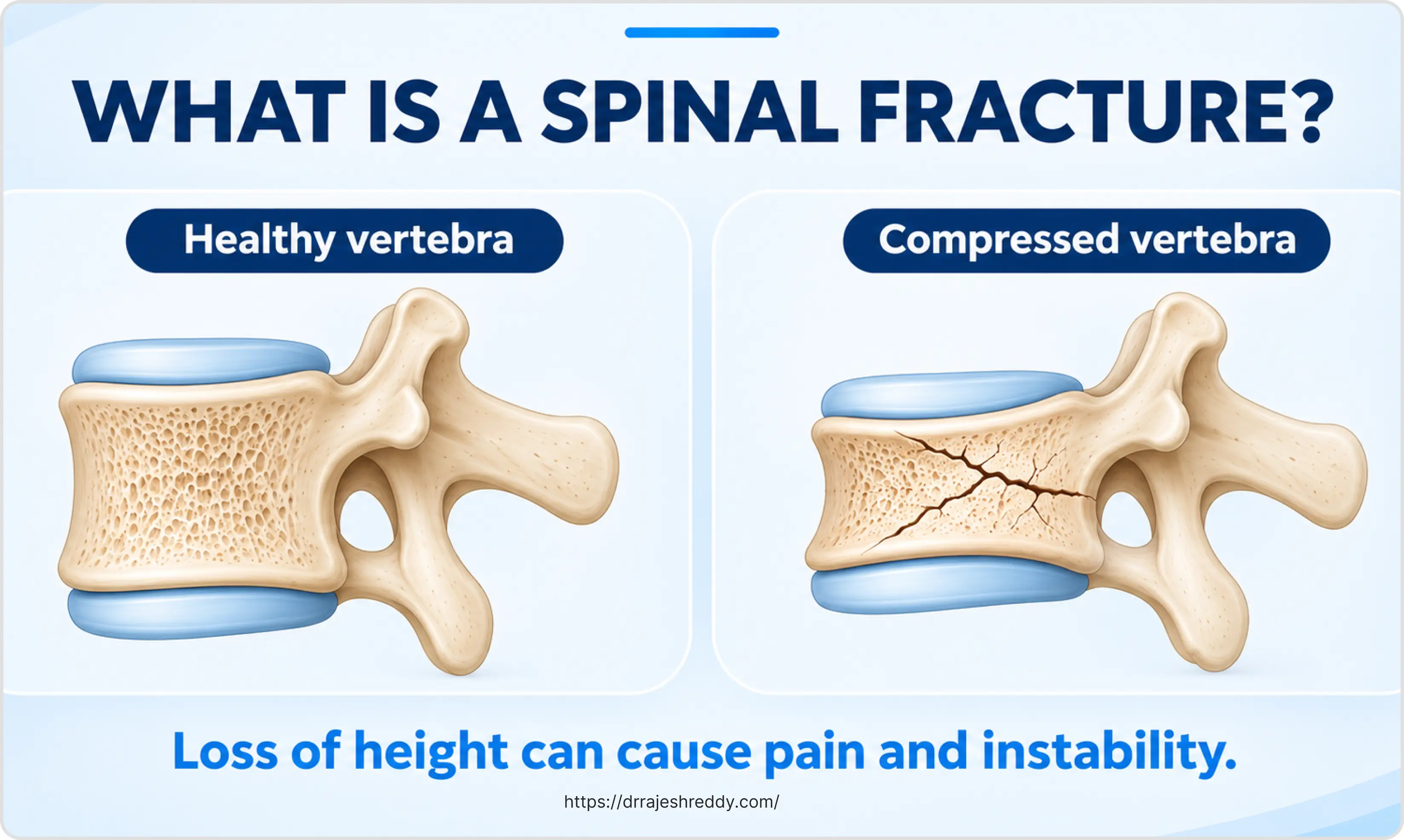
The term minimally invasive spine surgery describes a group of techniques that share a common anatomical strategy: accessing the spine while causing the least possible damage to the muscles, ligaments, and soft tissues that surround it. In conventional open spine surgery, these soft tissues are stripped away from the bony spine and retracted to give the surgeon a direct view. In MIS, they are instead dilated — gently separated along their natural fibre planes — using progressively larger tubular dilators until a working channel is established.
Through this tube — typically 18 to 22 mm in diameter — the surgeon operates using a microscope, an endoscope, or fluoroscopic guidance, depending on the procedure. The paraspinal muscles are never cut; they spring back to their original position when the tube is removed. The bone, disc, and nerve structures are addressed as needed, and the instruments are withdrawn.
The price of this tissue preservation is a steeper technical learning curve and the need for high-quality intraoperative imaging — fluoroscopy or three-dimensional navigation — to compensate for the reduced direct visualisation. In experienced hands, however, MIS procedures are as safe and reliable as their open equivalents, with a substantially better recovery profile.
MIS vs. Open Spine Fracture Surgery: A Direct Comparison
Both approaches have their place in spine fracture management. The table below compares them across the parameters that matter most to patients:
| Parameter | Minimally Invasive Surgery | Open Surgery |
|---|---|---|
| Skin incision | 1–3 cm per level (multiple small incisions) | 10–20 cm midline incision |
| Muscle handling | Tubular retractor — muscle dilated, not cut | Bilateral muscle stripping from spinous processes |
| Blood loss | 50–150 mL (typical) | 300–800 mL (typical) |
| Fluoroscopy / imaging | Intensive intraoperative fluoroscopy or navigation | Moderate fluoroscopy |
| Operating time | Often longer (2–4 hrs) | Often shorter for simple cases |
| Post-op pain | Significantly less | More; requires stronger analgesia |
| Hospital stay | 1–3 days | 4–7 days |
| Return to activity | 2–4 weeks for sedentary work | 6–8 weeks |
| Infection risk | Lower (smaller wound surface) | Higher |
| Scar | Minimal — multiple small punctures | Long midline scar |
| Best for | Stable fractures, older/frail patients, single-level disease | Complex fractures, multilevel disease, revision surgery |
The choice between minimally invasive and open surgery is never simply about which technique is newer or better — it is about which approach best matches the individual fracture pattern, the patient's neurological status, their medical fitness, and the goals of surgery. Dr. Rajesh Reddy will discuss both options transparently, explain why one is preferred for your specific situation, and give you a realistic expectation of the recovery.
Minimally Invasive Procedures for Spine Fractures
The following procedures represent the MIS toolkit for spine fracture management. Several are available at Dr. Rajesh Reddy's centre in Hyderabad:
| MIS Procedure | Fracture Type / Indication | Key Features |
|---|---|---|
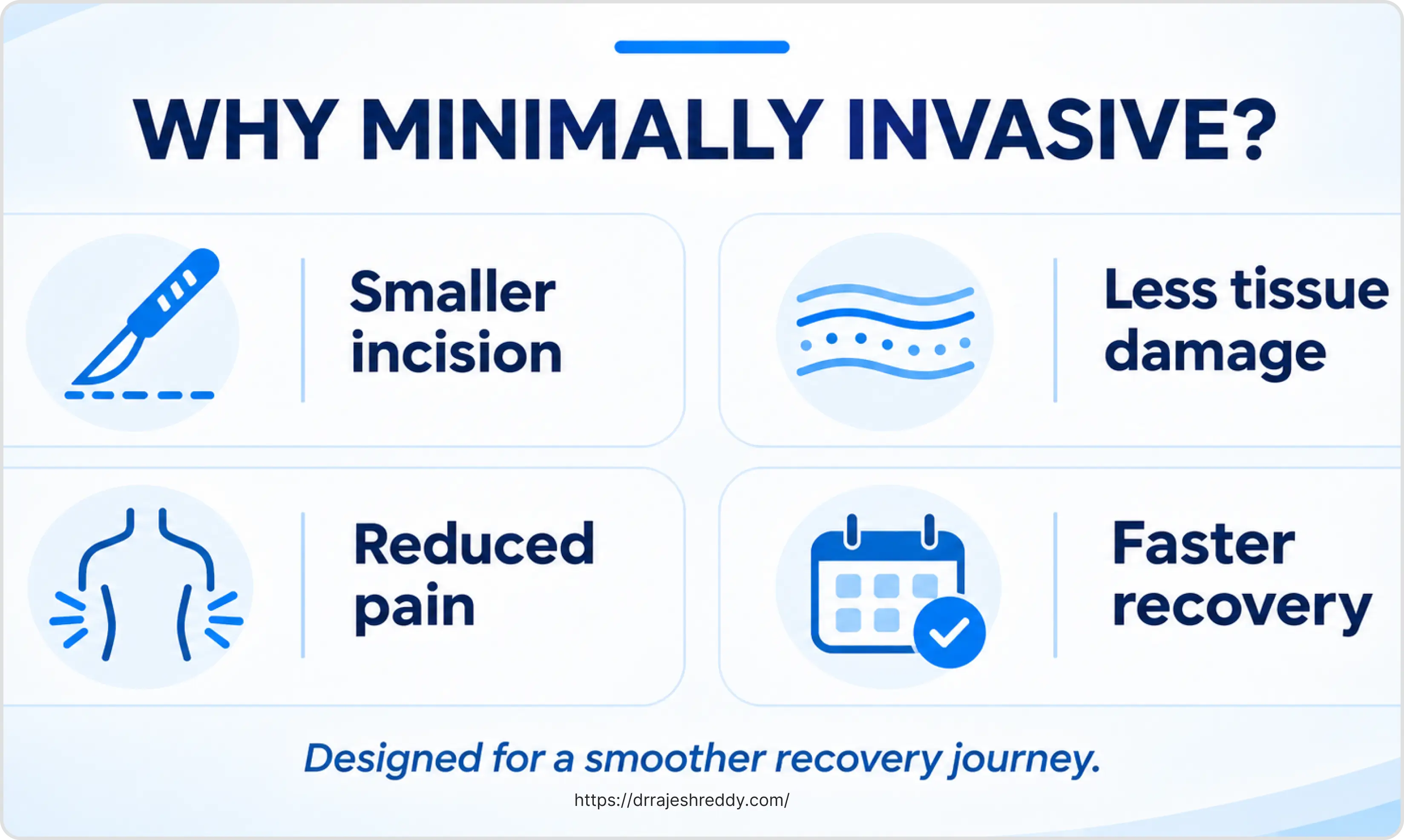
| Percutaneous pedicle screw fixation | Thoracolumbar burst fractures (neurologically intact); fracture-dislocations requiring stabilisation without decompression | 4–6 small stab incisions; screws placed under fluoroscopy or navigation; connecting rod passed percutaneously; no muscle stripping; direct visualisation of anatomy not required |
| Balloon kyphoplasty | Vertebral compression fractures (osteoporotic, traumatic); painful fractures not responding to conservative care; progressive kyphosis | Balloon tamp inflates inside collapsed vertebra to restore height; cavity filled with PMMA bone cement; bilateral transpedicular approach; immediate pain relief in most patients |
| Vertebroplasty | Painful vertebral compression fractures; pathological fractures from metastases or myeloma; when kyphoplasty not indicated | Direct cement injection without balloon; faster; lower cost; less height restoration than kyphoplasty; real-time fluoroscopic monitoring essential |
Who Is a Good Candidate for MIS Spine Fracture Surgery?
Patient selection is the single most important determinant of a good outcome in minimally invasive spine surgery. Not every fracture is suitable, and not every patient who wants MIS will benefit from it.
| Good candidates for MIS | Unsuitable for MIS (open surgery preferred) |
|---|---|
| Neurologically intact patients with stable or mildly unstable fractures | Neurological deficit requiring extensive decompression under direct vision |
| Elderly or medically frail patients who cannot tolerate lengthy open procedures | Three-column instability (fracture-dislocation) needing direct reduction |
| Osteoporotic vertebral compression fractures causing pain and disability | Infection or tumour requiring extensive debridement |
| Single-level or two-level disease without complex deformity | Prior failed surgery at the same level (revision surgery) |
| Patients who need to return to work or activity quickly | Active systemic infection or severe coagulopathy |
| Metastatic vertebral fractures with good expected survival (> 3 months) | Patient unable to tolerate prone positioning (severe cardiac/respiratory disease) |
Clinical Perspective: A patient with an osteoporotic vertebral compression fracture causing severe pain, significant height loss, or progressive kyphotic deformity is an excellent candidate for balloon kyphoplasty — even if they are in their seventies or eighties with multiple medical comorbidities. The procedure takes 45–60 minutes, is performed under sedation or light general anaesthesia, and typically produces dramatic pain relief within 24–48 hours. It is one of the clearest examples of MIS providing a transformative benefit to a population that would otherwise struggle to tolerate open surgery.
Percutaneous Pedicle Screw Fixation: The Workhorse of MIS Fracture Surgery
For traumatic thoracolumbar fractures that require stabilisation — burst fractures, flexion-distraction injuries, fracture-dislocations without major neurological compromise — percutaneous pedicle screw fixation is the most commonly performed MIS procedure. It achieves the same mechanical goal as open posterior fixation (immobilising the fractured segment to allow healing) through four to six stab incisions of 1–2 cm each, with no muscle retraction and no direct exposure of the bony spine.
The Step-by-Step Surgical Technique:
- Positioning: Patient positioned prone on a radiolucent frame; biplanar fluoroscopy confirms fracture alignment and anatomical landmarks.
- Jamshidi Access: Jamshidi needle advanced through the pedicle under biplanar fluoroscopic guidance; guide wire accurately placed through the needle into the vertebral body.
- Serial Dilation: Sequential dilators passed over the guide wire to gently separate muscle fibers; cannulated pedicle screw inserted smoothly through the dilator set.
- Construct Assembly: Steps repeated at each instrumented level — typically one level above and one level below the fractured vertebra.
- Subfascial Rod Insertion: Connecting rod passed percutaneously through the screw tulips using a specialised subfascial rod-passing device, then locked securely with set screws.
- Closure: Fascial and skin closure completed with small absorbable sutures; no wound drain required in most cases.
Patients are typically mobilised the same evening or the following morning. The instrumentation can be removed at 12–18 months once the fracture has healed, also percutaneously, if the patient wishes.
Balloon Kyphoplasty and Vertebroplasty for Compression Fractures
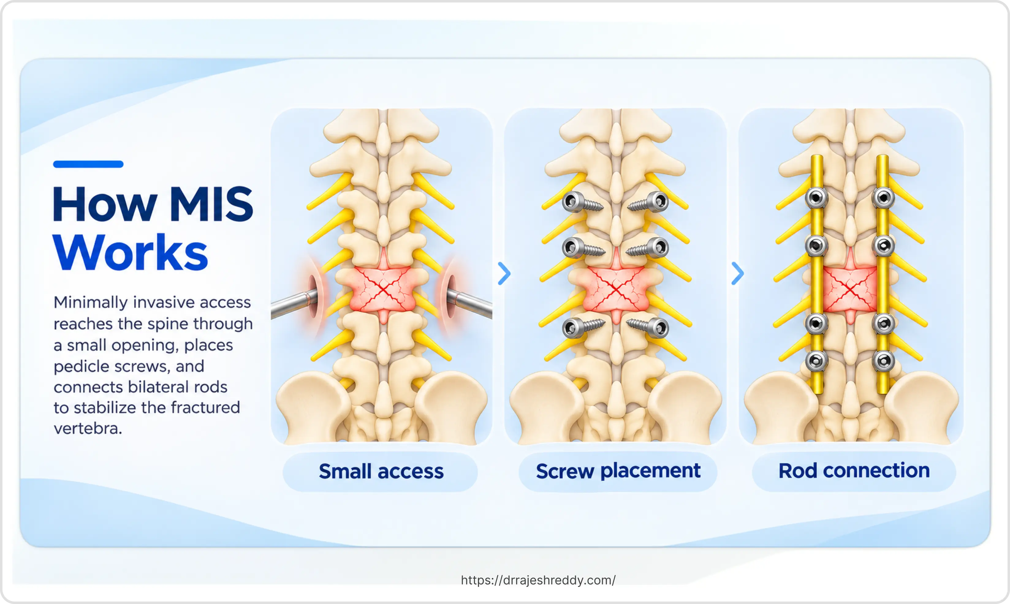
Vertebral compression fractures — the most common fracture type in patients over 60 — compress the front (anterior) column of the vertebra, producing a wedge-shaped deformity, height loss, and often severe axial back pain. When conservative management (rest, analgesia, bracing) fails to control pain, or when the fracture is progressive, cement augmentation procedures are highly effective.
How Balloon Kyphoplasty Works
In kyphoplasty, a working cannula is placed into the collapsed vertebra through a transpedicular approach — through the pedicle, which is the bony bridge between the posterior arch and the vertebral body — under fluoroscopic guidance. A deflated balloon tamp is advanced through the cannula into the fractured bone. The balloon is slowly inflated under pressure, compacting the cancellous bone of the vertebral body and — in recently fractured vertebrae — partially restoring the lost vertebral height. The cavity created by the balloon is then filled with polymethylmethacrylate (PMMA) bone cement, which sets within minutes to provide immediate mechanical stability.
The procedure is performed bilaterally (through both pedicles) at the fractured level. Duration is typically 45–75 minutes. Most patients notice a substantial reduction in pain within 24–48 hours.
Vertebroplasty vs. Kyphoplasty: Which Is Used When?
Vertebroplasty injects cement directly without balloon pre-treatment. It is faster and less expensive, but provides less height restoration and operates at higher injection pressure — which slightly increases the risk of cement extravasation. Kyphoplasty's balloon-created low-pressure cavity significantly reduces extravasation risk and allows better height restoration in acute fractures. Dr. Rajesh Reddy uses kyphoplasty as the preferred technique for acute osteoporotic fractures with height loss, and vertebroplasty selectively for chronic fractures or metastatic lesions where height restoration is less critical.
Recovery After MIS Spine Fracture Surgery
Recovery timelines vary by procedure, but the general pattern after MIS spine surgery is substantially better than open surgery:
Day of Surgery
Theatre in the morning; mobilised with physiotherapist by the afternoon; clear liquids and then diet as tolerated; pain managed with oral analgesics.
Day 1–2 (Hospital Ward)
Physiotherapy gait and stair training; wound check (1–3 small dressings); most patients discharged home on day 1–2 after kyphoplasty, day 2–3 after percutaneous fixation.
Week 1–2 (Home Recovery)
Light activity; no lifting or heavy bending; gentle wound care; oral pain medication; short walks encouraged.
Week 2–6
Progressive increase in walking; physiotherapy sessions for core strengthening and posture; most patients with desk or sedentary work return at 2–4 weeks.
Week 6 (First Major Review)
MRI or CT and standing X-ray; clinical review; physiotherapy programme adjusted; manual or physical work return typically at 6–8 weeks.
Month 3–6
Full return to normal activities for most patients; bone healing confirmed on imaging; consideration of implant removal for younger patients.
Instrumentation Removal Considerations: Instrumentation removal is a consideration unique to younger patients with traumatic fractures who have had percutaneous pedicle screws placed. Once the fracture has healed solidly — typically at 12–18 months — the screws can be removed through the same small incisions, under a short general anaesthetic. Most patients in their thirties and forties opt for removal to avoid long-term implant-related discomfort; older patients and those with osteoporotic fractures typically retain their hardware permanently.

Frequently Asked Questions About MIS Spine Fracture Surgery
For carefully selected patients, the clinical outcomes of MIS spine fracture surgery are equivalent to open surgery in terms of fracture healing, deformity correction, and neurological preservation — with significantly better recovery metrics: less blood loss, shorter hospital stay, less post-operative pain, and faster return to function. The key word is 'selected': not all fractures are suitable for MIS, and attempting a minimally invasive approach on a fracture that requires open surgery would compromise the surgical result. Dr. Rajesh Reddy will honestly advise whether MIS is appropriate for your specific fracture pattern.
Vertebroplasty is typically performed under sedation with local anaesthesia, while kyphoplasty is commonly performed under general anaesthesia. The choice depends on the patient's medical fitness, procedural requirements, and the number of levels treated. Most patients recover within a few hours.
Pedicle screws used in percutaneous fixation are titanium alloy and are MRI-compatible. They do not set off airport metal detectors. For most traumatic fractures in younger patients, the screws can be removed once the fracture has fully healed — typically at 12–18 months — through the original small incisions, in a 30-minute procedure. For older patients, those with osteoporotic bone, or those with instrumented fusion, the hardware is generally left permanently. Retained titanium hardware is safe and does not cause long-term problems in the vast majority of patients.
Many stable vertebral fractures — particularly osteoporotic compression fractures — do not require surgery and can be managed with a short period of rest, analgesics, and a back brace. Surgery (kyphoplasty or vertebroplasty) is considered when: pain is severe and not controlled by conservative measures after 4–6 weeks; the fracture is progressive (losing further height); the kyphotic deformity is significant; or the fracture is causing neurological symptoms. An X-ray taken lying down and standing can identify dynamic instability not visible on a single view. Dr. Rajesh Reddy will review your imaging and advise whether the fracture pattern warrants intervention.
Yes. Metastatic spine fractures are one of the most important indications for MIS techniques — particularly because cancer patients are often medically unfit for open surgery and need to minimise the interval between spine intervention and the continuation of systemic cancer treatment. Kyphoplasty or vertebroplasty provides pain relief and mechanical stability in metastatic vertebral fractures. Percutaneous fixation can stabilise impending or actual pathological fractures in patients with good expected survival. The choice of technique is individualised and made in collaboration with the patient's oncologist.
Cement extravasation — leakage of PMMA bone cement outside the vertebral body — is the most discussed risk of augmentation procedures. With kyphoplasty, the balloon-created cavity allows cement to be injected at lower pressure, reducing extravasation rates to approximately 5–10% compared with 20–40% for vertebroplasty. The vast majority of extravasation events are small and clinically silent — detected on post-procedural imaging but not causing symptoms. Symptomatic extravasation (cement migrating into the spinal canal or venous system) is rare in experienced hands. Real-time fluoroscopic monitoring during injection, using high-viscosity cement, and stopping injection at the first sign of leakage are the primary preventive measures.
Consult Dr. Rajesh Reddy for Minimally Invasive Spine Surgery in Hyderabad
Whether you have sustained a traumatic spine fracture, are suffering from a painful osteoporotic compression fracture, or are seeking a second opinion on whether open surgery is truly necessary, Dr. Rajesh Reddy Sannareddy provides expert minimally invasive spine consultations at Medicover Hospital - Financial District, Hyderabad. Book via the Dr. Reddy's Neuro Care app on Google Play or call 9000224265.
Scientific References
The clinical statements on this page are supported by peer-reviewed neurosurgical and spinal literature:
- Ringel F, et al. Clinical evidence for superior surgical outcome after minimally invasive versus open lumbar spinal fusion: a systematic review. World Neurosurg. 2017;101:414–422. [PubMed]
- Verlaan JJ, et al. Surgical treatment of traumatic fractures of the thoracic and lumbar spine: a systematic review of the literature on techniques, complications, and outcome. Spine. 2004;29(7):803–814. [PubMed]
- Kallmes DF, et al. A randomized trial of vertebroplasty for osteoporotic spinal fractures. N Engl J Med. 2009;361:569–579. [PubMed]
- Wardlaw D, et al. (FREE Trial investigators). Efficacy and safety of balloon kyphoplasty compared with non-surgical care for vertebral compression fracture (FREE): a randomised controlled trial. Lancet. 2009;373(9668):1016–1024. [PubMed]