
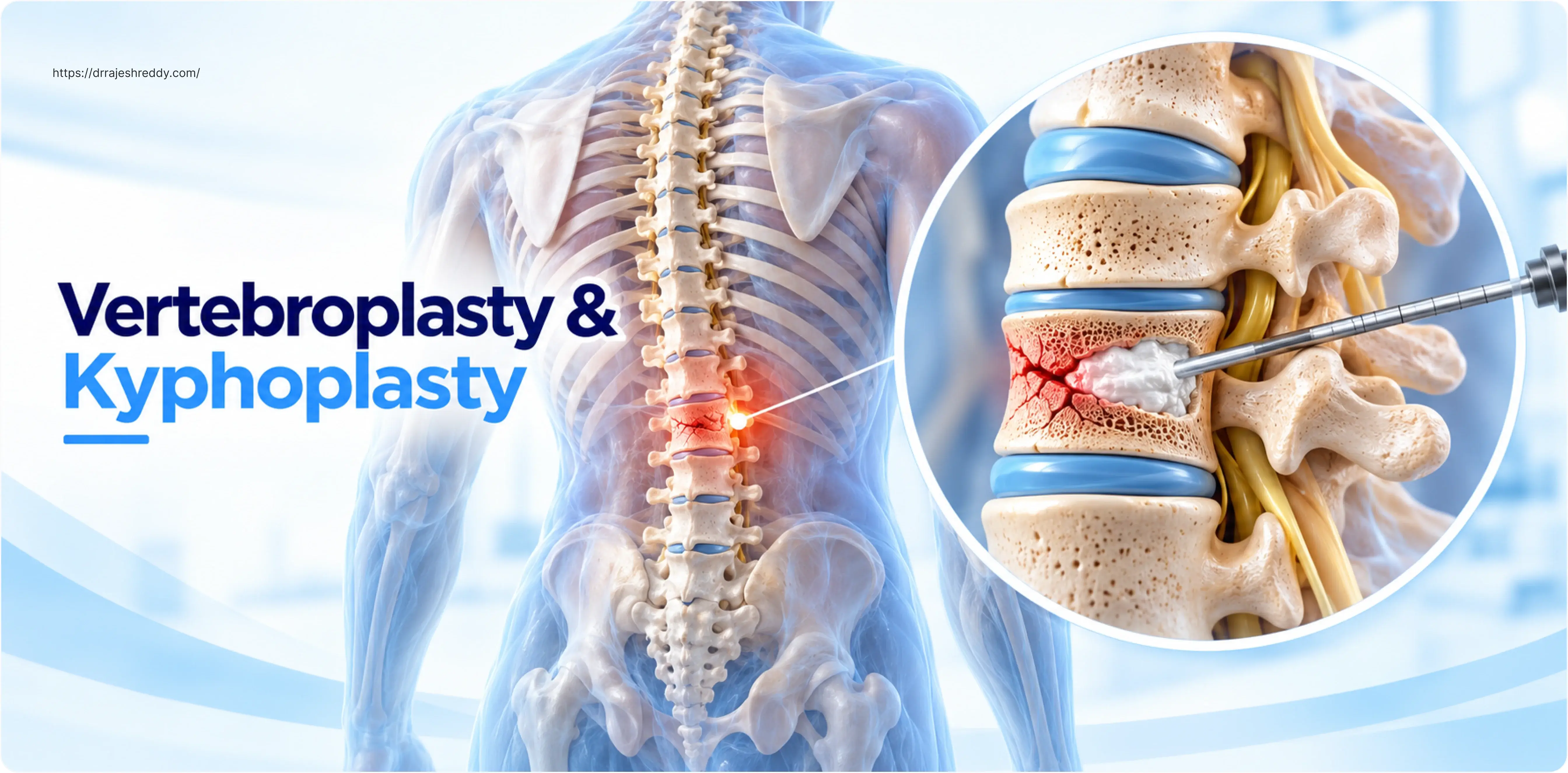
A spinal compression fracture can reduce a previously active, independent person to near-immobility within hours. The sudden, sharp back pain — often following a seemingly minor event such as bending forward, lifting a light object, or even coughing — is the presenting symptom of a vertebral body that has collapsed under loads it can no longer withstand. In osteoporotic patients, that threshold has been progressively lowered over years of silent bone loss, and the fracture is the point at which the bone simply gives way.
For decades, the only treatment options were bed rest, pain medication, and a brace — a conservative programme that offered gradual pain relief but did nothing to address the structural failure of the vertebra, the progressive kyphotic deformity (the forward stooping posture that follows multiple compression fractures), or the prolonged period of immobility that significantly worsens the osteoporosis itself. For elderly patients, extended bed rest carries its own serious risks: pneumonia, deep vein thrombosis, pressure ulcers, deconditioning, and accelerated bone loss.
Vertebroplasty and kyphoplasty changed this paradigm. Both procedures involve the percutaneous (through-the-skin) injection of bone cement into the fractured vertebral body under image guidance — stabilising the fracture, relieving pain, and in the case of kyphoplasty, restoring some of the lost vertebral height. The procedures are minimally invasive, typically performed under sedation or general anaesthesia, and most patients walk within hours and go home the same day or the following morning.
Dr. Rajesh Reddy Sannareddy, Senior Consultant Neurosurgeon in Hyderabad, performs vertebroplasty and kyphoplasty as part of his comprehensive spine fracture service — offering patients with osteoporotic and pathological compression fractures a precise, image-guided approach to pain relief and vertebral stabilisation.
What Is a Spinal Compression Fracture?
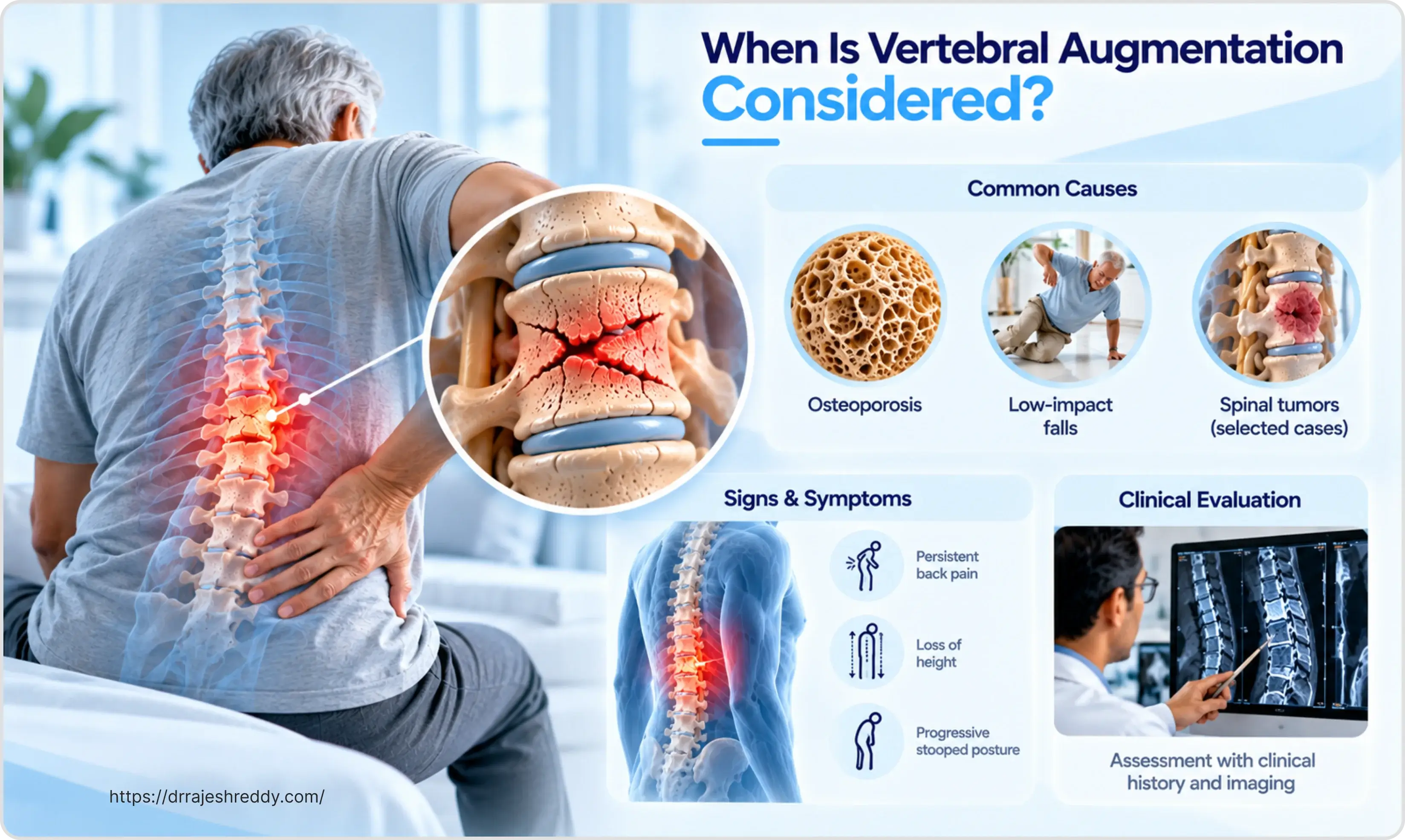
A vertebral compression fracture (VCF) occurs when the vertebral body — the large, block-shaped anterior part of each vertebra that bears axial load — collapses under compressive forces. Unlike a traumatic burst fracture (where high-energy impact shatters the vertebra), a compression fracture typically involves failure of the anterior column only: the front of the vertebral body wedges downward while the posterior wall (the back of the bone adjacent to the spinal canal) remains intact.
The result is a wedge-shaped deformity: the front of the vertebra becomes shorter than the back, tilting the spine forward and contributing to thoracic kyphosis (the hunched posture associated with osteoporosis). When multiple levels fracture over time, the cumulative kyphosis can become significant — reducing height by several centimetres, compressing the abdominal cavity, impairing lung function, and causing chronic pain.
Who Gets Compression Fractures?
Osteoporosis is by far the most common underlying cause. It is estimated that osteoporotic vertebral fractures affect approximately 1.4 million people worldwide each year, making them the most common osteoporotic fracture — more common than hip or wrist fractures. In India, the scale of the problem is large but substantially underdiagnosed: many patients accept back pain and height loss as an inevitable part of ageing rather than recognising these as the sequelae of vertebral fractures.
Primary risk groups include:
- Post-menopausal women: Oestrogen withdrawal accelerates bone resorption, and the majority of osteoporotic compression fractures occur in this group.
- Elderly men: Testosterone decline and age-related bone loss lead to increasing fracture risk after age 70.
- Patients on long-term corticosteroids: Glucocorticoid-induced osteoporosis is the most common secondary cause of compression fractures.
- Cancer patients: Vertebral metastases weaken bone and cause pathological fractures at loads that would not injure normal vertebrae.
- Patients with multiple myeloma: Diffuse bone involvement makes multiple simultaneous vertebral fractures common.
- Metabolic bone diseases: Patients with hyperparathyroidism, renal osteodystrophy, or osteomalacia.
Vertebroplasty: The Procedure Explained
Vertebroplasty was first performed in France in 1984 for the treatment of a painful vertebral haemangioma, with the initial clinical report published in 1987 by Galibert et al. (Galibert and colleagues). It was subsequently adopted for osteoporotic compression fractures in the 1990s and has since become one of the most widely performed minimally invasive spine procedures worldwide.
How Vertebroplasty Works
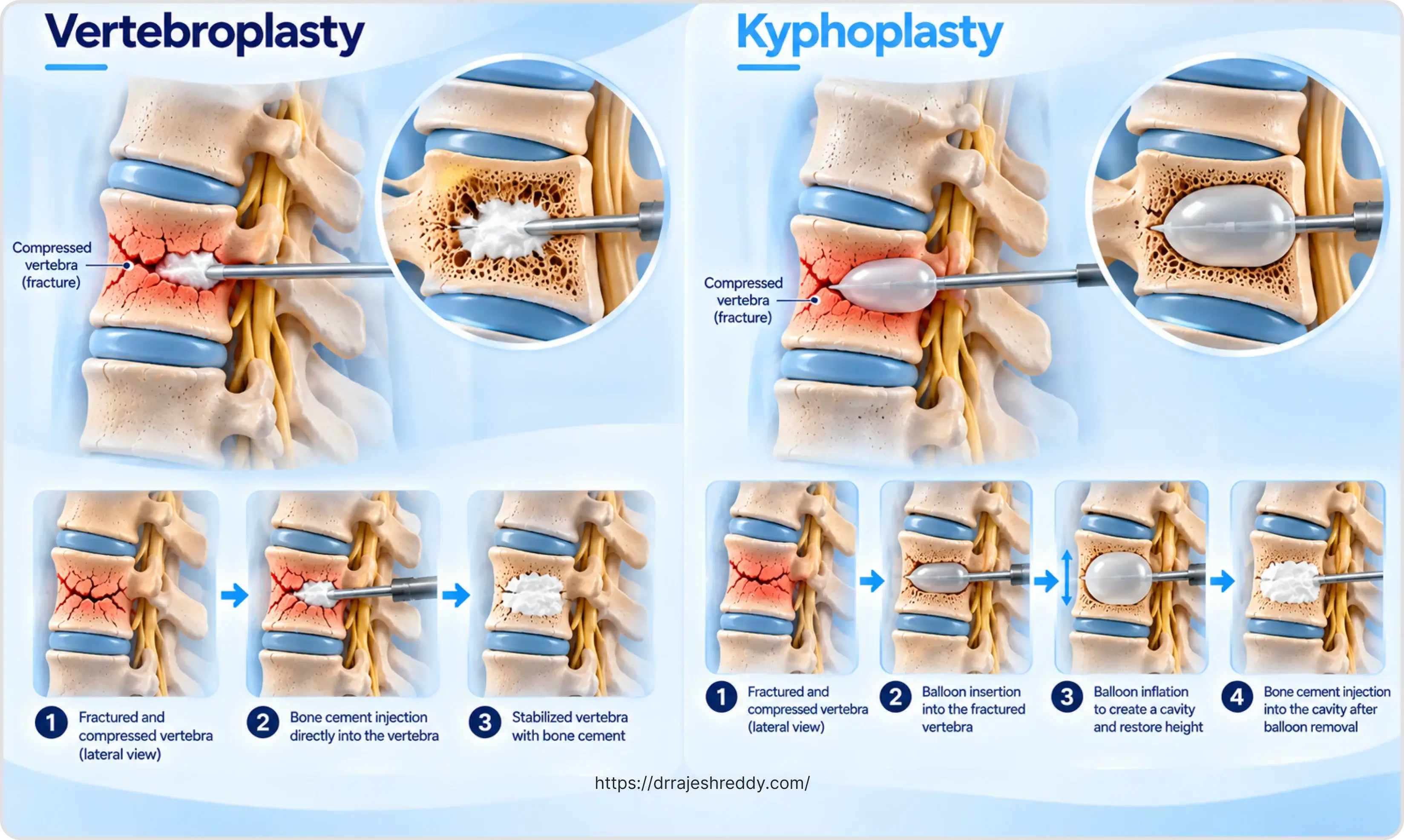
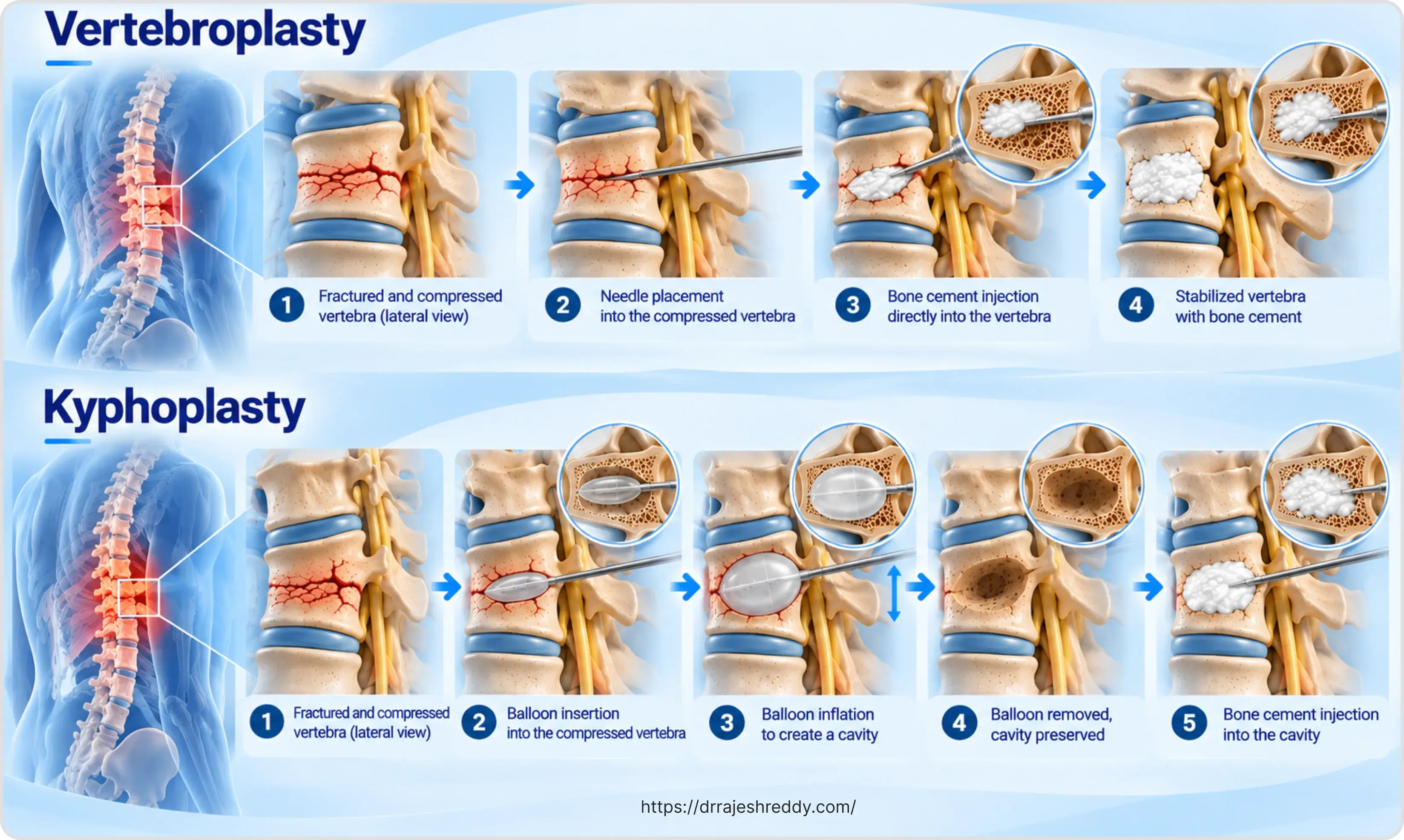
Under fluoroscopic (real-time X-ray) or CT guidance, with the patient positioned prone (face down), a hollow needle — a trocar — is advanced through the pedicle of the fractured vertebra (the bony bridge connecting the back of the vertebra to its body) and positioned within the collapsed vertebral body. This transpedicular approach keeps the needle safely away from the spinal canal.
Once the needle tip is correctly positioned within the fractured bone, polymethylmethacrylate (PMMA) bone cement — the same acrylic cement used in hip and knee arthroplasty — is mixed to a toothpaste-like consistency and injected under fluoroscopic guidance. The cement flows into the trabecular spaces of the fractured vertebra, fills the fracture lines, and polymerises (hardens) within minutes, rigidly stabilising the fracture.
The mechanism of pain relief is twofold: mechanical stabilisation of the fracture eliminates the micro-motion at the fracture site that generates pain with every breath and movement; and the exothermic reaction of cement polymerisation may also ablate pain-sensitive nerve endings within the fractured bone. The result is typically rapid — many patients report significant pain reduction within hours of the procedure.
Kyphoplasty (Balloon Kyphoplasty): Restoring Vertebral Height
Kyphoplasty adds a critical additional step to the vertebroplasty technique: before cement injection, an inflatable bone tamp (a small balloon) is inserted into the fractured vertebra and gradually inflated. The expanding balloon compacts the surrounding cancellous bone, creates a cavity within the vertebral body, and — most importantly — can restore some of the lost vertebral height by pushing the collapsed endplates back toward their original position.
Once the desired height restoration is achieved (or the balloon reaches its maximum safe inflation volume), the balloon is deflated and removed, leaving a well-defined cavity within the bone. Cement is then injected at low pressure to fill this cavity — the contained, pre-formed space means cement fills under lower pressure than vertebroplasty, reducing the risk of cement leakage.
The Height Restoration Advantage
In acute fractures — those treated within 6 weeks of the injury — kyphoplasty can restore 30–60% of the lost vertebral height on average. This is clinically significant for several reasons:
- Kyphotic deformity is reduced: The patient's posture improves and the progressive forward stooping of the thoracic spine is partially corrected.
- Adjacent segment stress is reduced: A kyphotic deformity concentrates mechanical stress on the vertebrae immediately above and below the fractured level, increasing the risk of adjacent fractures; height restoration reduces this.
- Thoracic and abdominal capacity improves: Lung and abdominal function may improve in patients with severe thoracic kyphosis, for whom the deformity was compressing the thoracic cavity.
In subacute or chronic fractures, the fractured bone has begun to heal in its collapsed position and height restoration becomes progressively less achievable. This is why early intervention — in the acute phase of pain — offers the best combination of pain relief and height restoration.
Vertebroplasty vs. Kyphoplasty: A Direct Comparison
The choice between vertebroplasty and kyphoplasty depends on the fracture characteristics, the degree of height loss, the patient's age and bone quality, and the goals of treatment. The table below compares the two procedures directly:
| Feature | Vertebroplasty | Kyphoplasty (Balloon Kyphoplasty) |
|---|---|---|
| Procedure | Cement injected directly under pressure into fractured vertebra | Balloon inflated first to create cavity and restore height; cement fills cavity |
| Height restoration | Minimal — does not restore lost vertebral height | Moderate — balloon inflation can restore 30–60% of lost height |
| Kyphosis correction | Limited | Better — partial correction of kyphotic deformity possible |
| Cement pressure | Higher injection pressure | Lower injection pressure (fills pre-formed cavity) |
| Cement leakage risk | Higher (cement injected under pressure into open bone) | Lower (cement fills a contained cavity at lower pressure) |
| Procedure time | 30–45 minutes | 45–60 minutes (additional balloon step) |
| Cost | Lower | Higher (balloon device cost) |
| Best suited for | Acute osteoporotic fractures without significant height loss | Fractures with height loss, kyphotic deformity, or in younger patients |
| Anaesthesia | Local + sedation or general | Local + sedation or general |
| Hospital stay | Same-day or overnight | Same-day or overnight |
In clinical practice, kyphoplasty is generally preferred for patients with significant vertebral height loss, kyphotic deformity, younger patients where preserving height and alignment has long-term significance, and in pathological fractures where the goal is both pain relief and structural restoration. Vertebroplasty remains an effective option for acute osteoporotic fractures without significant height loss, and in settings where the additional cost of the balloon device is a consideration.
Patient Selection: Who Is a Candidate?
Not every compression fracture requires or benefits from vertebroplasty or kyphoplasty. Careful patient selection — based on clinical presentation, fracture age, MRI findings, and the patient's overall health — is essential for achieving good outcomes and avoiding complications.
| Factor | Suitable for Procedure | Not Suitable / Caution Required |
|---|---|---|
| Fracture age | Acute (< 6 weeks) — best results; subacute (6–12 weeks) — still beneficial | Chronic (> 3 months) — fracture healed; cement will not benefit |
| Fracture type | Osteoporotic compression fracture; pathological fracture (cancer) | Burst fracture with posterior wall involvement — cement may retropulse |
| Neurological status | Neurologically intact | Any neurological deficit — decompression surgery required first |
| MRI findings | Bone marrow oedema on STIR sequence — indicates acute/subacute fracture | No oedema — fracture healed; procedure will not relieve pain |
| Vertebral body integrity | Posterior wall intact | Posterior wall fractured — cement leakage into canal risk |
| Pain level | Severe (VAS ≥ 7/10) limiting mobility | Mild pain — conservative management may suffice |
| Response to bed rest | Minimal improvement with rest | Significant improvement with rest — may heal conservatively |
| Bone quality | Any — procedure works in osteoporotic bone | N/A — procedure designed for low-density bone |
| Coagulation | Normal or correctable | Uncorrectable coagulopathy — bleeding risk |
The Procedure: Step-by-Step
1. Pre-Procedure Preparation
Routine blood investigations including coagulation profile and renal function; current medications reviewed — blood thinners (warfarin, clopidogrel, rivaroxaban) are held for the appropriate washout period before the procedure; nil by mouth for 4–6 hours if general anaesthesia or deep sedation is planned; consent discussion covering procedure risks, cement leakage, adjacent fracture risk, and the possibility of incomplete pain relief.
2. Positioning and Access
The patient is positioned prone on the operating table. For vertebroplasty, local anaesthetic is infiltrated into the skin, soft tissues, and the periosteum of the pedicle through which the needle will pass. For kyphoplasty or in anxious patients, intravenous sedation (monitored anaesthesia care) or general anaesthesia is used. Two approaches are available: transpedicular (the standard approach — through the pedicle on one or both sides) and extrapedicular (a lateral approach used for upper thoracic vertebrae where the pedicles are small).
3. Needle Placement
Under continuous fluoroscopic guidance — with anteroposterior (AP) and lateral X-ray views confirming needle position at each step — the trocar is advanced through the pedicle and positioned in the anterior one-third to one-half of the vertebral body. Correct positioning is confirmed before any cement or balloon is introduced. CT guidance is used in cases where fluoroscopic anatomy is unclear or the pedicle is particularly narrow.
4. Balloon Inflation (Kyphoplasty Only)
For kyphoplasty, the inflatable bone tamp is inserted through the working cannula and positioned within the vertebral body. The balloon is gradually inflated using a contrast-filled syringe under fluoroscopic monitoring — the height restoration and balloon position are assessed in real time. Inflation continues until: the endplate begins to lift (height is being restored); the balloon reaches the cortical margin of the vertebral body; or the maximum safe inflation volume is reached. The balloon is then deflated and removed.
5. Cement Injection
PMMA cement is mixed and monitored for viscosity — it must be injected at the correct consistency (neither too liquid, which risks leakage, nor too stiff, which risks inadequate filling). The cement is injected slowly under continuous fluoroscopic guidance, watching carefully for any leakage into the venous system, the disc, or the epidural space. Injection is immediately stopped if any cement approaches the posterior vertebral wall. The procedure is typically complete within 30–60 minutes.
Pathological Fractures: Vertebroplasty in Cancer
Vertebral metastases — bone deposits from cancers of the breast, lung, prostate, kidney, and thyroid, as well as multiple myeloma — are a major cause of pathological compression fractures. The pain from metastatic vertebral disease can be severe and refractory to analgesics, and the associated fracture significantly impairs the patient's quality of life and functional independence.
Vertebroplasty and kyphoplasty are highly effective for pain palliation in patients with pathological compression fractures from metastatic disease, where the goals are different from osteoporotic fractures:
- Rapid pain relief: Allowing significant reduction in opioid analgesia and improved quality of life.
- Spine stabilisation: Preventing further collapse and progressive neurological risk.
- Enabling radiotherapy: A stabilised vertebra allows radiotherapy to proceed to the lesion without the risk of further collapse during treatment.
- Enabling continuation of systemic therapy: Patients who are bedridden by vertebral pain cannot receive chemotherapy safely; vertebroplasty restores mobility and allows cancer treatment to continue.
In patients with cancer, vertebroplasty is typically combined with a multidisciplinary oncology approach — the procedure addresses the structural failure while radiotherapy, systemic therapy, and bone-modifying agents (bisphosphonates, denosumab) address the underlying disease. Dr. Rajesh Reddy works closely with oncology teams in Hyderabad to provide integrated vertebral stabilisation as part of a comprehensive cancer treatment plan.
Recovery After Vertebroplasty or Kyphoplasty
One of the most significant advantages of these procedures over both conservative management and open surgery is the speed of recovery. Most patients experience meaningful pain relief within hours and are mobilised on the day of the procedure.
| Timeframe | What to Expect |
|---|---|
| Immediately post-procedure | Transferred to recovery room; vital signs monitored; groin or back access site checked; pain assessment at 30 minutes |
| 2–4 hours | Most patients are sitting up and walking short distances; pain is typically significantly reduced compared to pre-procedure |
| Day 0–1 | Same-day discharge for most; overnight observation if general anaesthesia used, elderly, or significant comorbidities; oral analgesia only |
| Week 1 | Resume light household activities; avoid bending, twisting, and lifting > 2–3 kg; walking encouraged; driving from day 3–5 if pain controlled |
| Week 2–4 | Gradual increase in activity; physiotherapy referral for core strengthening; osteoporosis treatment commenced or optimised |
| Month 1–3 | Most patients have returned to previous activity level; X-ray at 4–6 weeks to confirm cement position and fracture healing |
| Month 3–6 | Assessment of adjacent level fracture risk; DEXA scan if not yet performed; bisphosphonate or denosumab therapy review |
| Long-term | Annual follow-up; osteoporosis management is lifelong — the procedure treats the fracture, not the underlying bone fragility |

Osteoporosis Treatment: The Essential Next Step
Vertebroplasty and kyphoplasty stabilise the fractured vertebra but do not treat the underlying osteoporosis. Without addressing the bone fragility, the risk of adjacent vertebral fractures — which occurs in approximately 20% of patients within the first year after a treated compression fracture — remains high.
All patients who undergo these procedures should receive a comprehensive osteoporosis assessment and treatment plan:
- DEXA scan: To quantify bone mineral density and establish a baseline for monitoring treatment response.
- Calcium and Vitamin D3: Calcium (1,200 mg/day) and Vitamin D3 (800–2,000 IU/day) supplementation as the foundation of metabolic bone support.
- Anti-resorptive medications: Bisphosphonates (alendronate, zoledronic acid) or denosumab reduce future vertebral fracture risk by 40–70%.
- Anabolic bone-forming therapy: Teriparatide for severe osteoporosis, particularly valuable in patients with multiple fractures.
- Fall prevention programme: Home safety assessment, balance training, and hip protectors for high-fall-risk patients.
The fracture that brought the patient to vertebroplasty is often not their first — studies show that up to 60% of osteoporotic patients have had prior silent vertebral fractures. A compression fracture is a sentinel event: it signals severe systemic bone fragility that requires urgent medical therapy, not just management of the immediate injury.
Frequently Asked Questions
The majority of patients — approximately 80–90% in published studies — experience meaningful pain relief after vertebroplasty or kyphoplasty, typically rated as a reduction of at least 4 points on a 10-point pain scale. Complete pain relief is achieved in a significant proportion, but not all patients. Pain that persists after the procedure may relate to coexisting degenerative disc disease, adjacent facet joint arthritis, or muscle spasm — which the cement does not address. Dr. Rajesh Reddy will set realistic expectations during the consultation, based on your imaging and the precise source of your pain.
PMMA bone cement is a permanent material — it does not reabsorb or degrade over time. Once hardened within the vertebral body, it provides durable stabilisation indefinitely. The procedure itself does not need to be repeated for the treated vertebra. However, because the underlying osteoporosis is not cured by the procedure, other vertebrae remain at risk of fracturing — and approximately 20% of patients sustain a new compression fracture at an adjacent or distant level within a year. Each new fracture would be assessed for potential treatment on its own merits. This is why osteoporosis treatment after the procedure is not optional — it is essential.
Vertebroplasty and kyphoplasty are specifically designed for this patient population — they are minimally invasive, short-duration procedures that avoid the physiological stress of general anaesthesia (sedation or local anaesthesia is often sufficient) and the prolonged recovery of open surgery. Patients in their 80s and 90s with cardiac, respiratory, and renal comorbidities routinely undergo these procedures safely. The key assessment is whether any blood-thinning medications need to be temporarily held, and whether the patient's overall medical condition allows safe positioning prone for 30–60 minutes. Dr. Rajesh Reddy's team will co-ordinate with the patient's treating physicians as needed.
Yes — vertebroplasty and kyphoplasty are valuable tools for pain palliation and stabilisation in patients with vertebral metastases or multiple myeloma. The goals are different from osteoporotic fractures (palliation and maintaining function rather than curative intent) but the procedure is equally effective at relieving pain and stabilising the fractured vertebra. The procedure does not treat the underlying cancer — this is done through radiotherapy, systemic therapy, and bone-modifying agents in parallel. Dr. Rajesh Reddy works alongside the patient's oncology team to plan vertebral stabilisation as part of the overall cancer management strategy.
Cement leakage is the most important procedural risk — cement that enters the spinal canal, epidural space, or venous system can cause neurological injury or pulmonary embolism. However, clinically significant leakage is uncommon in experienced hands: the large majority of leaks are small, contained within the disc or paravertebral tissues, and cause no symptoms. The risk is minimised by careful patient selection (intact posterior wall on CT), correct cement viscosity, continuous fluoroscopic monitoring during injection, and immediate cessation if leakage is detected. The overall risk of a clinically significant neurological complication from cement leakage is less than 1% in published series.
Most patients are sitting and walking within 2–4 hours of the procedure ending. This is one of the most striking contrasts with conservative management — where weeks of relative immobility may be required for pain to settle — and with open spine surgery, where mobilisation takes much longer. Physiotherapy supervises the first walk to ensure safety and proper movement patterns. The majority of patients are discharged the same day or the following morning.
Consult Dr. Rajesh Reddy for Vertebroplasty and Kyphoplasty in Hyderabad
If you or a family member is suffering from severe back pain following a compression fracture — from osteoporosis, cancer, or other causes — Dr. Rajesh Reddy Sannareddy provides vertebroplasty and kyphoplasty consultations in Hyderabad with same-day or next-day procedure scheduling for appropriate cases. Book via the Dr. Reddy's Neuro Care app on Google Play or call 079951 11695.