A spine fracture is one of the most consequential injuries the human body can sustain. At its mildest, it is a painful compression fracture in an elderly osteoporotic patient that responds to minimally invasive cement injection and early mobilisation. At its most severe, it is a fracture-dislocation of the cervical spine following a road traffic accident, with the spinal cord crushed between displaced vertebrae and the patient paralysed from the neck down — a situation in which the speed and quality of neurosurgical intervention determines whether that paralysis is temporary or permanent.
Between these extremes lies the full spectrum of spinal fractures — burst fractures from falls from height, pathological fractures from metastatic cancer collapsing a vertebra, osteoporotic wedge fractures in post-menopausal women, Chance fractures from seat-belt injuries, and unstable cervical fractures from sports and diving accidents. Each requires a precise diagnostic assessment, an understanding of spinal biomechanics and neurological risk, and the surgical skill to stabilise the spine while protecting or recovering the neural elements within it.
Dr. Rajesh Reddy Sannareddy, Senior Consultant Neurosurgeon in Hyderabad, has extensive experience across the full spectrum of spinal trauma and pathological fractures — from minimally invasive percutaneous fixation of thoracolumbar fractures to complex anterior-posterior reconstructions, vertebroplasty and kyphoplasty for osteoporotic collapse, and emergency decompression for spinal cord injury. His approach integrates the latest minimally invasive spine surgery (MISS) techniques with the judgement to know when open surgery provides a better outcome.
What Is a Spine Fracture?
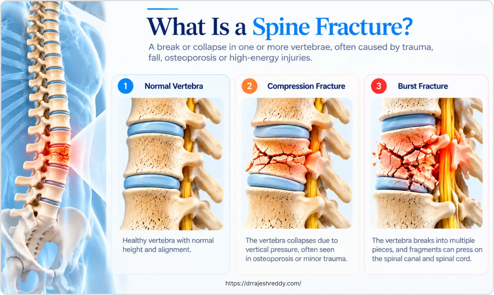
The spine is a column of 33 vertebrae stacked from the skull to the pelvis, connected by intervertebral discs, ligaments, and facet joints. Each vertebra consists of a body (the weight-bearing anterior block), a posterior arch (protecting the spinal cord within the spinal canal), and bony processes for muscle attachment and articulation with adjacent vertebrae. A spine fracture occurs when one or more of these structures breaks — from traumatic force, pathological weakening, or a combination of both.
The critical question with any spinal fracture is not just whether the bone is broken, but whether the fracture is stable or unstable, and whether the spinal cord, nerve roots, or cauda equina are at risk. A stable fracture — one that does not threaten to shift further and compress neural structures — may be managed without surgery. An unstable fracture — one that is likely to displace or has already caused neurological compromise — requires surgical stabilisation, decompression, or both.
The Three-Column Model of Spinal Stability
Denis's three-column model provides a practical framework for assessing spinal fracture stability. The spine is divided into three longitudinal columns:
- Anterior column: anterior two-thirds of the vertebral body and the anterior longitudinal ligament
- Middle column: posterior one-third of the vertebral body, posterior wall, and posterior longitudinal ligament
- Posterior column: pedicles, facet joints, laminae, spinous processes, and posterior ligamentous complex
A fracture involving two or more columns is considered unstable and typically requires surgical treatment. Compression fractures involving only the anterior column are generally stable; burst fractures involving the anterior and middle columns with retropulsion of bone into the spinal canal are unstable and carry neurological risk.
Types of Spine Fractures: A Complete Overview
The table below summarises the main types of spinal fractures, their causes, neurological risk, and typical treatment approach:
| Fracture Type | Common Cause | Neurological Risk | Typical Treatment |
|---|---|---|---|
| Compression fracture | Osteoporosis; minor fall in elderly | Low if stable; no cord involvement | Bracing; vertebroplasty / kyphoplasty for pain relief |
| Burst fracture | High-energy trauma (RTA, fall from height) | Moderate-high; bone fragments may enter canal | Surgical stabilisation if unstable or neurological deficit |
| Flexion-distraction (Chance fracture) | Seat-belt injury; forward flexion under restraint | Moderate; ligamentous instability common | Posterior instrumented fusion |
| Fracture-dislocation | Severe trauma; high-energy polytrauma | High; often associated with cord injury | Emergency surgical reduction and stabilisation |
| Odontoid (C2) fracture | Elderly fall; high-speed trauma | Variable; atlantoaxial instability risk | Collar / halo (Type I, III); surgery (Type II displaced) |
| Hangman's fracture (C2 pedicle) | Hyperextension injury (motor vehicle, diving) | Variable; often neurologically intact | Cervical collar; surgery if severe displacement |
| Jefferson fracture (C1 burst) | Axial load (diving, fall on head) | Variable; unstable if transverse ligament torn | Halo vest; surgery if ligament disrupted |
| Pathological fracture | Metastatic cancer; myeloma; infection; osteoporosis | Variable; depends on degree of collapse | Treat underlying cause; decompression + stabilisation if cord threatened |
Not every spine fracture requires surgery. The treatment decision is based on the fracture's stability, the presence or absence of neurological deficit, the patient's age, bone quality, general health, and activity level. Dr. Rajesh Reddy will assess all of these factors and provide a clear, evidence-based recommendation — which may include observation, bracing, minimally invasive intervention, or open surgery.
Spine Fractures by Region
Cervical Spine Fractures (C1–C7)
Fractures of the cervical spine — the seven vertebrae between the skull and the upper thorax — carry the highest neurological risk of any spinal injury. The cervical spinal cord lies within a relatively narrow canal, and displacement of a fractured cervical vertebra can cause tetraplegia (paralysis of all four limbs) or death from respiratory failure if the injury is at or above C4, where the phrenic nerve (which drives breathing) originates.
Upper cervical injuries (C1–C2) are anatomically distinct. Jefferson fractures of C1 (burst fracture of the atlas) and odontoid fractures of C2 carry instability risk at the craniocervical junction. Hangman's fractures — bilateral pedicle fractures of C2 from hyperextension — are paradoxically often neurologically intact because the canal widens at C2. Subaxial cervical fractures (C3–C7) are more commonly associated with disc injury, cord compression, and neurological deficit.
- Emergency stabilisation of the cervical spine — in a rigid collar or on a spine board — is mandatory from the scene of injury until imaging confirms stability or surgical fixation is performed
- MRI is essential in any cervical fracture with neurological symptoms — to assess cord signal change (oedema or contusion), disc herniation into the canal, and ligamentous injury not visible on CT
- High-dose methylprednisolone (within 8 hours of injury) is controversial and institution-specific; discuss with Dr. Rajesh Reddy
Thoracic Spine Fractures (T1–T12)
The thoracic spine is naturally more rigid than the cervical or lumbar spine — braced by the rib cage — making it more resistant to fracture but also less tolerant of displaced fractures, as the thoracic spinal canal is narrower relative to the cord. The thoracic cord has a relatively poor blood supply compared to the cervical and lumbar regions, making it more vulnerable to ischaemic injury when compressed.
Thoracic fractures most commonly occur at the thoracolumbar junction (T11–L2) — the transition zone between the rigid thoracic spine and the more mobile lumbar spine, where stress concentrates during forward flexion and axial load. Burst fractures of the thoracolumbar junction are among the most common surgically treated spinal injuries in young adults following road traffic accidents and falls from height.
Lumbar Spine Fractures (L1–L5)
Lumbar fractures below L1 involve the cauda equina — the bundle of nerve roots descending from the conus medullaris — rather than the spinal cord itself. Cauda equina injury from a lumbar burst fracture may cause lower limb weakness and numbness, bladder and bowel dysfunction, and sexual dysfunction — but the potential for neurological recovery is generally better than with cord injuries, as peripheral nerve roots have greater regenerative capacity than spinal cord.
L4–L5 and L5–S1 fractures — particularly in the context of pre-existing degenerative disease — may cause significant nerve root compression requiring decompression at the time of stabilisation.
Spinal Cord Injury: Assessing Neurological Status
When a spinal fracture has caused or may be causing spinal cord injury, a standardised neurological assessment is performed using the ASIA (American Spinal Injury Association) Impairment Scale. This scale classifies the completeness and level of the spinal cord injury, guides prognosis, and is used to track neurological recovery over time:
| ASIA Grade | Classification | Clinical Description |
|---|---|---|
| A | Complete | No motor or sensory function preserved in sacral segments S4–S5 |
| B | Sensory Incomplete | Sensory but no motor function preserved below neurological level, including S4–S5 |
| C | Motor Incomplete | Motor function preserved below neurological level — more than half of key muscles below level have < Grade 3 strength |
| D | Motor Incomplete | Motor function preserved below neurological level — at least half of key muscles have Grade 3 or greater strength |
| E | Normal | Motor and sensory function normal |
The distinction between complete (ASIA A) and incomplete (ASIA B–D) cord injury is the most clinically important — incomplete injuries retain some potential for neurological recovery with timely decompression and rehabilitation, while complete injuries have very limited recovery potential regardless of intervention. However, the surgical goal in both — to decompress the cord and stabilise the spine — remains the same: preventing further injury and creating the best possible environment for whatever recovery is biologically possible.
Neurogenic Shock
High thoracic or cervical spinal cord injuries can cause neurogenic shock — a combination of hypotension (low blood pressure) from loss of sympathetic tone and bradycardia (slow heart rate) from unopposed vagal activity. Neurogenic shock is distinct from haemorrhagic shock and requires vasopressors (noradrenaline) rather than aggressive fluid resuscitation to maintain spinal cord perfusion pressure. Management of neurogenic shock in the ICU — targeting mean arterial pressure > 85 mmHg for 7 days after SCI — is a critical determinant of neurological outcome.
Diagnosing a Spine Fracture: The Workup
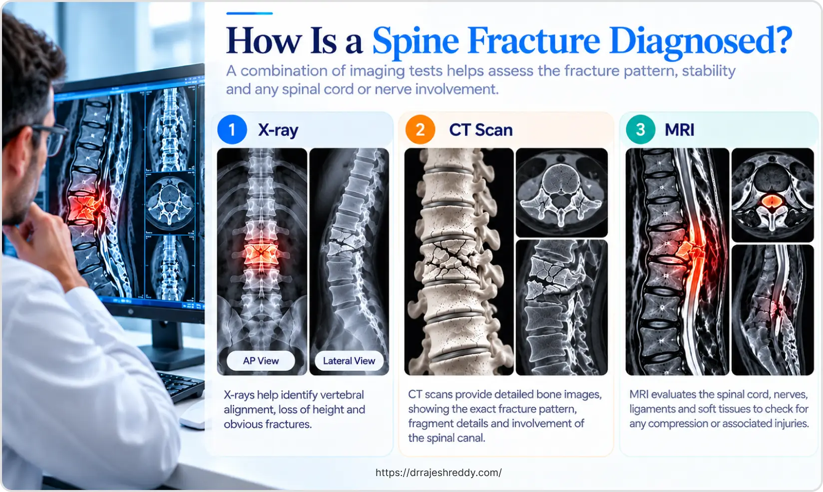
Plain X-Rays
Anteroposterior and lateral plain X-rays of the symptomatic spinal region are the first-line investigation in the emergency setting — particularly for cervical spine clearance after trauma. They identify gross fractures, dislocations, and loss of vertebral height, but miss many fractures — particularly those involving the posterior elements or at complex anatomical sites (C1–C2, cervicocranial junction).
CT Scan — The Gold Standard for Fracture Characterisation
CT of the spine with sagittal and coronal reconstructions provides the most detailed three-dimensional picture of the bony injury — fracture pattern, degree of comminution, canal compromise from retropulsed fragments, and involvement of the posterior elements and facet joints. Thin-slice CT (1mm) is essential for upper cervical fractures. CT angiography of the neck vessels (CTA) should be performed whenever a cervical fracture involves the transverse foramen, as vertebral artery injury occurs in approximately 20–30% of such cases.
MRI — Essential for Neurological Assessment
MRI of the spine is mandatory whenever there is a neurological deficit, or whenever the extent of ligamentous injury is uncertain. It uniquely shows: spinal cord signal change (oedema on T2 — indicating cord injury and guiding prognosis), disc herniation into the canal, posterior ligamentous complex disruption (a marker of instability), haematoma, and epidural compression. STIR sequences are particularly sensitive for ligamentous and soft tissue injury.
CT Myelography
In patients who cannot undergo MRI (pacemaker, certain implants), CT myelography — injection of contrast into the CSF followed by CT — provides detailed imaging of the neural elements within the spinal canal and identifies sites of cord or root compression.
Spine Fracture Treatment Options: A Complete Overview
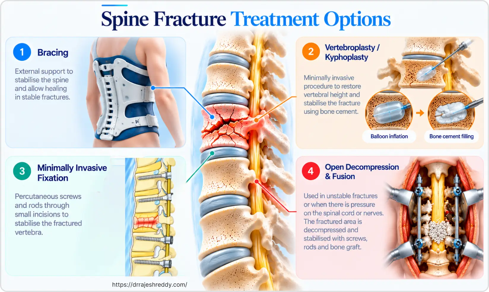
The treatment of spinal fractures spans from entirely non-surgical management to complex multi-level reconstructive surgery. The right approach is determined by fracture type, stability, neurological status, the patient's age and health, and the urgency of the clinical situation:
| Treatment | Best Suited For | Key Advantage | Typical Recovery |
|---|---|---|---|
| Cervical collar / TLSO brace | Stable fractures without neurological deficit; elderly low-demand patients | Non-invasive; avoids surgical risk | 6–12 weeks immobilisation; gradual mobilisation |
| Halo vest | Unstable upper cervical fractures (C1, C2) in selected patients | Non-invasive cervical stabilisation; avoids surgery in elderly | 8–12 weeks; significant patient burden |
| Vertebroplasty / Kyphoplasty | Osteoporotic or pathological compression fractures with pain; no cord compression | Minimally invasive; immediate pain relief; same-day or overnight | Pain relief often within 24–48 hrs; rapid return to activity |
| Anterior cervical decompression and fusion (ACDF) | Cervical burst fractures; disc-bone fragments compressing cord from front | Direct cord decompression; anterior column reconstruction | Collar 6 weeks; return to activity 8–12 weeks |
| Posterior cervical fusion | Cervical fracture-dislocations; posterior ligamentous instability | Reduces and stabilises the fracture; corrects alignment | Mobilise day 1–2; collar 6–12 weeks |
| Thoracolumbar MISS (percutaneous pedicle screws) | Thoracolumbar burst and compression fractures — neurologically intact | Minimally invasive; small incisions; less blood loss; faster recovery | Hospital 2–4 days; return to light activity 4–6 weeks |
| Open decompression + instrumented fusion | Burst fractures with cord compression; fracture-dislocations; failed conservative | Complete decompression + multi-level stabilisation | Hospital 5–7 days; rehabilitation 3–6 months |
| Corpectomy + cage reconstruction | Severe burst fractures; tumour-related collapse; multi-level pathology | Restores anterior column height and alignment with cage and plate | Longer recovery; combined anterior-posterior approach often needed |
Conservative Treatment: When Surgery Is Not Needed
A significant proportion of spinal fractures — particularly stable compression fractures in the elderly and minor cervical fractures in younger patients — can be managed without surgery. Conservative treatment is appropriate when: the fracture is stable (involves only the anterior column); there is no neurological deficit; the patient has good bone quality and healing potential; and the degree of deformity is acceptable.
Bracing and Immobilisation
- Cervical collar (soft or rigid): Used for minor cervical fractures without instability — such as isolated spinous process fractures or minor compression fractures. A rigid Philadelphia collar or Miami J collar provides more support than a soft collar.
- TLSO (Thoracolumbar Sacral Orthosis): A custom-moulded or prefabricated brace that limits thoracolumbar flexion and rotation. Used for stable thoracolumbar compression fractures or as post-operative support after surgical fixation.
- Halo vest: A halo ring bolted to the skull and attached to a rigid vest — provides rigid immobilisation of the entire cervical spine. Used for unstable upper cervical fractures (Type II odontoid fractures in elderly, Jefferson fractures) where surgery carries high risk. Significant patient discomfort; now used less frequently with improvements in minimally invasive cervical fixation.
Vertebroplasty and Kyphoplasty
For osteoporotic and pathological compression fractures that are causing severe pain but have not caused neurological compression, vertebroplasty and kyphoplasty offer rapid, minimally invasive pain relief:
- Vertebroplasty: bone cement (PMMA) injected percutaneously through a needle under fluoroscopic guidance directly into the fractured vertebral body — stabilises the fracture, reduces pain in approximately 80–90% of patients within 24–48 hours
- Kyphoplasty: a balloon is first inflated inside the fractured vertebra to restore height, then deflated and the cavity filled with cement — achieves better height restoration than vertebroplasty and reduces cement leak risk
- Both are day or overnight procedures performed under local anaesthesia with sedation or light general anaesthesia
- Not appropriate where there is epidural extension of cement risk, significant spinal canal compromise, or where the fracture is acute and unstable beyond compression
Vertebroplasty and kyphoplasty are highly effective for pain from osteoporotic compression fractures — but they do not treat the underlying osteoporosis. All patients with osteoporotic fractures should be assessed and treated for osteoporosis (bisphosphonates, calcium, vitamin D, DEXA scan) to prevent future vertebral collapse.
Surgical Treatment: Stabilisation, Decompression, and Reconstruction
Surgery for spinal fractures aims to achieve three goals — in order of priority: (1) protect or recover neurological function through decompression, (2) restore and maintain spinal alignment, and (3) stabilise the spine to allow early mobilisation and prevent progressive deformity. The surgical approach — anterior, posterior, or combined — depends on where the pathology lies and what needs to be done to achieve these goals.
Minimally Invasive Spine Surgery (MISS) for Spinal Fractures
The development of percutaneous pedicle screw fixation has transformed the surgical management of thoracolumbar fractures in neurologically intact patients. Rather than making a large midline incision and stripping paravertebral muscles from the spine — as in traditional open fixation — percutaneous screws are placed through small skin punctures (typically 4 x 1.5 cm incisions) under fluoroscopic or CT-navigation guidance, connected by rods passed through subcutaneous tunnels.
The advantages over open fixation are substantial: dramatically reduced blood loss, shorter operating time, minimal muscle damage, significantly less post-operative pain, shorter hospital stay (typically 2–4 days versus 5–7 for open), and faster return to activity. For neurologically intact patients with unstable thoracolumbar burst fractures or compression fractures causing progressive kyphosis, percutaneous fixation is now the preferred approach at experienced centres.
Anterior Cervical Decompression and Fusion (ACDF)
For cervical burst fractures or fracture-dislocations where bone or disc material is compressing the cord from the front, anterior decompression provides the most direct relief of cord compression. Through a small incision on the side of the neck, the surgeon removes the fractured vertebral body (corpectomy) or the damaged disc and posterior fragments, places a cage or bone graft to restore height, and fixes the construct with an anterior plate and screws. This approach directly removes the compressive pathology without manipulating the already-injured cord.
Posterior Decompression and Instrumented Fusion
When the primary instability is posterior — fracture-dislocations, facet joint fractures, posterior ligamentous disruption — a posterior approach with pedicle screw-rod fixation corrects the dislocation, restores alignment, and decompresses the canal through laminectomy or foraminotomy. Posterior fixation provides multi-level stability across the fracture — typically 2 levels above and 2 below the fracture — through large-diameter pedicle screws connected by rigid rods.
Combined Anterior-Posterior Surgery
Complex fractures — particularly thoracolumbar burst fractures with severe comminution, loss of anterior column height > 50%, or associated posterior ligamentous disruption — require anterior column reconstruction (corpectomy and cage) combined with posterior instrumented fusion for complete stability. These combined procedures are performed in a single operation at experienced centres or as staged procedures 2–5 days apart, depending on the patient's overall condition.
Emergent Surgical Decompression
When a patient has an incomplete spinal cord injury (ASIA B–D) and imaging shows reducible cord compression — from a displaced fracture fragment, herniated disc, or extradural haematoma — emergent surgical decompression within 8–24 hours of injury has been shown to improve neurological outcomes. The STASCIS trial demonstrated that early (< 24 hours) decompression for cervical SCI was associated with significantly better 6-month neurological outcomes compared to delayed decompression. Dr. Rajesh Reddy accepts emergency spinal cord decompression cases transferred from across the region.
Pathological Spine Fractures: Cancer, Myeloma, and Osteoporosis
A significant proportion of spinal fractures in adults over 50 are pathological — caused by weakening of the vertebral body by an underlying disease process rather than by trauma alone. The most common causes are:
- Metastatic cancer — breast, prostate, lung, kidney, and thyroid cancers frequently metastasise to the spine, causing vertebral collapse and potential cord compression (metastatic epidural spinal cord compression, MESCC)
- Multiple myeloma — a plasma cell malignancy that causes diffuse lytic bone disease; spinal fractures and cord compression are common presenting features
- Osteoporosis — age-related bone loss; even minor falls or coughing can cause vertebral compression fractures in severely osteoporotic bone
- Spinal infection (discitis/osteomyelitis) — bacterial or tuberculous infection destroys vertebral bone and disc, causing progressive kyphosis and potential cord compression
Metastatic Epidural Spinal Cord Compression (MESCC)
MESCC is a neurosurgical emergency. When metastatic disease or pathological fracture compresses the spinal cord, the patient may develop rapidly progressive weakness, sensory loss, and loss of bladder/bowel function — a window of hours to days exists before the deficit becomes permanent. The PATCHELL trial demonstrated that circumferential decompressive surgery followed by radiotherapy produced significantly better ambulatory outcomes than radiotherapy alone for selected patients with MESCC. Dr. Rajesh Reddy provides emergency assessment and surgical decompression for MESCC cases from across Telangana and Andhra Pradesh.
Recovery After Spine Fracture Treatment
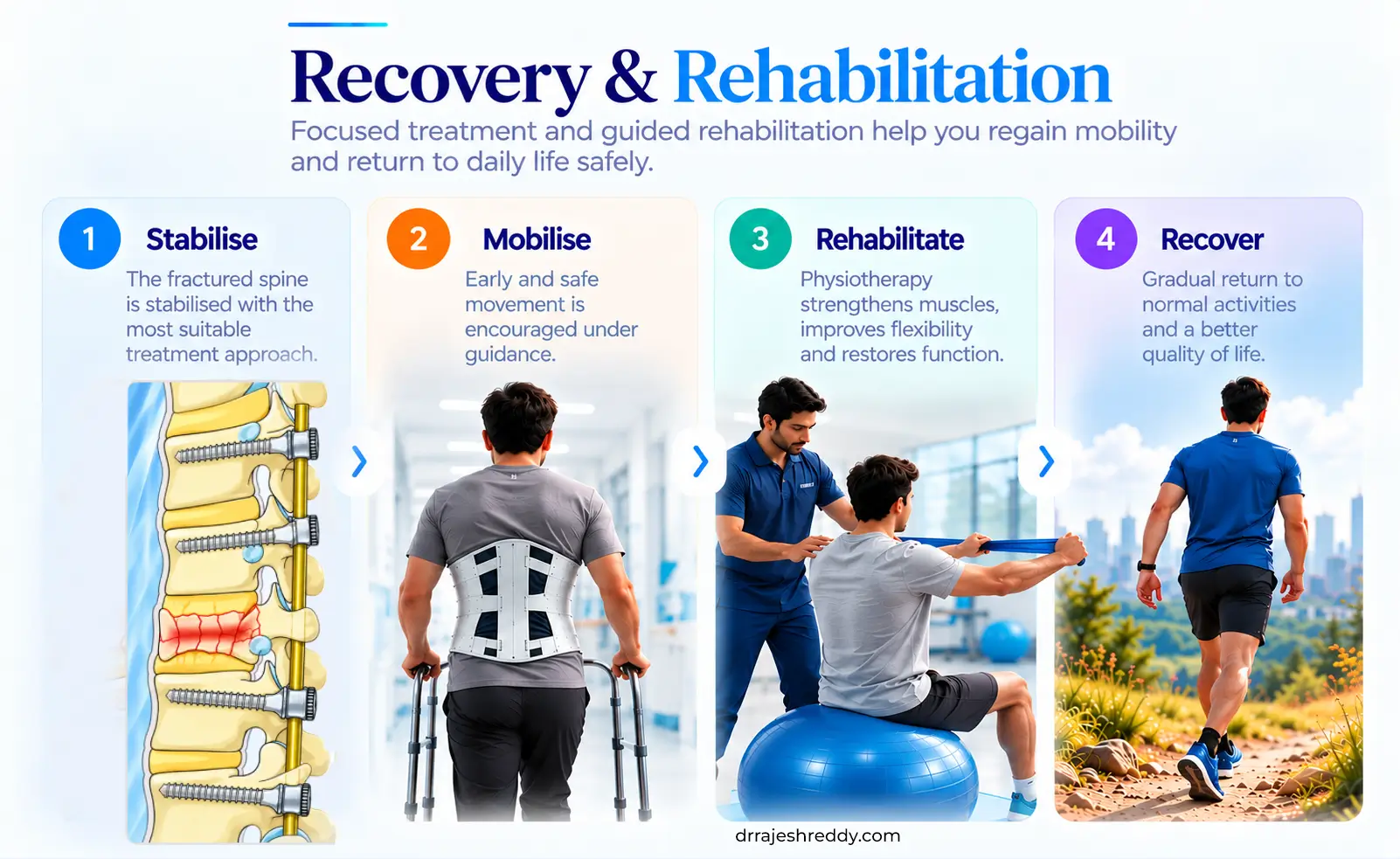
After Conservative Management (Bracing)
- Immobilisation period: 6–12 weeks depending on fracture type and location
- Serial X-rays at 6 weeks and 12 weeks to confirm healing and absence of progressive deformity
- Physiotherapy commences from week 2–4 — initially gentle core activation and mobilisation within brace
- Return to light activity: 6–8 weeks; strenuous activity and heavy lifting: 3–6 months
After Vertebroplasty / Kyphoplasty
- Hospital stay: day case or overnight
- Pain relief: often dramatic — within 24–48 hours of cement injection
- Mobilisation: walking permitted the same day or next morning
- Return to normal activity: 1–2 weeks; avoid heavy lifting for 4–6 weeks
- Osteoporosis treatment: commenced or optimised before or at the time of the procedure
After Minimally Invasive Fixation (MISS)
- Hospital stay: 2–4 days
- Mobilisation: walking with physiotherapy support from day 1–2
- Brace: TLSO brace for 6–12 weeks depending on fracture type
- Return to desk work: 4–6 weeks; physically demanding work: 3–6 months
- Implant removal: generally not required; screws are typically left permanently
After Open Decompression and Fusion
- Hospital stay: 5–7 days; longer after combined anterior-posterior procedures
- Mobilisation with physiotherapy: from day 1–2
- Post-operative brace: typically 6–12 weeks
- Neurological recovery: incomplete SCI — improvement may occur over 12–24 months; rehabilitation is the mainstay
- Return to work: 3–6 months for desk work; 6–12 months for physical work
Why Choose Dr. Rajesh Reddy for Spine Fracture Treatment in Hyderabad?
Spine fracture management — particularly in the context of neurological injury — demands a neurosurgeon who understands both the spinal biomechanics and the neurological consequences of injury, and who can offer the full spectrum of surgical approaches without defaulting to a single technique. Dr. Rajesh Reddy's training and practice span the complete range:
Spine Fracture Expertise — Dr. Rajesh Reddy
- MCh Neurosurgery (Exam Topper), NIMS Hyderabad; DNB Neurosurgery — Double board certified
- Visiting Scholar — Barrow Neurological Institute, Phoenix (advanced spine trauma); Okayama University, Japan; Klinikum Stuttgart, Germany (minimally invasive spine techniques)
- Minimally invasive spine surgery — percutaneous pedicle screw fixation, endoscopic discectomy, vertebroplasty and kyphoplasty
- Open spine surgery — anterior cervical decompression and fusion, posterior cervical and thoracolumbar fixation, corpectomy and cage reconstruction, laminectomy and decompression
- Emergency spinal cord decompression — accepts acute SCI transfers from hospitals across Telangana, Andhra Pradesh, Karnataka, Maharashtra, and Chhattisgarh
- Pathological fracture management — combined surgical and oncological approach for metastatic cord compression
- Intraoperative neurophysiological monitoring — MEPs and SSEPs used routinely during spinal cord surgery to protect neural function
- Over 5,000 neurosurgical procedures across a 15+ year career
- Clinical acumen and judgement gained through years of training with leading global experts
Consult Dr. Rajesh Reddy for Spine Fracture Treatment in Hyderabad
Whether you are managing a recent spinal fracture, seeking a second opinion on treatment options, or require emergency assessment for spinal cord injury or metastatic cord compression — Dr. Rajesh Reddy Sannareddy provides comprehensive spinal fracture consultations with the full range of surgical and non-surgical treatment options.
Book via the Dr. Reddy's Neuro Care app on Google Play or call 079951 11695.
Spinal Cord Injury Self Help Group
Are you interested in volunteering to support patients and families navigating a spinal cord injury or spinal fracture diagnosis? Reach out to learn how you can make a difference at a patient's most vulnerable moment.