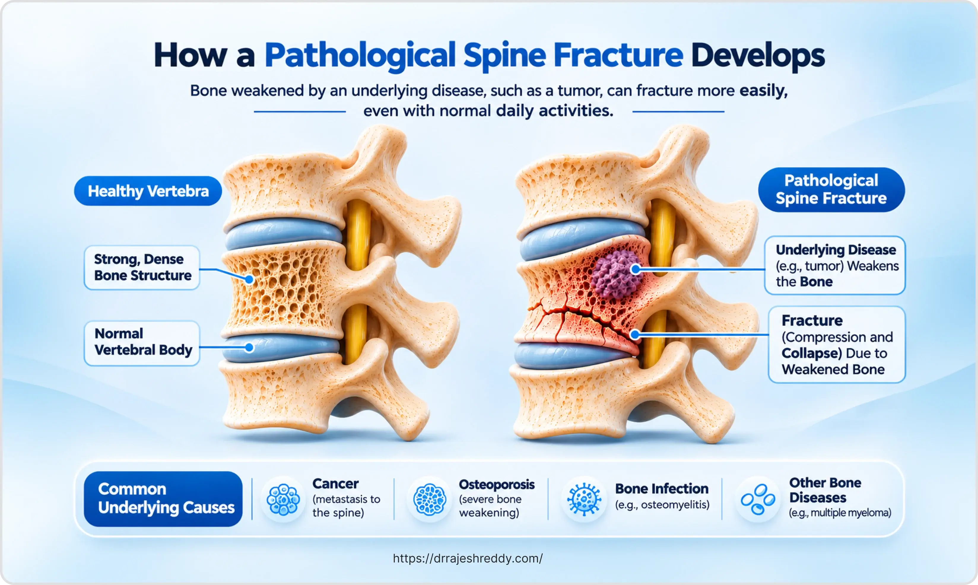
Not every broken vertebra is the result of a fall or a road accident. A significant proportion of spine fractures occur in bone that has been weakened by an underlying disease — most commonly osteoporosis or cancer — where normal daily activities, minor trips, or even a cough or sneeze can be enough to cause a vertebral body to collapse. These are pathological fractures: fractures occurring through abnormal bone, where the real problem is not the fracture itself but the condition that made the bone vulnerable.
The implications of a pathological fracture are fundamentally different from those of a traumatic fracture. Treatment cannot be directed at the fracture in isolation — the underlying disease must be identified, staged, and addressed alongside any surgical intervention. A patient with a painful vertebral compression fracture from osteoporosis requires not just cement augmentation to stabilise the vertebra, but investigation and treatment of the osteoporosis itself to prevent further fractures. A patient with a metastatic vertebral fracture requires not only spine stabilisation but integration of systemic oncological treatment and, in most cases, stereotactic radiotherapy.
Dr. Rajesh Reddy Sannareddy, Senior Consultant Neurosurgeon in Hyderabad, manages pathological spine fractures across the full spectrum — from osteoporotic compression fractures treated with minimally invasive kyphoplasty to complex metastatic spinal cord compression requiring emergency decompression, fixation, and coordination with radiation and medical oncology. His approach recognises that the spine surgeon's role in pathological fracture management is one part of a multidisciplinary treatment plan, not a standalone intervention.
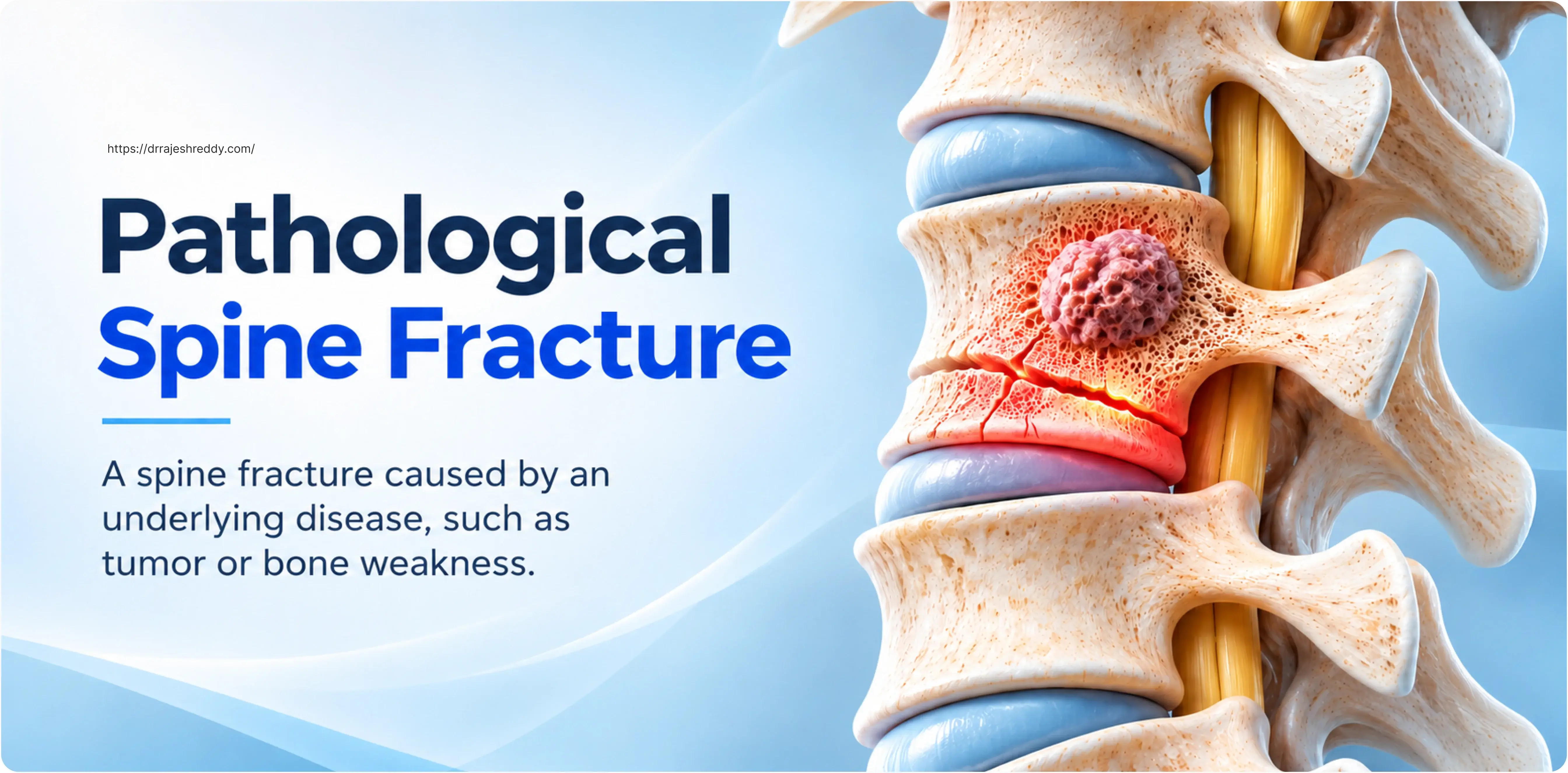
What Is a Pathological Fracture?
A pathological fracture is a fracture that occurs through bone that has been weakened by an underlying pathological process — as opposed to a traumatic fracture, which occurs through previously normal bone subjected to a force exceeding its structural limit. The distinction matters clinically because:
- The fracture may occur with minimal or no trauma — sometimes spontaneously.
- The fractured bone does not heal normally without addressing the underlying cause.
- Multiple vertebral levels may be affected simultaneously or sequentially.
- Treatment must address both the mechanical failure and the underlying disease.
- Prognosis depends heavily on the nature and stage of the underlying condition.
The spine is the most common site of pathological fracture in the body. This is partly because vertebrae are composed largely of cancellous (spongy) bone — the trabecular architecture of which is highly vulnerable to both the trabecular thinning of osteoporosis and the osteolytic destruction of metastatic disease — and partly because the axial skeleton bears the body's gravitational load throughout the day.
Causes of Pathological Spine Fractures
Tuberculosis of the spine — Pott's disease — remains one of the most important causes of pathological vertebral fracture in India and across South Asia. It typically involves two adjacent vertebrae and the intervening disc, with bone destruction, disc space collapse, and the formation of a paravertebral 'cold abscess'. Neurological complications, including spinal cord compression and paraplegia, occur in up to 30% of cases without adequate treatment. Dr. Rajesh Reddy manages Pott's disease with anti-tubercular chemotherapy as the primary treatment, reserving surgery for cases with neurological deficit, structural instability, or failure to respond to medical management.
Recognising a Pathological Spine Fracture: Symptoms and Red Flags
Pathological vertebral fractures often present differently from traumatic fractures, and a high index of suspicion is essential — particularly in patients with known risk factors.
Symptoms
- Sudden-onset severe back or neck pain, often without a significant injury.
- Progressive back pain that worsens over days to weeks — unlike mechanical back pain, which is typically episodic.
- Pain that is worse at rest or at night — a classic feature of bone pain from malignancy, unlike degenerative pain which is worse with activity.
- Neurological symptoms — leg weakness, numbness, difficulty walking, or bladder/bowel dysfunction — suggesting spinal cord or nerve root compression.
- Height loss and increasing thoracic kyphosis (dowager's hump) in elderly women — suggesting multiple osteoporotic compression fractures.
- Constitutional symptoms in oncological fractures — weight loss, fatigue, loss of appetite — suggesting active malignancy.
Red Flags That Should Prompt Urgent Assessment
Any patient over 50 presenting with new-onset back pain and no clear mechanical cause, any patient with a known cancer history developing new spinal pain, and any patient with progressive neurological symptoms should be assessed urgently with MRI of the spine. A 'simple' vertebral fracture on X-ray in a middle-aged or elderly patient without a significant fall is a pathological fracture until proven otherwise.
Diagnosis: What Imaging Is Needed?
- Plain X-ray: First-line; detects vertebral height loss, endplate fractures, and deformity. Insensitive for early metastatic disease.
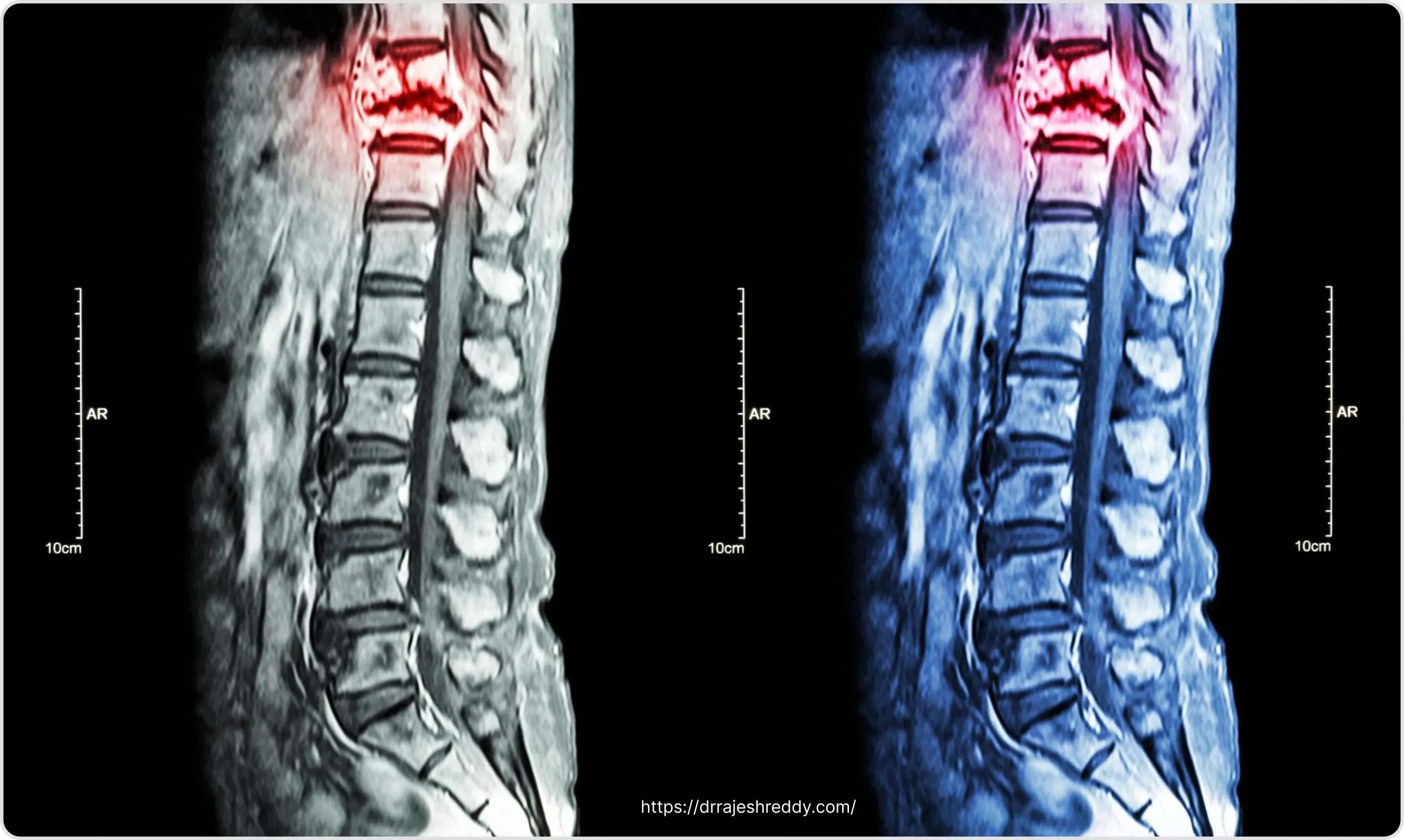
- MRI spine: The gold standard for pathological fractures — identifies bone marrow infiltration, epidural compression, cord signal change, and distinguishes acute from chronic fractures. Whole-spine MRI is essential when malignancy is suspected.
- CT spine: Better than MRI for assessing bony architecture — pedicle integrity, cortical destruction, and fracture morphology for surgical planning.
- Bone scan (99mTc scintigraphy) / PET-CT: Detects additional skeletal metastases; PET-CT is superior for staging and detecting metabolically active disease.
- DEXA scan: Measures bone mineral density (BMD) and is essential for diagnosing osteoporosis and monitoring treatment response.
- Biopsy: Required when the primary tumour is unknown; CT-guided percutaneous biopsy of the vertebral lesion provides tissue diagnosis.
Assessing Spinal Instability: The SINS Score
In pathological fractures from cancer, a critical clinical question is whether the spine is mechanically unstable — at risk of progressive collapse that will cause pain, deformity, or neurological injury if not surgically stabilised. The Spinal Instability Neoplastic Score (SINS) was developed by the Spine Oncology Study Group to standardise this assessment and guide surgical referral decisions.
| SINS Component | Score | Clinical Relevance |
|---|---|---|
| Spinal location | Junctional = 3; Mobile = 2; Semirigid = 1; Rigid = 0 | Junctional zones bear greater biomechanical stress and are inherently less stable when diseased |
| Pain | Mechanical = 3; Occasional non-mechanical = 1; None = 0 | Mechanical pain indicates the vertebra is failing under load |
| Bone lesion quality | Lytic = 2; Mixed = 1; Blastic = 0 | Lytic lesions destroy load-bearing bone more severely |
| Radiographic spinal alignment | Subluxation = 4; De novo deformity = 2; Normal = 0 | Deformity indicates progressive collapse likely without intervention |
| Vertebral body collapse | >50% = 3; <50% = 2; No collapse but >50% body involved = 1; None = 0 | Collapse quantifies structural failure already present |
| Posterolateral element involvement | Bilateral = 3; Unilateral = 1; None = 0 | Posterior element involvement removes the secondary stabilising structures |
Total SINS Score Interpretation: 0–6 = Stable; 7–12 = Potentially unstable; 13–18 = Unstable. The SINS score is completed by the treating clinician or radiologist reviewing the patient's MRI and CT, and provides a structured basis for surgical consultation. A score of 7 or above warrants referral to a spine surgeon for assessment of the need for stabilisation, even if the patient is neurologically intact.
The NOMS Framework: Decision-Making for Spinal Metastases
For metastatic spine fractures specifically, the Neurological-Oncological-Mechanical-Systemic (NOMS) framework — developed at Memorial Sloan Kettering Cancer Center — provides the most comprehensive decision-making structure. It integrates four domains:
- Neurological (N): Is there spinal cord or nerve root compression? High-grade epidural compression causing myelopathy requires emergency surgical decompression; low-grade compression may be managed with radiotherapy alone.
- Oncological (O): How radiosensitive is the tumour? Highly radiosensitive tumours respond to conventional radiotherapy; radioresistant tumours require high-dose stereotactic radiotherapy (SBRT) or surgical resection.
- Mechanical (M): Is the spine mechanically stable? SINS score guides this assessment — instability requires surgical fixation regardless of neurological or oncological status.
- Systemic (S): What is the patient's overall health, performance status, and systemic disease burden? A patient with poor performance status may not be a surgical candidate.
Dr. Rajesh Reddy applies the NOMS framework in collaboration with the patient's oncologist, radiation oncologist, and palliative care team in a multidisciplinary tumour board setting. This ensures that each patient receives a coherent, integrated treatment plan rather than a series of disconnected interventions.
Treatment Options for Pathological Spine Fractures
The available treatments span a spectrum from non-invasive to highly complex surgical procedures. The appropriate choice depends on the cause of the fracture, the degree of instability, the presence and severity of neurological compromise, and the patient's overall condition and expected survival.
| Treatment | Best Indication | What It Achieves |
|---|---|---|
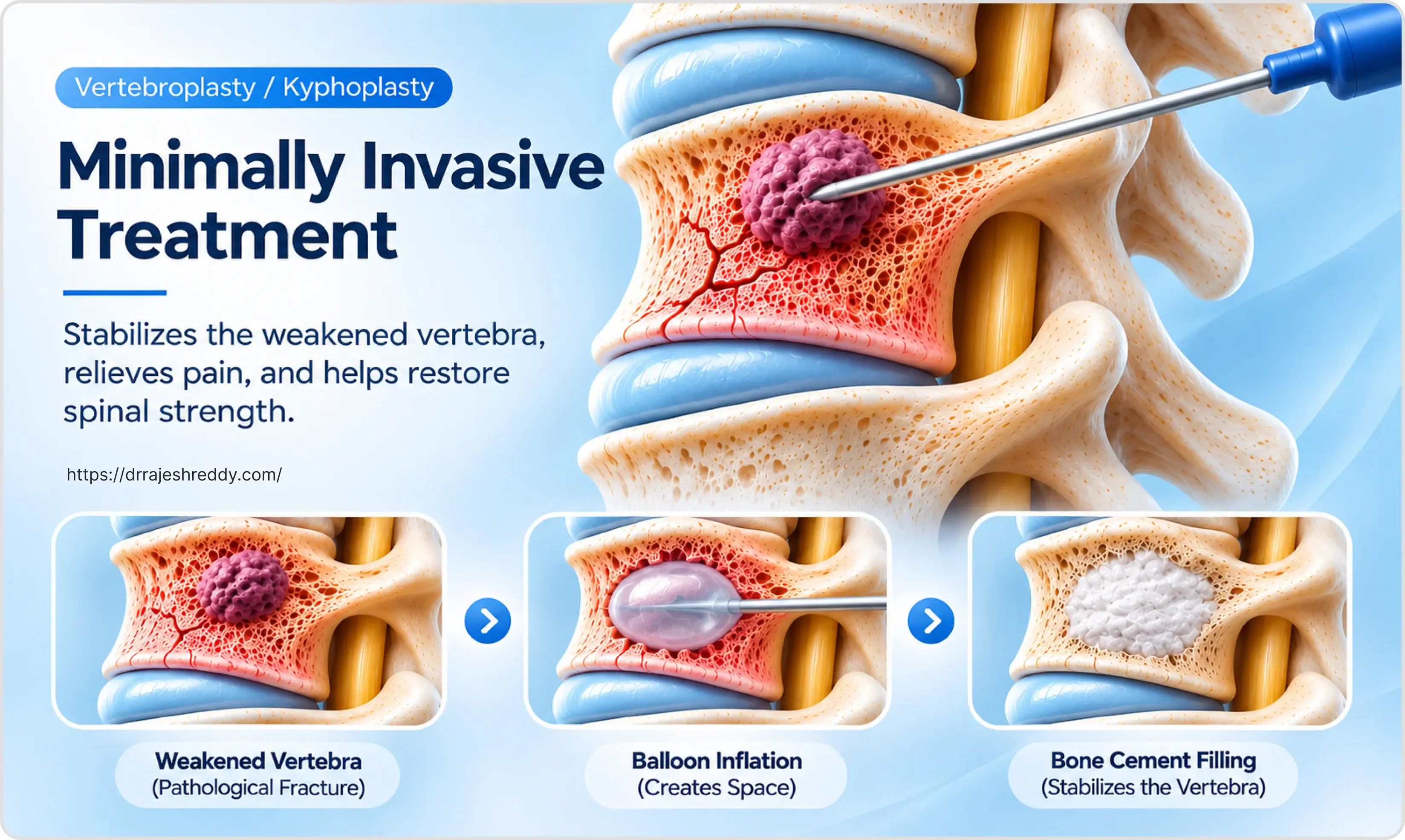
| Balloon kyphoplasty / vertebroplasty | Painful osteoporotic or metastatic compression fracture; stable neurological status; good cement fill expected | Pain relief, height restoration, mechanical stabilisation, rapid recovery |
| Percutaneous pedicle screw fixation | Metastatic fracture with instability; osteoporotic fracture with posterior column disruption | Immediate mechanical stability across fracture, low blood loss, short hospital stay |
| Separation surgery (decompression + posterior fixation) | Epidural spinal cord compression from metastasis; neurological deficit; planned SBRT | Direct decompression and a safe separation between tumour and cord for high-dose radiotherapy |
| En bloc spondylectomy | Solitary spinal metastasis with long expected survival; selected primary spinal tumours | Complete resection of involved vertebra in one piece; potentially curative for primary tumours |
| Stereotactic body radiotherapy (SBRT) | Post-operative tumour control; patients unfit for surgery; oligometastatic disease | Ablative high-dose radiotherapy to tumour while sparing spinal cord |
| Systemic treatment | All patients with osteoporosis or bone metastases | Reduces further fracture risk and treats the underlying disease |
| Bracing / orthotics | Mild osteoporotic fractures; post-operative support | Reduces pain by limiting spinal movement, but not curative |
For patients with metastatic spinal cord compression — the most urgent presentation in spinal oncology — the landmark Patchell trial (Lancet, 2005) demonstrated that direct decompressive surgery followed by radiotherapy was significantly superior to radiotherapy alone: patients treated with surgery were more likely to regain and retain the ability to walk and had better continence outcomes and quality of life. This evidence forms the basis for the modern surgical approach to metastatic epidural cord compression.
Osteoporotic Spine Fractures: A Specific Focus
Osteoporosis — a systemic condition of low bone mass and structural deterioration of bone tissue — affects an estimated 200 million people worldwide and is the leading cause of vertebral compression fractures in patients over 50. In India, the burden is substantial: estimates suggest that 46 million women over 50 have osteoporosis or osteopenia, and vertebral fractures are significantly underdiagnosed because many are attributed to 'normal ageing' without specific investigation.
Why Osteoporotic Fractures Are Underdiagnosed
The majority of osteoporotic vertebral fractures — estimated at 70% — are not brought to medical attention. Patients often attribute their pain to arthritis or muscle strain; many fractures are identified incidentally on imaging performed for another indication. This underdiagnosis has serious consequences: each vertebral fracture increases the risk of a subsequent fracture fivefold in the following year — a phenomenon known as the 'fracture cascade'. A patient who suffers one osteoporotic vertebral fracture without receiving anti-osteoporosis treatment is highly likely to suffer further fractures, progressive kyphosis, height loss, and functional decline.
The Fracture Liaison Service Principle
Best-practice management of osteoporotic fractures goes beyond treating the immediate fracture — it involves identifying and treating the osteoporosis to prevent the next fracture. Dr. Rajesh Reddy's management of osteoporotic vertebral fractures therefore includes:
- DEXA scan to quantify bone mineral density (T-score) and confirm osteoporosis.
- Investigation for secondary causes — vitamin D deficiency, calcium malabsorption, hyperparathyroidism, thyroid disease, hypogonadism, and steroid use.
- Anti-osteoporosis pharmacotherapy — bisphosphonates, denosumab, teriparatide, or romosozumab for severe or refractory cases.
- Calcium and vitamin D supplementation as standard adjunct.
- Falls prevention programme — physiotherapy, environmental modification, and review of medications that increase fall risk.
Frequently Asked Questions
Yes — back pain in a person with osteoporosis should be taken seriously, even without a history of a fall or injury. Osteoporotic vertebral fractures can occur with minimal provocation — lifting a bag, twisting suddenly, or even just sitting up from a chair. The key symptoms that should prompt an MRI are pain that is worse on standing and moving, pain at a specific spinal level that is tender to pressure, or any neurological symptoms such as leg weakness or numbness. An X-ray may show the fracture, but MRI is more sensitive for detecting recent fractures and ruling out malignancy as a cause.
New back pain in a patient with a known cancer history should be investigated urgently, as spinal metastases are common in many cancer types — particularly lung, breast, prostate, kidney, and thyroid cancer. The pain may be from bone involvement, a fracture, or early spinal cord compression. MRI of the whole spine is the preferred investigation. Early diagnosis and treatment of spinal metastases — before neurological deficit occurs — is associated with much better outcomes, including the ability to walk. Please do not delay seeking assessment.
Kyphoplasty and vertebroplasty are cement augmentation procedures — they stabilise the fractured vertebra by filling it with bone cement. They are highly effective for osteoporotic fractures and for select metastatic fractures where the goal is pain relief and mechanical stabilisation. They do not remove tumour or provide decompression of the spinal cord. Cancer fractures with neurological compromise or significant spinal cord compression require surgical decompression and fixation — a much more involved procedure. The choice between the two depends on the neurological status, the degree of instability (SINS score), and the overall oncological situation.
Kyphoplasty is typically performed as a day procedure or with one night in hospital. Most patients notice a significant reduction in pain within 24–48 hours — often described as dramatic. Light activity can resume within a few days; return to normal daily activities typically occurs within 1–2 weeks. The treated vertebra is permanently stabilised by the cement, so the fracture does not recur. However, adjacent vertebrae remain at risk of fracture if the osteoporosis is not treated — which is why medical treatment of the osteoporosis is just as important as the procedural intervention.
Yes, in many cases — but the outcome depends critically on how quickly it is identified and treated. Patients with mild neurological symptoms who receive prompt decompression and radiotherapy have the best chance of neurological recovery. Patients who have been completely paralysed for more than 24–48 hours before decompression have a much lower chance of meaningful recovery. This is why spinal cord compression from cancer is a medical emergency — any patient with cancer who develops leg weakness, difficulty walking, or bladder dysfunction should seek emergency assessment without delay.
It depends on the patient's overall condition, performance status, and expected survival. For patients with good performance status and reasonable expected survival, spine surgery — particularly minimally invasive stabilisation or separation surgery — is well-tolerated and significantly improves quality of life by relieving pain and preserving neurological function. For patients with end-stage disease, multiple organ involvement, or very poor performance status, the risks of surgery may outweigh the benefits, and palliative management with pain medication, bracing, and radiotherapy may be the most appropriate approach.
Consult Dr. Rajesh Reddy for Pathological Spine Fracture Management in Hyderabad
Whether you have been diagnosed with an osteoporotic vertebral fracture, have a cancer history and are experiencing spinal pain, or have been referred for assessment of spinal instability, Dr. Rajesh Reddy Sannareddy provides expert pathological fracture consultations in Hyderabad.
Book via the Dr. Reddy's Neuro Care app on Google Play or call 9000224265.
References
The clinical statements on this page are supported by peer-reviewed literature. Links open the publisher record for verification.
- Fisher CG, et al. A novel classification system for spinal instability in neoplastic disease: an evidence-based approach and expert consensus from the Spine Oncology Study Group. Spine. 2010;35(22):E1221–E1229. → https://doi.org/10.1097/BRS.0b013e3181e16ae2
- Patchell RA, et al. Direct decompressive surgical resection in the treatment of spinal cord compression caused by metastatic cancer: a randomised trial. Lancet. 2005;366(9486):643–648. → https://doi.org/10.1016/S0140-6736(05)66954-1
- Wardlaw D, et al. Efficacy and safety of balloon kyphoplasty compared with non-surgical care for vertebral compression fracture (FREE): a randomised controlled trial. Lancet. 2009;373(9668):1016–1024. → https://pubmed.ncbi.nlm.nih.gov/19246088/
- Laufer I, et al. The NOMS framework: approach to the treatment of spinal metastatic tumors. Oncologist. 2013;18(6):744–751. → https://pmc.ncbi.nlm.nih.gov/articles/PMC4063402/
- Kanis JA, et al. European guidance for the diagnosis and management of osteoporosis in postmenopausal women. Osteoporos Int. 2019;30(1):3–44. → https://pmc.ncbi.nlm.nih.gov/articles/PMC7026233/
- Fehlings MG, et al. A clinical practice guideline for the management of patients with degenerative cervical myelopathy and cervical radiculopathy. Global Spine J. 2017;7(3 Suppl):1S–4S. → https://pubmed.ncbi.nlm.nih.gov/29164035/